. 2024 Dec 19:2024:1672096.
doi: 10.1155/ije/1672096. eCollection 2024.

Angiotensin (1-7) Improves Pancreatic Islet Function via Upregulating PDX-1 and GCK: A Dose-Dependent Study in Mice

Affiliations

Angiotensin (1-7) Improves Pancreatic Islet Function via Upregulating PDX-1 and GCK: A Dose-Dependent Study in Mice

Ziwei Lin et al. Int J Endocrinol. .

Abstract

Purpose: This study aimed to verify the effect of angiotensin (1-7) on improving islet function and further explore the signaling pathway that may be involved in this improvement. It also aimed to explore the effects of angiotensin (1-7) on blood glucose levels, islet function, and morphological changes in db/db mice and its potential signal pathway. Methods: Forty-five db/db mice were divided randomly into a model control group and different doses of angiotensin (1-7) intervention groups (0, 150, 300, and 600 μg/kg/d), while seven db/m mice were assigned as the normal control group. The angiotensin (1-7) intervention groups received daily intraperitoneal administration for 8 weeks, whereas the normal control group was injected intraperitoneally with an equal volume of normal saline every day for 8 weeks. Changes in weight and food intake of mice were detected. Effect of angiotensin (1-7) on lipid metabolism, islet function, the morphology of pancreatic islets, and β-cell mass on mice were evaluated. The expression of PDX-1 and GCK in pancreatic tissue was verified. Results: The group receiving angiotensin (1-7) at a dosage of 600 μg/kg/d showed a significant decrease in body weight, triglyceride levels, and fasting blood glucose, along with an improvement in glucose tolerance. In the 300 μg/kg/d group, angiotensin (1-7) tended to increase the total volume of islets. Moreover, the intervention groups exhibited a significant increase in the ratio of β cells, small islets (30-80 μm in diameter), as well as the expression levels of PDX-1 and GCK in pancreatic tissue. Conclusion: Angiotensin (1-7) could improve glucose and lipid metabolism and islet function by promoting the expression of PDX-1 and GCK genes in the pancreas of db/db mice.

Keywords: GCK; PDX-1; angiotensin (1–7); diabetes mellitus; islet cells.


Conflict of interest statement

The authors declare no conflicts of interest.

Figures

Figure 1
Figure 1
Changes in weight and food intake of mice. (a) Line chart of food intake; (b) histogram total food intake; (c) line chart of body weight; (d) histogram of body weight. Bars represent the mean ± SD. Compared to control: ⁣p < 0.05, ⁣∗∗∗p < 0.001. n = 7–11 for each group.
Figure 2
Figure 2
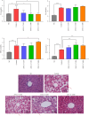
Effect of angiotensin (1–7) on lipid metabolism in db/db mice. (a) Serum triglycerides (TG) in different groups after intervention. (b) Serum total cholesterol (TC) in different groups after intervention. (c) Serum high-density lipoprotein (HDL) in different groups after intervention. (d) Serum low-density lipoprotein (LDL) in different groups after intervention. Each bar represents the mean ± SD. Compared to control: ⁣p < 0.05, ⁣∗∗p < 0.01, ⁣∗∗∗p < 0.001. n = 4 for each group. (e) Representative image of liver tissue stained with HE from NC group, control group, ANG-(1–7)150 group, ANG-(1–7)300 group, and ANG-(1–7)600 group. Original magnification ×400.
Figure 3
Figure 3
The effect of angiotensin (1–7) on islet function in db/db mice. (a) The fasting blood glucose of mice under different interventions. n = 7–11 for each group. (b) The intraperitoneal insulin tolerance test (IPITT) results and the area under the curve (AUC) of IPITT. n = 7–11 for each group. (c) The insulin tolerance test (ITT) results and the area under the curve (AUC) of ITT. n = 7–11 for each group. (d) Quantification of serum insulin in db/db mice under different interventions. n = 3 for each group. (e) The homeostasis model assessment of insulin resistance (Homa-IR). n = 3 for each group. Bars represent the mean ± SD. Compared to control: ⁣p < 0.05, ⁣∗∗p < 0.01, ⁣∗∗∗p < 0.001.
Figure 4
Figure 4
The effect of angiotensin on the morphology of pancreatic islets in db/db mice. (a) Representative image of pancreas tissues stained with HE from the control group, ANG-(1–7)150 group, ANG-(1–7)300 group, and ANG-(1–7)600 group. Original magnification ×100. (b) The counts of pancreatic islets under different interventions. (c) Quantification of total islet mass under different interventions. (d) The distribution of islet size in db/db mice with different interventions. Bars represent the mean ± SD. Compared to control: ⁣p < 0.05, ⁣∗∗p < 0.01, ⁣∗∗∗p < 0.001. n = 5 for each group.
Figure 5
Figure 5
Angiotensin (1–7) increased β-cell mass in db/db mice. (a) Quantification of the β-cell ratio of islets in db/db mice; (b) histogram of β-cell mass; (c) immunofluorescence staining of insulin (green) was performed on the β-cells. Original magnification ×400. Each bar represents the mean ± SD. Compared to control: ⁣p < 0.05, ⁣∗∗∗p < 0.001. n = 5 for each group.
Figure 6
Figure 6
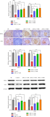
The expression of PDX-1 and GCK in db/db mice. (a) Relative expression of PDX-1 and GCK mRNA in the pancreas. n = 3 for each group. (b) Immunohistochemical staining of PDX-1, GCK in islet cells of mice. Original magnification ×400. n = 5 for each group. (c) Semiquantitative comparison of staining intensity of PDX-1 and GCK in islet cells of mice, which was determined by Image-Pro Plus. n = 5 for each group. (d) The expression of PDX-1 and GCK protein in the pancreas. (e) Semiquantification comparison of relative expression of PDX-1 and GCK, which was determined by Image-Pro Plus. n = 3 for each group. Bars represent the mean ± SD. Compared to control: ⁣p < 0.05, ⁣∗∗p < 0.01, ⁣∗∗∗p < 0.001.

References

    1. Hill M. A., Yang Y., Zhang L., et al. Insulin Resistance, Cardiovascular Stiffening and Cardiovascular Disease. Metabolism Clinical and Experimental . 2021;119:p. 154766. doi: 10.1016/j.metabol.2021.154766. - DOI - PubMed
    1. Demir S., Nawroth P. P., Herzig S., Ekim Üstünel B. Emerging Targets in Type 2 Diabetes and Diabetic Complications. Advanced Science . 2021;8(18):p. e2100275. doi: 10.1002/advs.202100275. - DOI - PMC - PubMed
    1. Head G. A. Cardiovascular and Metabolic Consequences of Obesity. Frontiers in Physiology . 2015;6:p. 32. doi: 10.3389/fphys.2015.00032. - DOI - PMC - PubMed
    1. Regoli M., Bendayan M., Fonzi L., Sernia C., Bertelli E. Angiotensinogen Localization and Secretion in the Rat Pancreas. Journal of Endocrinology . 2003;179(1):81–89. doi: 10.1677/joe.0.1790081. - DOI - PubMed
    1. Leung P. S., Chappell M. C. A Local Pancreatic Renin-Angiotensin System: Endocrine and Exocrine Roles. The International Journal of Biochemistry & Cell Biology . 2003;35(6):838–846. doi: 10.1016/s1357-2725(02)00179-6. - DOI - PubMed

LinkOut - more resources