

Severe Diffuse Ulcerative Esophagitis Caused by Epstein-Barr Virus/Cytomegalovirus Coinfection in an Immunocompetent Individual
- PMID: 39734390
- PMCID: PMC11671085
- DOI: 10.14309/crj.0000000000001578
Severe Diffuse Ulcerative Esophagitis Caused by Epstein-Barr Virus/Cytomegalovirus Coinfection in an Immunocompetent Individual
Abstract
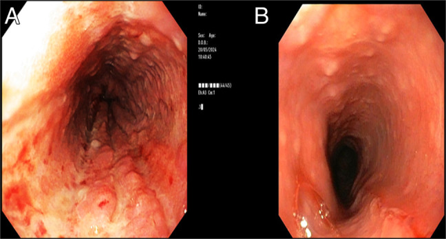
Cytomegalovirus (CMV) and Epstein-Barr virus (EBV) are important causes of viral esophagitis mainly in immunocompromised individuals. Both viruses lead to development of focal ulcerations in the esophagus. While there have been rare case reports of esophagitis in immunocompetent individuals, there has not been a single reported case of coinfection with both CMV and EBV in an immunocompetent individual and presenting with diffuse esophageal ulceration. We report a case of severe diffuse ulcerative esophagitis caused by EBV/CMV coinfection in an immunocompetent individual.
Keywords: Epstein-Barr virus; cytomegalovirus; esophagitis; infection; viral esophagitis.
© 2024 The Author(s). Published by Wolters Kluwer Health, Inc. on behalf of The American College of Gastroenterology.
Figures

Similar articles
-
Epidemiological and Liver Biomarkers Profile of Epstein-Barr Virus Infection and Its Coinfection with Cytomegalovirus in Patients with Hematological Diseases.Biomolecules. 2021 Aug 4;11(8):1151. doi: 10.3390/biom11081151. Biomolecules. 2021. PMID: 34439817 Free PMC article.
-
Unusual Epstein-Barr esophageal infection in an immunocompetent patient: a case report.J Med Case Rep. 2009 Jun 29;3:7314. doi: 10.4076/1752-1947-3-7314. J Med Case Rep. 2009. PMID: 19830180 Free PMC article.
-
[Viral Infection in Upper Gastrointestinal Tract].Korean J Helicobacter Up Gastrointest Res. 2024 Jun;24(2):122-126. doi: 10.7704/kjhugr.2024.0027. Epub 2024 Jun 10. Korean J Helicobacter Up Gastrointest Res. 2024. PMID: 40502839 Free PMC article. Review. Korean.
-
Evaluation of a multiplex PCR assay for detection of cytomegalovirus in stool samples from patients with ulcerative colitis.World J Gastroenterol. 2015 Nov 28;21(44):12667-75. doi: 10.3748/wjg.v21.i44.12667. World J Gastroenterol. 2015. PMID: 26640344 Free PMC article.
-
Epstein-Barr Virus and Cytomegalovirus Infections of the Liver.Gastroenterol Clin North Am. 2020 Jun;49(2):331-346. doi: 10.1016/j.gtc.2020.01.008. Gastroenterol Clin North Am. 2020. PMID: 32389366 Review.
References
-
- Wang HW, Kuo CJ, Lin WR, et al. . The clinical characteristics and manifestations of cytomegalovirus esophagitis. Dis Esophagus. 2016;29(4):392–9. - PubMed
-
- Hoversten P, Katzka D, Halland M. Infections of the esophagus: An update on risk factors, diagnosis, and management. Dis Esophagus. 2018;31(Suppl 1):65. - PubMed
-
- Hoversten P, Kamboj AK, Wu TT, Katzka DA. Risk factors, endoscopic features, and clinical outcomes of cytomegalovirus esophagitis based on a 10-year analysis at a single center. Clin Gastroenterol Hepatol. 2020;18(3):736–8. - PubMed
-
- Iwamuro M, Kondo E, Tanaka T, et al. . Endoscopic manifestations and clinical characteristics of cytomegalovirus infection in the upper gastrointestinal tract. Acta Med Okayama. 2017;71(2):97–104. - PubMed
-
- Cohen JI. Epstein–barr virus infection. N Engl J Med. 2000;343(7):481–92. - PubMed
Publication types
LinkOut - more resources
Full Text Sources
