. 2024 Dec 26;16(12):1022-1046.
doi: 10.4252/wjsc.v16.i12.1022.

Exosomes derived from microRNA-540-3p overexpressing mesenchymal stem cells promote immune tolerance via the CD74/nuclear factor-kappaB pathway in cardiac allograft

Affiliations

Exosomes derived from microRNA-540-3p overexpressing mesenchymal stem cells promote immune tolerance via the CD74/nuclear factor-kappaB pathway in cardiac allograft

Ji-Gang He et al. World J Stem Cells. .

Abstract

Background: Heart transplantation is a crucial intervention for severe heart failure, yet the challenge of organ rejection is significant. Bone marrow mesenchymal stem cells (BMSCs) and their exosomes have demonstrated potential in modulating T cells, dendtitic cells (DCs), and cytokines to achieve immunomodulatory effects. DCs, as key antigen-presenting cells, play a critical role in shaping immune responses by influencing T-cell activation and cytokine production. Through this modulation, BMSCs and their exosomes enhance graft tolerance and prolonging survival.

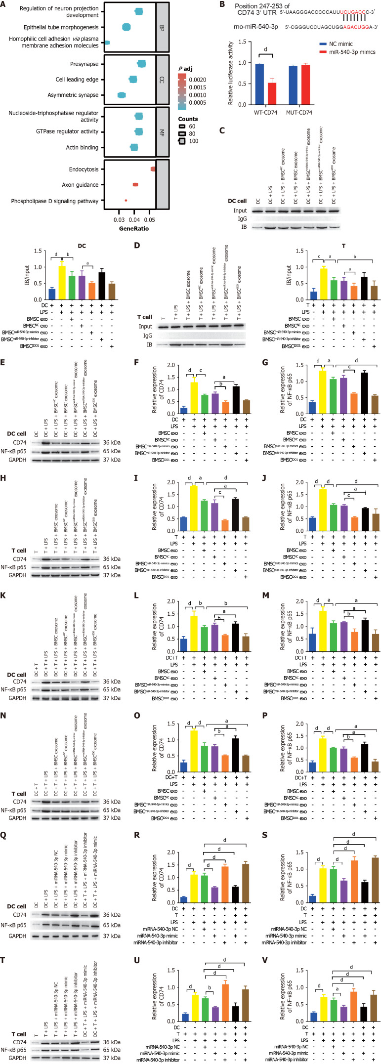
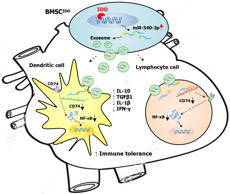
Aim: To explore the immunomodulatory effects of exosomes derived from BMSCs overexpressing microRNA-540-3p (miR-540-3p) on cardiac allograft tolerance, focusing on how these exosomes modulating DCs and T cells activity through the CD74/nuclear factor-kappaB (NF-κB) pathway.

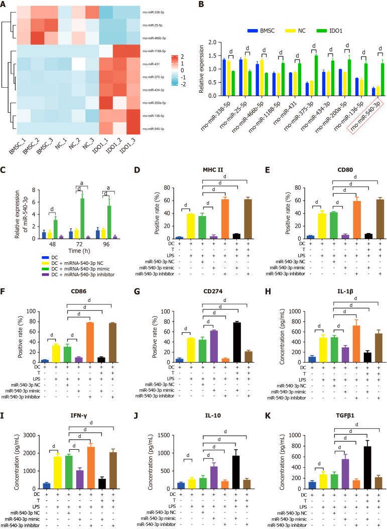
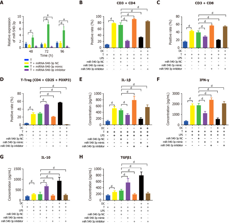
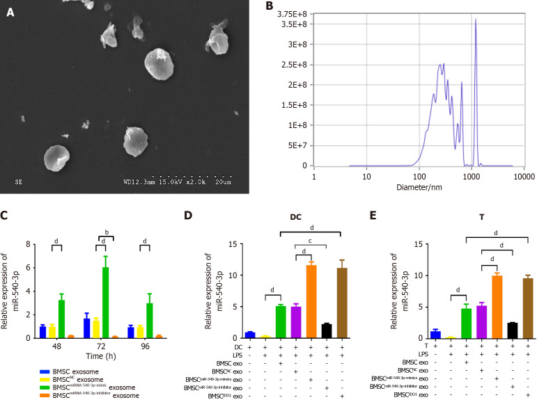
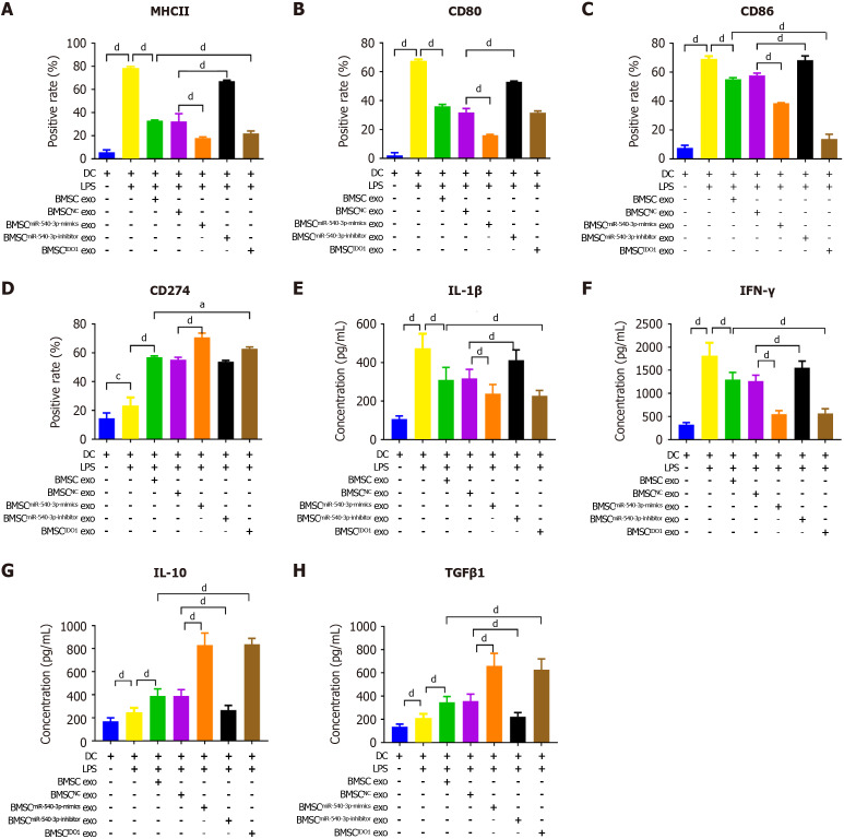
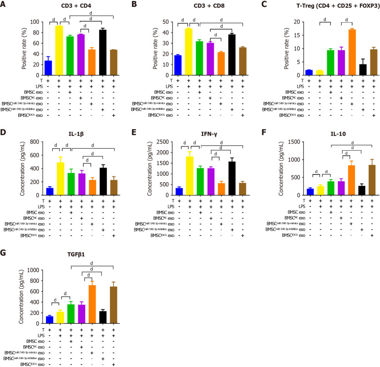
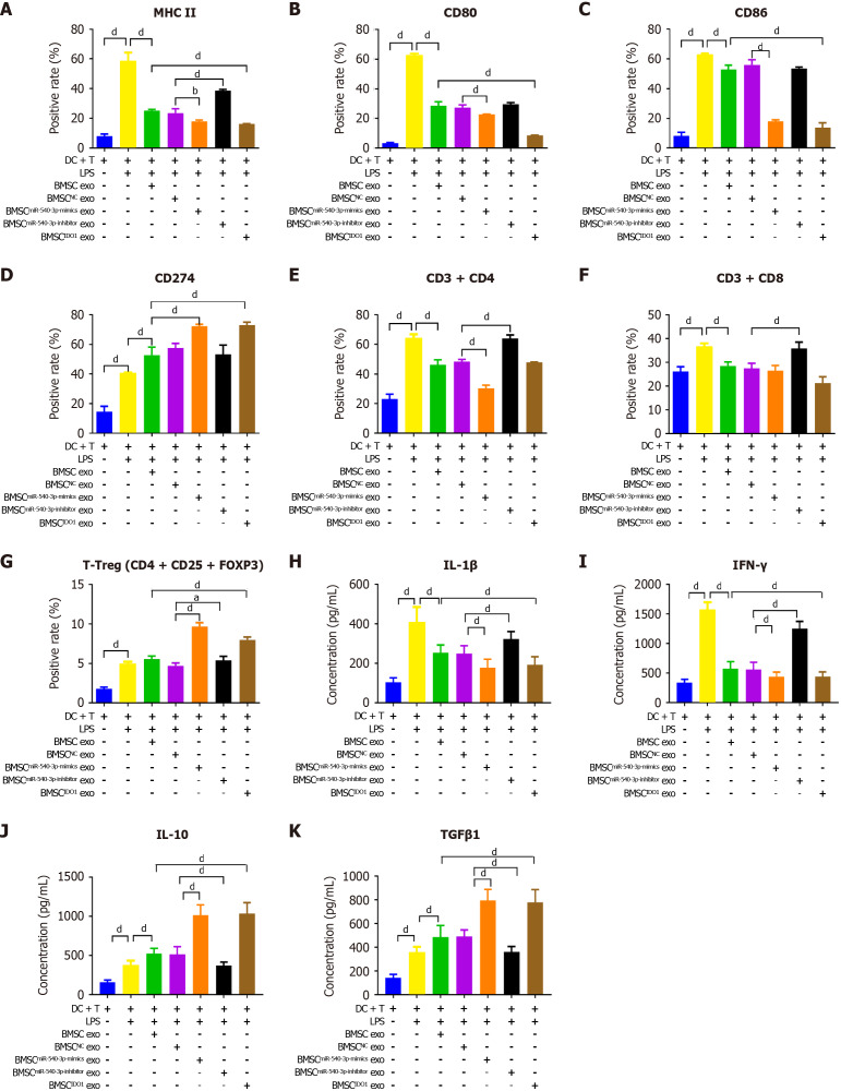
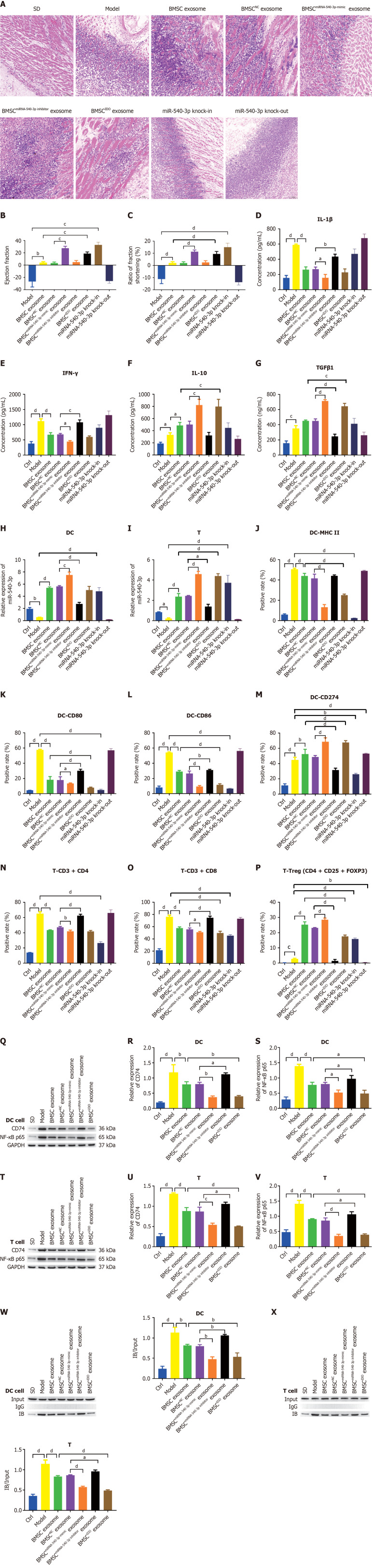
Methods: Rat models were used to assess the impact of miR-540-3p-enhanced exosomes on immune tolerance in cardiac allografts. MiR-540-3p expression was manipulated in BMSCs, and derived exosomes were collected and administered to the rat models post-heart transplantation. The study monitored expression levels of major histocompatibility complex II, CD80, CD86, and CD274 in DCs, and quantified CD4+ and CD8+ T cells, T regulatory cells, and cytokine profiles.

Results: Exosomes from miR-540-3p-overexpressing BMSCs lead to reduced expression of immune activation markers CD74 and NF-κB p65 in DCs and T cells. Rats treated with these exosomes showed decreased inflammation and improved cardiac function, indicated by lower levels of pro-inflammatory cytokines (interleukin-1β, interferon-γ) and higher levels of anti-inflammatory cytokines (interleukin-10, transforming growth factor β1). Additionally, miR-540-3p skewed the profiles of DCs and T cells towards immune tolerance, increasing the ratio of T regulatory cells and shifting cytokine secretion to favor graft acceptance.

Conclusion: Exosomes derived from BMSCs overexpressing miR-540-3p significantly enhance immune tolerance and prolong cardiac allograft survival by modulating the CD74/NF-κB pathway, which regulates activities of DCs and T cells. These findings highlight a promising therapeutic strategy to improve heart transplantation outcomes and potentially reduce the need for prolonged immunosuppression.

Keywords: Bone marrow mesenchymal stem cells; Cardiac allograft; Exosomes; Immune tolerance; MicroRNA-540-3p.


Conflict of interest statement

Conflict-of-interest statement: The Yunan Labreal Biotech Ltd, co. provided the research materials in the animal experiment. Although the animal experiment was performed in the platform of a private company, they did not aware of or interfere the experimental propose and not participate in the writing, review, and publication of this manuscript. No economic interests were appeared.

Figures

Figure 1
Figure 1
Overexpression of microRNA-540-3p in dendritic cells modulates surface markers and cytokine production. A: Ten immune-related microRNAs (miRNAs) in indoleamine 2,3-dioxygenase-overexpressing bone marrow mesenchymal stem cell exosomes compared to those in bone marrow mesenchymal stem cell exosomes; B: Quantification of immune-related miRNA expression by quantitative real-time polymerase chain reaction; C: Expression of miR-540-3p in dendritic cells (DCs) at 48, 72, and 96 hours after transfection; D-G: Flow cytometry analysis of the positive rate of cell surface markers in DCs (major histocompatibility complex II, CD80, CD86, CD274) and DCs after co-culture with T cells for 72 hours; H-K: Enzyme-linked immunosorbent assay-based quantitative analysis of cytokine production (interleukin-1β, interferon-γ, interleukin-10, transforming growth factor β1) in DCs and DCs after co-culture with T cells for 72 hours. aP < 0.05, dP < 0.0001. BMSC: Bone marrow mesenchymal stem cell; NC: Negative control; IDO: Indoleamine 2,3-dioxygenase; DC: Dendritic cell; LPS: Lipopolysaccharide; IL: Interleukin; TGF: Transforming growth factor; IFN: Interferon.
Figure 2
Figure 2
Overexpression of microRNA-540-3p in T cells modulates cell subtypes and cytokine production. A: Expression of microRNA-540-3p in T cells at 48, 72, and 96 hours post-plasmid transfection; B-D: Flow cytometry analysis of the positive rate of T cells (CD4+ T, CD8+ T, and T regulatory cell) and T cells after co-culture with dendritic cells for 72 hours; E-H: Enzyme-linked immunosorbent assay-based quantitative analysis of cytokine production (interleukin-1β, interferon-γ, interleukin-10, and transforming growth factor β1) in T cells and T cells after co-culture with dendritic cells for 72 hours. aP < 0.05, dP < 0.0001. NC: Negative control; LPS: Lipopolysaccharide; IL: Interleukin; TGF: Transforming growth factor; IFN: Interferon.
Figure 3
Figure 3
Production and characterization of bone marrow mesenchymal stem cellmiR-540-3p-mimic exosomes. A: Transmission electron microscopy image of isolated exosomes; B: Nanosight analysis for exosome size and concentration; C: Expression of microRNA-540-3p (miR-540-3p) in bone marrow mesenchymal stem cell-derived exosomes at 48, 72, and 96 hours after plasmid transfection; D: Expression of miR-540-3p in dendritic cells; E: Expression of miR-540-3p in T cells. bP < 0.01, cP < 0.001, dP < 0.0001. BMSC: Bone marrow mesenchymal stem cell; IDO: Indoleamine 2,3-dioxygenase; DC: Dendritic cell; LPS: Lipopolysaccharide; exo: Exosomes.
Figure 4
Figure 4
Bone marrow mesenchymal stem cellmiR-540-3p-mimic exosomes reduces immune resistance in dendritic cells. A-D: Flow cytometry analysis of the positive rate of cell surface markers in dendritic cells (major histocompatibility complex II, CD80, CD86, and CD274) for 72 hours; E-H: Enzyme-linked immunosorbent assay-based quantitative analysis of cytokine production (interleukin-1β, interferon-γ, interleukin-10, transforming growth factor β1) in dendritic cells. aP < 0.05, dP < 0.0001. MHC: Major histocompatibility complex; BMSC: Bone marrow mesenchymal stem cell; IDO: Indoleamine 2,3-dioxygenase; DC: Dendritic cell; LPS: Lipopolysaccharide; exo: Exosomes; IL: Interleukin; TGF: Transforming growth factor; IFN: Interferon.
Figure 5
Figure 5
Bone marrow mesenchymal stem cellmiR-540-3p-mimic exosomes reduces immune resistance in T cells. A-C: Flow cytometry analysis of the positive rate of CD4+ T cells, CD8+ T cells, and regulatory T cells for 72 hours; D-G: Enzyme-linked immunosorbent assay-based quantitative analysis of cytokine production (interleukin-1β, interferon-γ, interleukin-10, and transforming growth factor β1) in T cells. dP < 0.0001. BMSC: Bone marrow mesenchymal stem cell; LPS: Lipopolysaccharide; exo: Exosomes; IL: Interleukin; TGF: Transforming growth factor; IFN: Interferon.
Figure 6
Figure 6
Effect of bone marrow mesenchymal stem cellmiR-540-3p-mimic exosomes on dendritic cells after co-culturing with T cells. A-D: Flow cytometry analysis of the positive rate of cell surface markers (major histocompatibility complex II, CD80, CD86, and CD274) in dendritic cells cultured with T cells for 72 hours; E-G: Flow cytometry analysis of the positive rate of T cells (CD4+ T, CD8+ T, and T regulatory cells) and T cells after co-culture with dendritic cells; H-K: Enzyme-linked immunosorbent assay-based quantitative analysis of cytokine production (interleukin-1β, interferon-γ, interleukin-10, and transforming growth factor β1) in dendritic cell-T cells co-cultures. aP < 0.05, dP < 0.0001. MHC: Major histocompatibility complex; BMSC: Bone marrow mesenchymal stem cell; IDO: Indoleamine 2,3-dioxygenase; DC: Dendritic cell; LPS: Lipopolysaccharide; exo: Exosomes; IL: Interleukin; TGF: Transforming growth factor; IFN: Interferon; Treg: T regulatory cell.
Figure 7
Figure 7
CD74 is a target of microRNA-540-3p and regulates P65 expression. A: Gene Ontology and Kyoto Encyclopedia of Genes and Genomes enrichment analyses of microRNA-540-3p (miR-540-3p) targets; B: A dual-luciferase assay was used to detect the binding of miR-540-3p to CD74; C: Co-IP was used to detect the binding of CD74 and nuclear factor-kappaB (NF-κB) in dendritic cells (DCs); D: Co-IP was used to detect the binding of CD74 and NF-κB in T cells; E-G: Western blot analysis of the expression of CD74 and NF-κB in DCs; H-J: Western blot analysis of the expression of CD74 and NF-κB in T cells; K-M: Western blot analysis of CD74 and NF-κB expression in DCs after co-culture with T cells; N-P: Western blot analysis of the expression of CD74 and NF-κB in T cells after co-culture with DCs; Q-S: Western blot analysis of the expression of CD74 and NF-κB in DCs after plasmid transfection; T-V: Western blot analysis of CD74 and NF-κB expression in T cells after plasmid transfection. aP < 0.05, bP < 0.01, cP < 0.001, dP < 0.0001. Mut: Mutant; BMSC: Bone marrow mesenchymal stem cell; IDO: Indoleamine 2,3-dioxygenase; DC: Dendritic cell; LPS: Lipopolysaccharide; exo: Exosomes; NF-κB: Nuclear factor-kappaB.
Figure 8
Figure 8
Exosome-derived miR-540-3p alleviates immune resistance after heterotopic heart transplantation. A: Histopathological observation of the heart after heterotopic heart transplantation using hematoxylin and eosin staining; B: Detection and calculation of the difference in ejection fraction before and after heterotopic heart transplantation; C: Detection and calculation of differences in the percentage of fractional shortening before and after heterotopic heart transplantation; D-G: Enzyme-linked immunosorbent assay-based quantitative analysis of cytokine production (interleukin-1β, interferon-γ, interleukin-10, transforming growth factor β1) in serum; H: Expression of microRNA-540-3p in extracted dendritic cells (DCs); I: Expression of microRNA-540-3p in extracted T cells; J-M: Flow cytometric analysis of the positive rate of cell surface markers in DCs (major histocompatibility complex II, CD80, CD86, and CD274); N-P: Flow cytometric analysis of the positive rate of T cells (CD4+ T, CD8+ T, and T regulatory cells); Q-S: Western blot analysis of the expression of CD74 and nuclear factor-kappaB (NF-κB) in extracted DCs; T-V: Western blot analysis of CD74 and NF-κB expression in the extracted T cells; W: Co-IP was used to detect the binding of CD74 and NF-κB in extracted DCs; X: Co-IP was used to detect the binding of CD74 and NF-κB in extracted T cells. aP < 0.05, bP < 0.01, cP < 0.001, dP < 0.0001. BMSC: Bone marrow mesenchymal stem cell; IDO: Indoleamine 2,3-dioxygenase; DC: Dendritic cell; LPS: Lipopolysaccharide; NF-κB: Nuclear factor-kappaB; MHC: Major histocompatibility complex; IL: Interleukin; TGF: Transforming growth factor; IFN: Interferon.
Figure 9
Figure 9
Exosomes derived from microRNA-540-3p overexpressing bone marrow mesenchymal stem cells promote immune tolerance to cardiac allograft via the CD74/nuclear factor-kappaB pathway. BMSC: Bone marrow mesenchymal stem cell; IDO: Indoleamine 2,3-dioxygenase; IL: Interleukin; TGF: Transforming growth factor; IFN: Interferon; NF-κB: Nuclear factor-kappaB.

Similar articles

Cited by

References

    1. Savarese G, Lund LH. Global Public Health Burden of Heart Failure. Card Fail Rev. 2017;3:7–11. - PMC - PubMed
    1. Katz JN, Waters SB, Hollis IB, Chang PP. Advanced therapies for end-stage heart failure. Curr Cardiol Rev. 2015;11:63–72. - PMC - PubMed
    1. Mcdermott JK. Complications of Immunosuppression. In: Bogar L, Stempien-Otero A. Contemporary Heart Transplantation. Organ and Tissue Transplantation. Cham: Springer, 2020.
    1. Hasegawa H, Matsumoto T. Mechanisms of Tolerance Induction by Dendritic Cells In Vivo. Front Immunol. 2018;9:350. - PMC - PubMed
    1. Zhang X, Li M, Lian D, Zheng X, Zhang ZX, Ichim TE, Xia X, Huang X, Vladau C, Suzuki M, Garcia B, Jevnikar AM, Min WP. Generation of therapeutic dendritic cells and regulatory T cells for preventing allogeneic cardiac graft rejection. Clin Immunol. 2008;127:313–321. - PubMed