

Case Reports
doi: 10.1093/omcr/omae163.
eCollection 2024 Dec.
Paradoxical coronary embolism in Erdheim-Chester disease: invasive assessment and multidisciplinary management
Affiliations
- PMID: 39734688
- PMCID: PMC11682485
- DOI: 10.1093/omcr/omae163
Item in Clipboard
Case Reports
Paradoxical coronary embolism in Erdheim-Chester disease: invasive assessment and multidisciplinary management
Oxf Med Case Reports.
.
Abstract
We report a case of non-ST elevation myocardial infarction in a 36-year-old man with Erdheim-Chester disease (ECD). Multimodality assessment revealed acute coronary thrombus with simultaneous recurrent pulmonary embolism in spite of compliance with a direct oral anticoagulant. Prior case reports of acute myocardial infarction in this population have not outlined the role of catheter based intravascular assessment and treatment in this rare clinical entity.
Keywords: Erdheim-Chester disease; coronary embolism; non-ST elevation myocardial infarction.
© The Author(s) 2024. Published by Oxford University Press.
Conflict of interest statement
No conflict of interest.
Figures

Computerized tomography (CT) coronary angiogram. 15 mm non-calcified lesion from ostial to proximal left anterior descending (LAD) coronary artery causing moderate to severe stenosis.

Initial angiogram. Ostial filling defect in the left anterior descending (LAD) coronary artery (arrow).

Repeat angiogram. Improvement to previous ostial left anterior descending (LAD) coronary artery lesion.

Intravascular ultrasound (IVUS). Features of thrombus (arrow).

PFO closure. A 25 mm amplatzer PFO occluder using a 8-French Trevision delivery system was used.

Repeat intravascular ultrasound (IVUS). Eccentric fatty/fibrotic plaque in proximal left anterior descending (LAD) coronary artery with mild calcification.
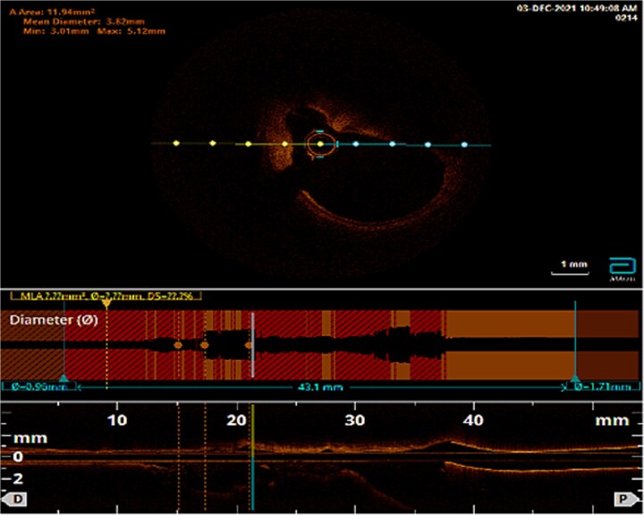
Repeat optical coherence tomography (OCT). Limited by dropout segment in proximal left anterior descending (LAD) coronary artery.
References
Publication types
LinkOut - more resources
Full Text Sources
