Review
. 2024 Sep 28;5(3):236-256.
doi: 10.12336/biomatertransl.2024.03.003. eCollection 2024.

The use of hydrogel microspheres as cell and drug delivery carriers for bone, cartilage, and soft tissue regeneration

Affiliations
Review

The use of hydrogel microspheres as cell and drug delivery carriers for bone, cartilage, and soft tissue regeneration

Chung-Hsun Lin et al. Biomater Transl. .

Abstract

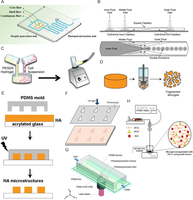
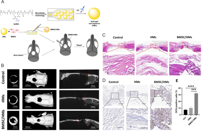
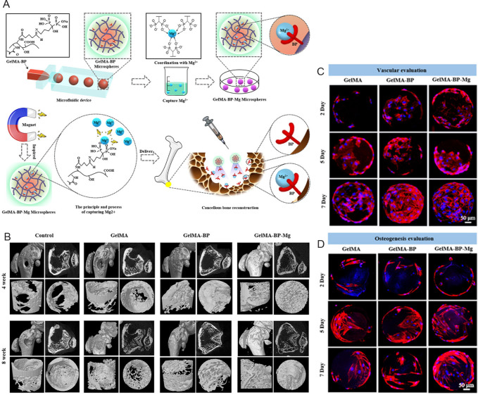
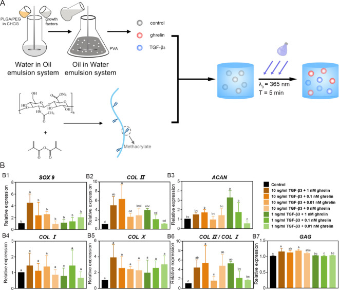
Bone, cartilage, and soft tissue regeneration is a complex process involving many cellular activities across various cell types. Autografts remain the "gold standard" for the regeneration of these tissues. However, the use of autografts is associated with many disadvantages, including donor scarcity, the requirement of multiple surgeries, and the risk of infection. The development of tissue engineering techniques opens new avenues for enhanced tissue regeneration. Nowadays, the expectations of tissue engineering scaffolds have gone beyond merely providing physical support for cell attachment. Ideal scaffolds should also provide biological cues to actively boost tissue regeneration. As a new type of injectable biomaterial, hydrogel microspheres have been increasingly recognised as promising therapeutic carriers for the local delivery of cells and drugs to enhance tissue regeneration. Compared to traditional tissue engineering scaffolds and bulk hydrogel, hydrogel microspheres possess distinct advantages, including less invasive delivery, larger surface area, higher transparency for visualisation, and greater flexibility for functionalisation. Herein, we review the materials characteristics of hydrogel microspheres and compare their fabrication approaches, including microfluidics, batch emulsion, electrohydrodynamic spraying, lithography, and mechanical fragmentation. Additionally, based on the different requirements for bone, cartilage, nerve, skin, and muscle tissue regeneration, we summarize the applications of hydrogel microspheres as cell and drug delivery carriers for the regeneration of these tissues. Overall, hydrogel microspheres are regarded as effective therapeutic delivery carriers to enhance tissue regeneration in regenerative medicine. However, significant effort is required before hydrogel microspheres become widely accepted as commercial products for clinical use.

Keywords: drug delivery; fabrication techniques; hydrogel microspheres; microgels; tissue regeneration.


Figures

Figure 1.
Figure 1.. Schematic diagrams of hydrogel microsphere processing routes. (A, B) Microfluidic: (A) The microfluidic chip has three chips to form a shell-like microsphere in a continuous oil flow. Reprinted from Wang et al. Copyright 2019 WILEY‐VCH Verlag GmbH & Co. KGaA, Weinheim. (B) A microcapillary device with a magnified image of the part where droplets formed. Reprinted from Martinez et al. Copyright 2012 WILEY‐VCH Verlag GmbH & Co. KGaA, Weinheim. (C) Batch emulsion: Cell encapsulated hydrogel microsphere made by mixing PEGDA hydrogel precursor solution and allogeneic skin fibroblasts in mineral oil. Reprinted from Sonnet et al. Copyright 2013 Orthopaedic Research Society. (D) Mechanical fragmentation: Fragmented microgels can be obtained by applying forces to bulk hydrogels using a fragmenting device like the tissue homogeniser. Reprinted from Widener et al. (E–G) Lithography: (E) Imprint lithography places PDMS moulds on a layer of hyaluronic acid and crosslinks the material by exposing it to UV light. Reprinted from Khademhosseini et al. Copyright 2006 Wiley Periodicals, Inc. (F) Photolithography uses a photomask to screen the UV light and crosslink the materials exposed to the UV light. (G) The flow lithography technique allows a continuous stream of material to pass through a region of UV light with specific shape. Reprinted from Laza et al. Copyright 2012 WILEY‐VCH Verlag GmbH & Co. KGaA, Weinheim. (H) Electrohydrodynamic spraying: A syringe pump sprays the hydrogel and cell precursor solution into the oil bath through a needle tip connected to a high-pressure source. Reprinted from Kim et al. HA: hyaluronic acid; HGF: hepatocyte growth factor; OECs: outgrowth endothelial cells; PDMS: polydimethylsiloxane; PEGDA: poly(ethylene glycol) diacrylate; UV: ultraviolet; VEGF: vascular endothelial growth factors.
Figure 2.
Figure 2.. Schematic diagram of the fabrication process of BMSCs-loaded GelMA HMs and their results of micromorphometric and histological analysis. (A) HMs produced from microfluidic methods are then crosslinked by UV light, seeded with BMSCs, and transplanted to the skull defect. (B) Micromorphometric analysis of the skull defect 8 weeks after transplantation. Images are superficial, three-dimensional, and sagittal views of microcomputed tomography images. Scale bars: 5 mm (left and right), 10 mm (middle). (C) HE staining in the skull defect area of control, HMs and BMSC/HMs groups at 8 weeks after transplantation. Scale bars: 50 μm. (D) Immunohistochemical staining of OCN-positive cells in the skull defect area 8 weeks after transplantation of HMs and BMSC/HMs. Scale bars: 50 μm (upper), 20 μm (lower). (E) Semi-quantitative analysis of the relative number of OCN-positive cells in the control, HMs and BMSC/HMs groups. Reprinted from Teng et al. BMSCs: bone marrow mesenchymal stem cells; GelMA: gelatin methacrylate; HE: hematoxylin-eosin; HMs: hydrogel microspheres; OCN: osteocalcin; UV: ultraviolet.
Figure 3.
Figure 3.. Schematic diagram of the fabrication process of GelMA-BP-Mg microspheres and their results of micromorphometric analysis and biocompatibility. (A) GelMA-BP microspheres were prepared by a microfluidic device and Mg was captured by Schiff alkali reactivity. GelMA-BP-Mg microspheres were then constructed by metal ion coordination ligands and delivered by injection. (B) Regeneration efficacy of the distal femur of rats with osteoporotic bone defects at 4 and 8 weeks after injection. Microcomputed tomography images show the results for control, GelMA, GelMA-BP, and GelMA-BP-Mg groups. (C, D) Proliferation of BMSCs on GelMA, GelMA-BP, and GelMA-BP-Mg microspheres after 2, 5, and 7 days. Scale bars: 50 μm. Red shows the skeleton; blue shows the nucleus. Reprinted with permission from Zhao et al. Copyright 2021, American Chemical Society. BMSC: bone marrow mesenchymal stem cells; BP: bisphosphonate; GelMA: gelatin methacrylate.
Figure 4.
Figure 4.. Schematic diagram of the fabrication process producing hydrogel microspheres with growth factor and chondrogenic differentiation results of hMSCs used for cartilage regeneration. (A) Fabrication of the PEG/PLGA microspheres containing TGF-β3 or ghrelin. (B) Results of the chondrogenic differentiation results of hMSCs with different concentrations of TGF-β3 and ghrelin after 21 days. (B1–7) The qRT-PCR analyses are done for SOX9, COL II, ACAN, COL I, COL X, COL II/COL I, and GAG. Reprinted from Lin et al. ACAN: aggrecan; COL I: type I collagen; COL II: type II collagen; COL X: type X collagen; GAG: glycosaminoglycan; hMSCs: human bone marrow mesenchymal stem cells; PEG: poly(ethylene glycol); PLGA: poly(lactic-co-glycolic acid); PVA: poly(vinyl alcohol); qRT-PCR: quantitative reserve transcription-polymerase chain reaction; SOX 9: Sry-type high-mobility-group box 9; TGF: transforming growth factor)
Figure 5.
Figure 5.. Schematic diagram of the fabrication process producing GMPs/GMPBs and results of in vivo and in vitro tests. (A) Microspheres were prepared by microfluidic method and loaded with PDGF-BB and BMSCs. (B) X-ray images of the knee joints of rats in the control group, GMs group, GMPs group, GMBs group, GMPBs group and Sham group at AP and LAT angles. (C) Cell migration images of control, GMs, GMPs, GMPBs groups at 0, 24 and 48 hours. Scale bars: 800 μm. Reprinted from Li et al. Copyright 2023 Wiley‐VCH GmbH. AP: anteroposterior; BMSCs: bone marrow mesenchymal stem cells; GelMA: gelatin methacrylate; GMs: GelMA microspheres; GMBs: BMSCs loaded GelMA microspheres; GMPs: PDGF-BB-loaded GelMA microspheres; GMPBs: BMSCs+PDGF-BB-loaded GelMA microspheres; LAT: lateral; MA: methacrylate; PDGF-BB: platelet-derived growth factors-BB; UV: ultraviolet.
Figure 6.
Figure 6.. Schematic diagram of the fabrication process of hydrogel microspheres with NSCs and results of using it on SCI. (A) PDGF-MPHM was formed using an electrospray device and implanted into the T10 SCI site of rats 1 day after SCI. (B) Representative fluorescence images of stained NSC cultured with basic medium, PDGF-MPH, and PDGF-MPHM. Green shows the phosphorylated platelet-derived growth factor receptor beta, and blue shows the nucleus. Scale bars: 50 μm. (C) Representative immunofluorescence images of cross-sections of the spinal cord of rats in the SCI group, NSCs grafting group, and PDGF-MPHM + NSCs group. Red shows the apoptosis cells, blue shows the nucleus, and green shows the grafted cells. Scale bars: 250 μm. (D) Representative immunofluorescence images of the SCI, NSCs graft, and PDGF-MPHM + NSCs groups 8 weeks after SCI. Scale bars: 1 mm (upper), 250 μm (lower). Reprinted with the permission from Wu et al. Copyright 2023, American Chemical Society. DAPI: 4′6-diamidino-2-phenylindole; GFAP: glial fibrillary acidic protein; GFP: green fluorescent protein; Nap-FFG: naphthalene acetic acid-phenylalanine-phenylalanine-glycine; NSCs: neural stem cells; PDGF-MPH: platelet-derived growth factor mimetic peptide hydrogel; PDGF-MPHM: platelet-derived growth factor mimetic peptide hydrogel microspheres; PDGFRβ: platelet-derived growth factor receptor β; SCI: spinal cord injury; Tuj1: beta tubulin III.
Figure 7.
Figure 7.. Schematic diagram of the synthesis process of mPDA-PEI@GelMA and wound healing results in diabetic mice in vivo. (A) The mixture of mPDA-PEI and GelMA was crosslinked under UV light after exiting the microfluidic device. (B) Representative images of wound healing in control, GelMA, mPDA@GelMA, and mPDA-PEI@GelMA groups. (C) Wound healing rate in four treatment groups on days 0, 3, 7 and 12. Reprinted from Xiao et al. GelMA: gelatin methacrylate; MA: methacrylate; mPDA: mesoporous polydopamine; mPDA NP: mesoporous polydopamine nanoparticle; PEI: polyethyleneimine; UV: ultraviolet.
Figure 8.
Figure 8.. Schematic diagram of the fabrication process of mECM@IL-4 + PM@IGF-1 composites and their muscle regeneration potential. (A) PLCL microspheres fabricated by microfluidics were modified with PDA-conjugated IGF-1 and complexed with mECM and IL-4 to form a composite material injected into the damaged area. (B) The representative images show the differentiation-promoting effects of control, composite, BMDMs and BMDMs/composite groups on injured muscle satellite cells. Green shows phalloidin staining area, red shows desmin staining area, grey shows the CD206 cells, and blue shows the nucleus. (C) Immunofluorescence images showing muscle regeneration at 2 and 8 weeks in a rat VML model. Green shows phalloidin staining area, and blue shows the nucleus. Scale bars: 50 μm. P represents the microspheres, and the white dashed line represents the border between the microspheres and the tissue. Reprinted from Li et al. BMDMs: bone marrow-derived macrophages; CTX: cardiotoxin; DAPI: 4′6-diamidino-2-phenylindole; dECM: decellularised extracellular matrix; IGF-1: growth factor-1; IL-4: interleukin-4; mECM: muscle-derived extracellular matrix; PDA: polydopamine; PLCL: poly(l-lactide-caprolactone); PVA: polyvinyl alcohol; PM: PLCL microsphere; VML: volumetric muscle loss.

Similar articles

Cited by

References

    1. Ye J., Xie C., Wang C., Huang J., Yin Z., Heng B. C., Chen X., Shen W. Promoting musculoskeletal system soft tissue regeneration by biomaterial-mediated modulation of macrophage polarization. Bioact Mater. 2021;6:4096–4109. - PMC - PubMed
    1. Xiong Y., Mi B. B., Lin Z., Hu Y. Q., Yu L., Zha K. K., Panayi A. C., Yu T., Chen L., Liu Z. P., Patel A., Feng Q., Zhou S. H., Liu G. H. The role of the immune microenvironment in bone, cartilage, and soft tissue regeneration: from mechanism to therapeutic opportunity. Mil Med Res. 2022;9:65. - PMC - PubMed
    1. Yu L., Cavelier S., Hannon B., Wei M. Recent development in multizonal scaffolds for osteochondral regeneration. Bioact Mater. 2023;25:122–159. - PMC - PubMed
    1. Nair A., Thevenot P., Dey J., Shen J., Sun M. W., Yang J., Tang L. Novel polymeric scaffolds using protein microbubbles as porogen and growth factor carriers. Tissue Eng Part C Methods. 2010;16:23–32. - PMC - PubMed
    1. Li Y., Liu W., Liu F., Zeng Y., Zuo S., Feng S., Qi C., Wang B., Yan X., Khademhosseini A., Bai J., Du Y. Primed 3D injectable microniches enabling low-dosage cell therapy for critical limb ischemia. Proc Natl Acad Sci U S A. 2014;111:13511–13516. - PMC - PubMed

LinkOut - more resources