Case Reports
. 2024 Dec 24:17:5803-5813.
doi: 10.2147/IDR.S485152. eCollection 2024.

First Documented Successful Treatment of Chronic Postoperative Fungal Endophthalmitis Induced by Trichosporon Inkin with Fluconazole

Affiliations
Case Reports

First Documented Successful Treatment of Chronic Postoperative Fungal Endophthalmitis Induced by Trichosporon Inkin with Fluconazole

Ning Fan et al. Infect Drug Resist. .

Abstract

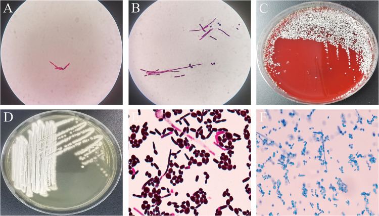
This report details an uncommon occurrence of chronic endophthalmitis following cataract surgery attributed to an infection by Trichosporon inkin (T. inkin). The infection was identified through MALDI-TOF mass spectrometry along with sequencing analysis. Although the patient exhibited a robust immune response, the infection escalated quickly from the right eye to the left. Treatment involved vitrectomy and peeling surgery on the right eye, paired with systemic fluconazole antifungal therapy and intravitreal injection, resulting in significant recovery. The visual acuity of the right eye enhanced from finger counting to 20/63. This account represents the inaugural documented instance of endophthalmitis caused by T. inkin that was effectively managed with fluconazole. This underscores the critical role of vitreous humor enrichment culture and antifungal susceptibility testing of T. inkin in the treatment of endophthalmitis.

Keywords: MALDI-TOF; Trichosporon inkin; endophthalmitis; fluconazole; matrix-assisted laser desorption ionization time of flight.


Conflict of interest statement

The authors state that the study was carried out without any commercial or financial ties that might be interpreted as a possible conflict of interest.

Figures

Figure 1
Figure 1
Patient’s diagnosis and treatment process.
Figure 2
Figure 2
OCT (optical coherence tomography) examination of the right eye’s macula. On the fifth day of hospitalization (January 5): white lesions are faintly visible in front of the retina (A). Post vitrectomy (January 18): white lesions visible in the preretina (B). At the follow-up visit (February 5): Preretinal white lesions become smaller and lighter in color (C).
Figure 3
Figure 3
Scanned laser ophthalmoscope (SLO) images of the patient. On the 5th day of admission, a significant number of floating cells were observed in the vitreous cavity of the right eye, exhibiting gray-white turbidity, with multiple white cotton ball-like lesions vaguely discernible. Following vitrectomy (4 days post fluconazole treatment), the scattered white cotton ball-like lesions became more apparent. The size of these lesions decreased after 20 days of fluconazole treatment (AC). On the fifth day of admission and four days post fluconazole treatment, two white cotton ball-like lesions were detected in the superior and inferior temporal retina of the left eye. After 20 days of fluconazole treatment, the diameter of these lesions reduced compared to before (DF).
Figure 4
Figure 4
Microbiological examination pictures of the patient. Direct smear microscopy of vitreous humor revealed the presence of hyphae (Gram stain, ×1000) (A). Following positive enrichment culture of vitreous humor, smear microscopy showed conidia and hyphae (Gram stain, ×1000) (B). White dry colonies cultured on Columbia blood agar at 35°C for 3 days (C). Incubate on Sabouraud dextrose agar (SDA) at 35°C for 3 days, white yeast-like colonies will appear (D). The strain shows blastocyst conidia, articular conidia and hyphae under a microscope (Gram stain, ×1000) (E). Stained with lactic acid phenol and cotton blue, blastocyst conidia, articular conidia and hyphae can be seen (×1000) (F).

Similar articles

References

    1. Jannic A, Lafaurie M, Denis B, et al. Trichosporon inkin causing invasive infection with multiple skin abscesses in a renal transplant patient successfully treated with voriconazole. JAAD Case Rep. 2017;4(1):27–29. doi:10.1016/j.jdcr.2017.10.008 - DOI - PMC - PubMed
    1. Madariaga MG, Tenorio A, Proia L. Trichosporon inkin peritonitis treated with caspofungin. J Clin Microbiol. 2003;41(12):5827–5829. doi:10.1128/JCM.41.12.5827-5829.2003 - DOI - PMC - PubMed
    1. Milan EP, Silva-Rocha WP, de Almeida JJS, et al. Trichosporon inkin meningitis in Northeast Brazil: first case report and review of the literature. BMC Infect Dis. 2018;18(1):470. doi:10.1186/s12879-018-3363-7 - DOI - PMC - PubMed
    1. Guo LN, Yu SY, Hsueh PR, et al. Invasive infections due to Trichosporon: species distribution, genotyping, and antifungal susceptibilities from a multicenter study in China. J Clin Microbiol. 2019;57(2):e01505–18. doi:10.1128/JCM.01505-18 - DOI - PMC - PubMed
    1. Nishimoto K. Remarkable works and cases in the history of medical mycology in Japan. Med Mycol J. 2017;58(2):J29–J33. doi:10.3314/mmj.17.002 - DOI - PubMed

Publication types

LinkOut - more resources