

Stalking the stalk: Isolated pituitary stalk thickening and predictive factors for proliferative disease
- PMID: 39734811
- PMCID: PMC11672106
- DOI: 10.1093/noajnl/vdae214
Stalking the stalk: Isolated pituitary stalk thickening and predictive factors for proliferative disease
Abstract
Background: Few studies have evaluated predictive factors of isolated pituitary stalk thickening (iPST) in children.
Methods: In this retrospective study, radiology, endocrinology, and neuro-oncology databases were interrogated to identify patients with iPST between January 2000 and June 2019. A blinded, longitudinal assessment of MRIs was performed using quantitative, semi-quantitative, and qualitative metrics. Neuroimaging parameters were correlated to clinical parameters.
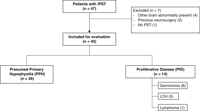
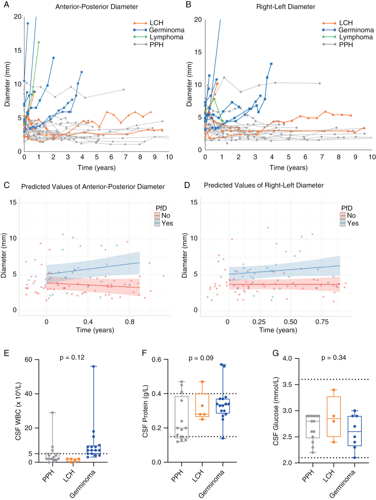
Results: Forty-seven patients were identified, with 40 meeting the inclusion criteria. Median age at baseline MRI was 9.6 years (range 0.9-17.5) with median follow-up of 5.2 years (range 0.3-18.6). Twenty-five (63%) were female. Thirty-four (85%) had pituitary dysfunction, including 31 with central diabetes insipidus (cDI). cDI was not predictive of proliferative disease (PfD): 69% of those with presumed primary hypophysitis (PPH) versus 93% with PfD (P = .1). Fourteen (35%) patients were diagnosed with PfD (germinoma = 8, Langerhans cell histiocytosis = 5, lymphoma = 1) at median of 1.3 years (range 0.3-4.0) after initial MRI. Progressive thickening of the stalk over time was associated with PfD (86% vs 4% in PPH, P < .0001), as was thickening of the entire stalk (56% in PfD vs 27% in PPH, P < .0001) with different imaging trends over time observed in PfD versus PPH. A "sack of marbles" appearance with heterogeneous enhancement on post-contrast imaging was associated with germinoma.
Conclusions: In this cohort, 35% of children with iPST were diagnosed with PfD. The association of cDI and PfD was not statistically significant. Progressive thickening of the entire stalk was predictive of PfD and a "sack of marbles" pattern was found to be highly suggestive of germinoma.
Keywords: CSF pleocytosis; LCH; germinoma; hypophysitis; pituitary stalk thickening.
© The Author(s) 2024. Published by Oxford University Press, the Society for Neuro-Oncology and the European Association of Neuro-Oncology.
Conflict of interest statement
None declared.
Figures

References
-
- Fujisawa I, Asato R, Okumura R, et al. Magnetic resonance imaging of neurohypophyseal germinomas. Cancer. 1991;68(5):1009–1014. - PubMed
-
- Tao Y, Lian D, Hui-juan Z, Hui P, Zi-meng J.. Value of brain magnetic resonance imaging and tumor markers in the diagnosis and treatment of intracranial germinoma in children. Zhongguo Yi Xue Ke Xue Yuan Xue Bao. 2011;33(2):111–115. - PubMed
-
- Marchand I, Barkaoui MA, Garel C, Polak M, Donadieu J; Writing Committee. Central diabetes insipidus as the inaugural manifestation of Langerhans cell histiocytosis: natural history and medical evaluation of 26 children and adolescents. J Clin Endocrinol Metab. 2011;96(9):E1352–E1360. - PubMed
-
- Fahrner B, Prosch H, Minkov M, et al. Long-term outcome of hypothalamic pituitary tumors in Langerhans cell histiocytosis. Pediatr Blood Cancer. 2012;58(4):606–610. - PubMed
-
- Capra M, Wherrett D, Weitzman S, et al. Pituitary stalk thickening and primary central nervous system lymphoma. J Neurooncol. 2004;67(1-2):227–231. - PubMed
LinkOut - more resources
Full Text Sources