

Extramedullary Intradural Primary Spinal Angiosarcoma: A Case Study
- PMID: 39734979
- PMCID: PMC11682733
- DOI: 10.7759/cureus.74767
Extramedullary Intradural Primary Spinal Angiosarcoma: A Case Study
Abstract
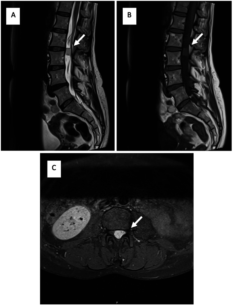
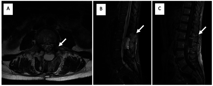
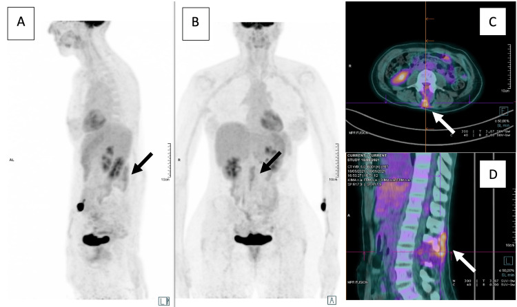
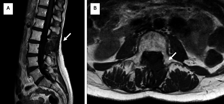
Angiosarcoma is a rare soft tissue sarcoma, namely when it presents as a primary intradural extramedullary spinal neoplasm, with only one case of non-vertebral origin reported in the literature. We present the case of a 51-year-old woman with neurological symptoms of paraparesis and constipation who underwent a magnetic resonance imaging (MRI) that revealed a well-demarcated, predominantly homogeneous, intensely enhancing intradural extramedullary lesion in T2-weighted images. Histology, immunohistochemistry, and genetics of the lesion showed an angiosarcoma. The patient received adjuvant radiation therapy (RT) and is currently stable, showing no signs of recurrence, in the third year of follow-up. Our work demonstrates a rare case of extramedullary intradural primary spinal angiosarcoma, highlighting the challenges associated with its diagnosis. The multidisciplinary approach, including surgery and adjuvant RT, has been shown to effectively control the disease. Although the prognosis is generally poor, the presented case shows apparent success.
Keywords: neuro radiology; neurosurgery; primary angiosarcoma; primary intradural extramedullary spinal tumor; radiation therapy.
Copyright © 2024, Catalo et al.
Conflict of interest statement
Human subjects: Consent for treatment and open access publication was obtained or waived by all participants in this study. Health Ethics Committee - Research Center of Instituto Português de Oncologia do Porto (CI-IPOP) issued approval CES 137-024. This HEC believes that there is no ethical impediment to publishing this study, safeguarding the guarantees requested by the data protection officer. Conflicts of interest: In compliance with the ICMJE uniform disclosure form, all authors declare the following: Payment/services info: All authors have declared that no financial support was received from any organization for the submitted work. Financial relationships: All authors have declared that they have no financial relationships at present or within the previous three years with any organizations that might have an interest in the submitted work. Other relationships: All authors have declared that there are no other relationships or activities that could appear to have influenced the submitted work.
Figures

Similar articles
-
Rare malignant primary spinal intradural extramedullary mesenchymal chondrosarcoma: a case report and literature review.Transl Cancer Res. 2022 Sep;11(9):3371-3378. doi: 10.21037/tcr-21-2703. Transl Cancer Res. 2022. PMID: 36237254 Free PMC article.
-
Primary intradural extramedullary spinal mesenchymal chondrosarcoma: case report and literature review.BMC Musculoskelet Disord. 2019 Sep 4;20(1):408. doi: 10.1186/s12891-019-2799-2. BMC Musculoskelet Disord. 2019. PMID: 31484514 Free PMC article. Review.
-
Intradural, extramedullary spinal sarcoidosis: report of a rare case and review of the literature.Spine J. 2006 Mar-Apr;6(2):204-10. doi: 10.1016/j.spinee.2005.06.009. Epub 2006 Feb 3. Spine J. 2006. PMID: 16517395
-
Intradural extramedullary spinal ependymoma: a benign pathology?Spine (Phila Pa 1976). 2005 May 1;30(9):E251-4. doi: 10.1097/01.brs.0000161008.13441.7b. Spine (Phila Pa 1976). 2005. PMID: 15864145
-
Primary spinal multifocal intradural-extramedullary Ewing sarcoma in children: presentation of a case and review of the literature.Turk J Pediatr. 2021;63(6):1084-1090. doi: 10.24953/turkjped.2021.06.018. Turk J Pediatr. 2021. PMID: 35023660 Review.
References
-
- Angiosarcoma. Young RJ, Brown NJ, Reed MW, Hughes D, Woll PJ. Lancet Oncol. 2010;11:983–991. - PubMed
-
- Spiker AM, Mangla A, Ramsey ML. Treasure Island (FL): StatPearls Publishing; [ Dec; 2023 ]. 2023. Angiosarcoma. - PubMed
-
- Primary spinal angiosarcoma. Liu ZH, Lee ST, Jung SM, Tu PH. J Clin Neurosci. 2010;17:387–389. - PubMed
-
- Case report: Angiosarcoma of the cervical spine. Pülhorn H, Elliot T, Clark J, Gonzalvo A. J Clin Neurosci. 2017;45:129–131. - PubMed
Publication types
LinkOut - more resources
Full Text Sources