Review
. 2024 Dec 13:15:1503130.
doi: 10.3389/fmicb.2024.1503130. eCollection 2024.

Advances in research on the impact and mechanisms of pathogenic microorganism infections on pyroptosis

Affiliations
Review

Advances in research on the impact and mechanisms of pathogenic microorganism infections on pyroptosis

Pan Shang et al. Front Microbiol. .

Abstract

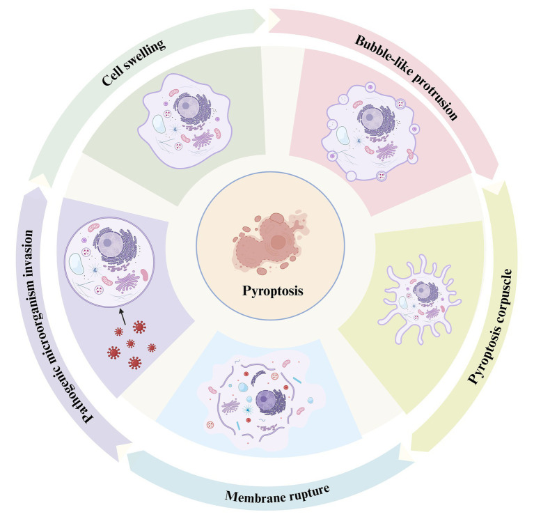
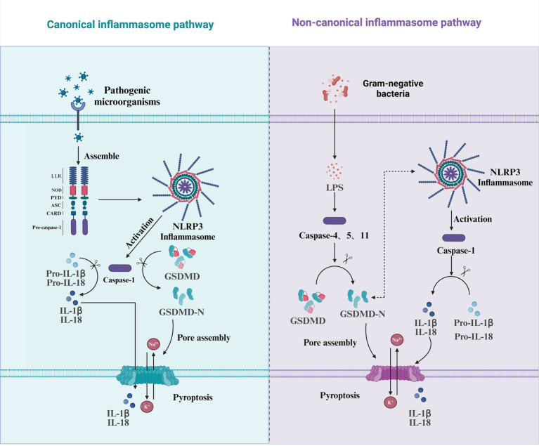
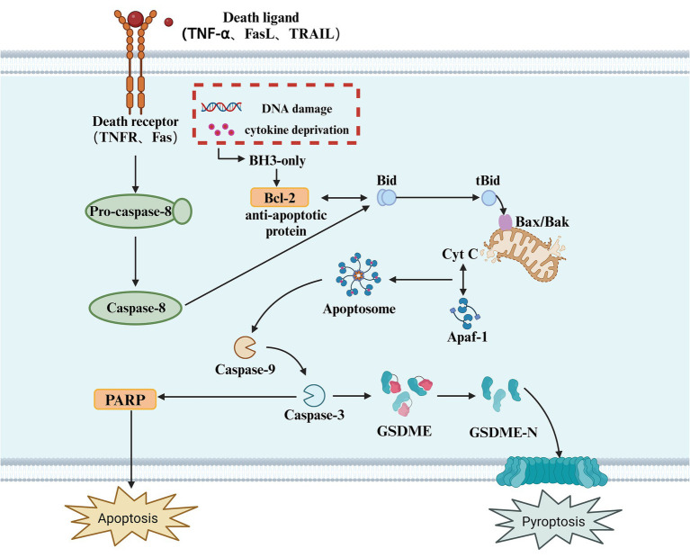
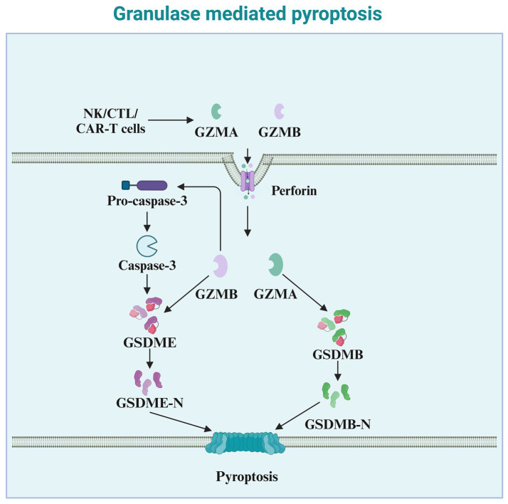
Pyroptosis, also known as inflammatory necrosis, is a form of programmed cell death characterized by the activation of gasdermin proteins, leading to the formation of pores in the cell membrane, continuous cell swelling, and eventual membrane rupture. This process results in the release of intracellular contents, including pro-inflammatory cytokines like IL-1β and IL-18, which subsequently trigger a robust inflammatory response. This process is a crucial component of the body's innate immune response and plays a significant role in combating infections. There are four main pathways through which pathogenic microorganisms induce pyroptosis: the canonical inflammasome pathway, the non-canonical inflammasome pathway, the apoptosis-associated caspase-mediated pathway, and the granzyme-mediated pathway. This article provides a brief overview of the effects and mechanisms of pathogen infections on pyroptosis.

Keywords: caspase; granzyme; inflammasome; pathogenic microorganisms; pyroptosis.


Conflict of interest statement

The authors declare that the research was conducted in the absence of any commercial or financial relationships that could be construed as a potential conflict of interest.

Figures

Figure 1
Figure 1
The process of pyroptosis. This diagram illustrates the sequential stages of pyroptosis, a form of programmed necrosis mediated by gasdermin. The process begins with the invasion of pathogenic microorganisms (left), during which the invading pathogens interact with the host cell, triggering an inflammatory response. Cell swelling (top left) follows as the cell undergoes structural changes in response to the infection. As the pyroptotic response intensifies, bubble-like protrusions (top right) begin to form on the cell membrane, which is a hallmark of this cell death process. Subsequently, the cell forms a pyroptosis corpuscle (bottom right), consisting of membrane-bound structures created during pyroptosis. Finally, the process concludes with membrane rupture (bottom left), where the cell membrane breaks apart, releasing pro-inflammatory cytokines such as IL-1β and IL-18, which contribute to inflammation and immune system activation.
Figure 2
Figure 2
Pyroptosis mediated by the canonical and non-canonical inflammasome pathways. This diagram illustrates the two major pathways involved in the induction of pyroptosis: the canonical inflammasome pathway and the non-canonical inflammasome pathway. In the canonical pathway (left), pathogenic microorganisms trigger the assembly of the NLRP3 inflammasome by activating pattern recognition receptors such as NOD, PYD, ASC, and CARD. This leads to the activation of Caspase-1, which cleaves pro-inflammatory cytokines (Pro-IL-1β and Pro-IL-18) into their active forms (IL-1β and IL-18) and also activates GSDMD, releasing its N-terminal domain (GSDMD-N), which then assembles pores in the membrane, culminating in pyroptosis. In the non-canonical pathway (right), gram-negative bacteria release LPS, which is recognized by Caspase-4/5/11. This activation also leads to the cleavage of GSDMD, resulting in the release of its active N-terminal domain (GSDMD-N) and pore formation. In this pathway, Caspase-1 is also activated but is involved in cleaving Pro-IL-1β and Pro-IL-18, promoting further inflammation and the onset of pyroptosis. Both pathways culminate in membrane rupture and the release of inflammatory cytokines, playing a crucial role in the host’s immune defense response.
Figure 3
Figure 3
Apoptotic caspase-mediated pyroptosis. This diagram illustrates the apoptotic signaling pathway, which can also lead to pyroptosis. The process is initiated by the binding of death ligands (such as TNF-α, FasL, or TRAIL) to their respective receptors (TNFR, Fas), triggering the activation of Caspase-8. In response to cellular stress, such as DNA damage or cytokine deprivation, BH3-only proteins are activated, which in turn modulate anti-apoptotic proteins like Bcl-2. The activation of Caspase-8 leads to the cleavage of Bid, generating tBid, which translocates to the mitochondria and interacts with Bax/Bak, facilitating the release of Cyt C from the mitochondria. This activation leads to the formation of the Apoptosome, which activates Caspase-9, and subsequently Caspase-3. Caspase-3 cleaves GSDME, releasing its active N-terminal fragment (GSDME-N), which forms pores in the cell membrane, ultimately resulting in pyroptosis. Additionally, in this pathway, PARP cleavage is also a key event in apoptosis, with its activation promoting cellular disassembly. Pyroptosis, as an inflammatory form of cell death, results from caspase activation, which causes the release of pro-inflammatory cytokines and the rupture of the cell membrane.
Figure 4
Figure 4
Granzyme-mediated pyroptosis. This diagram illustrates granule-mediated pyroptosis, a process involving cytotoxic immune cells such as NK cells, CTLs, and CAR-T cells. These cells release granzymes (GZMA and GZMB) and perforin to induce pyroptosis. Perforin facilitates the entry of granzymes into the target cell. Once inside the cell, GZMA and GZMB cleave Pro-caspase-3, leading to the activation of Caspase-3. Activated Caspase-3 then cleaves GSDME, producing the active GSDME-N fragment. This fragment inserts into the cell membrane, forming pores, which culminates in pyroptosis. Additionally, GZMB activates GSDMB, leading to the release of the active GSDMB-N fragment, which also contributes to membrane pore formation and pyroptotic cell death. The overall process plays a critical role in the immune defense against infected or cancerous cells.

Similar articles

Cited by

References

    1. Angosto-Bazarra D., Alarcon-Vila C., Hurtado-Navarro L., Banos M. C., Rivers-Auty J., Pelegrin P. (2022). Evolutionary analyses of the gasdermin family suggest conserved roles in infection response despite loss of pore-forming functionality. BMC Biol. 20:9. doi: 10.1186/s12915-021-01220-z - DOI - PMC - PubMed
    1. Balasubramanian A., Hsu A. Y., Ghimire L., Tahir M., Devant P., Fontana P., et al. . (2024). The palmitoylation of gasdermin d directs its membrane translocation and pore formation during pyroptosis. Sci. Immunol. 9:eadn1452. doi: 10.1126/sciimmunol.adn1452 - DOI - PMC - PubMed
    1. Beckwith K. S., Beckwith M. S., Ullmann S., Saetra R. S., Kim H., Marstad A., et al. . (2020). Plasma membrane damage causes nlrp3 activation and pyroptosis during mycobacterium tuberculosis infection. Nat. Commun. 11:2270. doi: 10.1038/s41467-020-16143-6, PMID: - DOI - PMC - PubMed
    1. Bedient L., Pokharel S. M., Chiok K. R., Mohanty I., Beach S. S., Miura T. A., et al. . (2020). Lytic cell death mechanisms in human respiratory syncytial virus-infected macrophages: roles of pyroptosis and necroptosis. Viruses 12:932. doi: 10.3390/v12090932 - DOI - PMC - PubMed
    1. Bertheloot D., Latz E., Franklin B. S. (2021). Necroptosis, pyroptosis and apoptosis: an intricate game of cell death. Cell. Mol. Immunol. 18, 1106–1121. doi: 10.1038/s41423-020-00630-3 - DOI - PMC - PubMed

LinkOut - more resources