. 2024 Dec 20:2024:7461604.
doi: 10.1155/sci/7461604. eCollection 2024.

Transient Receptor Potential Ankyrin 1 (TRPA1) Mediated LPS-Induced Inflammation in Periodontal Ligament Stem Cells by Inhibiting the Phosphorylation of JNK

Affiliations

Transient Receptor Potential Ankyrin 1 (TRPA1) Mediated LPS-Induced Inflammation in Periodontal Ligament Stem Cells by Inhibiting the Phosphorylation of JNK

Xian Wang et al. Stem Cells Int. .

Abstract

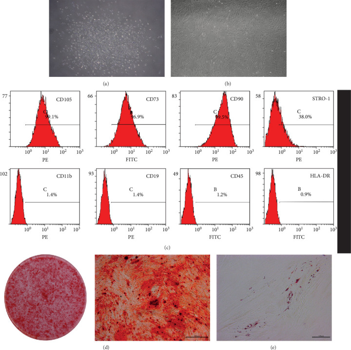
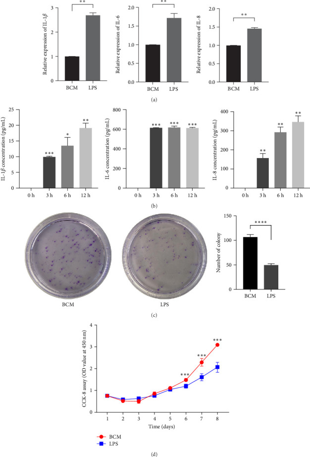
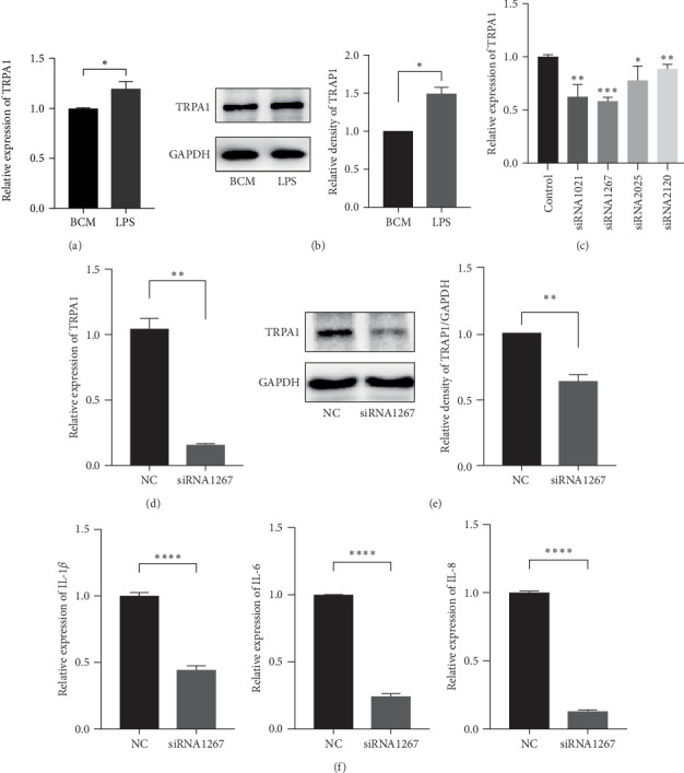
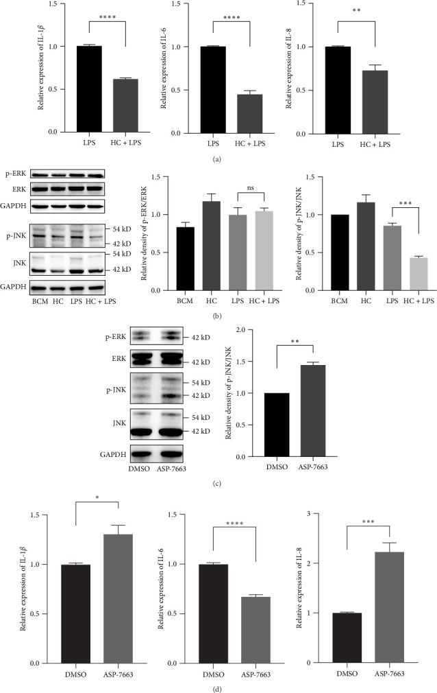
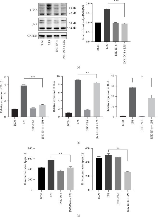
Transient receptor potential ankyrin 1 (TRPA1) molecule is an important type of transient receptor potential (TRP) cation channels, which can cause extracellular Ca2+ to flow into cells after activation. TRPA1 plays an important role in acute and chronic pain, inflammation, kidney disease, cough and asthma, osteoarthritis, cardiovascular disease, obesity, diabetes, and other diseases. In this study, the expression of interleukin (IL)-1β, IL-6, and IL-8 in periodontal ligament stem cells (PDLSCs) treated by lipopolysaccharide (LPS) and the effect of LPS on PDLSCS proliferation were detected. Meanwhile, the change in TRPA1 expression in PDLSCs treated by LPS was also assessed. By knocking down the expression of TRPA1 and using the TRPA1 antagonist HC-030031, the expression of IL-1β, IL-6, and IL-8 in PDLSCs treated by LPS was downregulated. After LPS stimulation, the proliferation ability of PDLSCs decreased, the gene expression and secretion of IL-1β, IL-6, and IL-8 increased and the gene and protein expression of TRPA1 were upregulated. Reducing the expression of TRPA1 can effectively inhibit the increase of gene expression of IL-1β, IL-6, and IL-8 after LPS stimulation, and pretreatment of PDLSCs with HC-030031 can also achieve the above effect. And research has found that HC-030031 can inhibit the phosphorylation level of JNK in PDLSCs treated by LPS. The use of JNK inhibitor JNK-IN-8 can also reduce the expression of IL-1β, IL-6, and IL-8 in PDLSCs. Finally, this study found LPS could cause the upregulation of TRPA1, and the inhibition of TRPA1 could produce an anti-inflammatory effect in PDLSCs treated by LPS due to its inhibition of JNK phosphorylation.

Keywords: JNK; PDLSCs; TRPA1.


Conflict of interest statement

The authors declare no conflicts of interest.

Figures

Figure 1
Figure 1
Isolation, cultivation, and identification of PDLSCs. P0 (A) and P3 (B) showed a typical fibroblast-like spindle appearance. Flow cytometry indicated that PDLSCs express markers of MSCs, CD73, CD90, CD105, and do not express CD11b, CD19, CD45, and HLA-DR (C). Alizarin red staining of PDLSCs under osteogenic induction medium for 21 (D). Oil red O staining of PDLSCs under adipogenic induction medium for 14 (E). B in panel (C) indicates the FITC channel of flow cytometry and C in panel (C) indicates the PE channel of flow cytometry. PDLSC, periodontal ligament stem cell.
Figure 2
Figure 2
LPS promotes the expression of proinflammatory cytokine IL-1β, IL-6, IL-8 and inhibits the proliferation of PDLSCs. Human PDLSCs were stimulated with a LPS (1 μg/mL) for 12 h, IL-1β, IL-6, and IL-8 mRNA was measured by real-time PCR (A). In addition, culture media of PDLSCs stimulated by LPS at different times were collected, IL-1β, IL-6, and IL-8 concentrations in the culture media were measured by immunoassay and the results are expressed as mean ± SEM (B). Image of crystal violet staining for the colony-forming unit assay after cultivation 7 days (C). Image of cell proliferation assay of PDLSCs treated by LPS for 8days (D). p  < 0.05, ∗∗p  < 0.01, ∗∗∗p  < 0.001,∗∗∗∗p  < 0.0001. IL, interleukin; LPS, lipopolysaccharide; PDLSC, periodontal ligament stem cell.
Figure 3
Figure 3
The expression of IL-1β, IL-6, and IL-8 were downregulated by gene silencing of TRPA1. PDLSCs were stimulated with LPS (1 μg/mL) for 12 h, the mRNA (A) and protein (B) expression of TRPA1 was measured by real-time PCR and western blot. Four siRNAs targeting TRPA1 were designed to transfect PDLSCs to lower TRPA1. After 1 day of transfection, the gene-silencing effect was measured by real-time PCR (C). PDLSCs were transfected by siRNA 1267 for 1 day, the mRNA and protein expression of TRPA1 were measured by real-time PCR and western blot (D and E). SiRNA 1267 PDLSCs and siNC PDLSCs were cultured with LPS 12 h after 2 days of transfection, IL-1β, IL-6, and IL-8 mRNA were measured by real-time PCR (F). The results are expressed as mean ± SEM. p  < 0.05, ∗∗p  < 0.01,∗∗∗p  < 0.001,∗∗∗∗p  < 0.0001. IL, interleukin; LPS, lipopolysaccharide; PDLSC, periodontal ligament stem cell.
Figure 4
Figure 4
Inhibition of TRPA1 represses the phosphorylation of JNK. PDLSCs were pretreated by HC-030031 with or without for 3 h, followed by incubation by LPS (1 μg/mL) for 12 h, the expression of IL-1β, IL-6, and IL-8 were measured by real-time PCR (A). Then the expression of p-ERK, ERK, p-JNK, and JNK in PDLSCs treated by LPS (1 μg/mL) for 3 h were measured by western blot (B). TRPA1 agonist ASP-7663 was used to stimulate PDLSCs for 3 h, the protein levels of p-ERK, ERK, p-JNK, and JNK were measured by western blot and the gene expressions of IL-1β, IL-6, and IL-8 were measured by real-time PCR (C and D). The results are expressed as mean ± SEM. p  < 0.05, ∗∗p  < 0.01, ∗∗∗p  < 0.001, ∗∗∗∗p  < 0.0001. IL, interleukin; LPS, lipopolysaccharide; PDLSC, periodontal ligament stem cell.
Figure 5
Figure 5
Inhibition of the phosphorylation of JNK can inhibit the expression of inflammatory cytokines in PDLSCs treated by LPS. PDLSCs were pretreated with or without JNK antagonist JNK-IN-8 for 3 h, followed by incubation by LPS (1 μg/mL) for 12 h, the protein levels of p-JNK and JNK were measured by western blot (A), the gene expressions of IL-1β, IL-6, and IL-8 were measured by real-time PCR and the concentrations of IL-6 and IL-8 in the culture media were measured by immunoassay (B and C). The results are expressed as mean ± SEM. p  < 0.05, ∗∗p  < 0.01, ∗∗∗p  < 0.001, ∗∗∗∗p  < 0.0001. IL, interleukin; LPS, lipopolysaccharide; PDLSC, periodontal ligament stem cell.

Similar articles

References

    1. Tonetti M. S., Jepsen Søren, Jin L., Otomo-Corgel J. Impact of the Global Burden of Periodontal Diseases on Health, Nutrition and Wellbeing of Mankind: A Call for Global Action. Journal of Clinical Periodontology . 2017;44(5):456–462. doi: 10.1111/jcpe.12732. - DOI - PubMed
    1. Kwon T. H., Lamster I. B., Levin L. Current Concepts in the Management of Periodontitis. International Dental Journal . 2021;71(6):462–476. doi: 10.1111/idj.12630. - DOI - PMC - PubMed
    1. McCulloch C. A. G. Origins and Functions of Cells Essential for Periodontal Repair: The Role of Fibroblasts in Tissue Homeostasis. Oral Diseases . 1995;1(4):271–278. doi: 10.1111/j.1601-0825.1995.tb00193.x. - DOI - PubMed
    1. Zhang Z., Deng M., Hao M., Tang J. Periodontal Ligament Stem Cells in the Periodontitis Niche: Inseparable Interactions and Mechanisms. Journal of Leukocyte Biology . 2021;110(3):565–576. doi: 10.1002/JLB.4MR0421-750R. - DOI - PubMed
    1. Jin S., Jiang H., Sun Y., et al. Osteogenic Differentiation of Periodontal Membrane Stem Cells in Inflammatory Environments. Open Life Sciences . 2022;17(1):1240–1248. doi: 10.1515/biol-2022-0474. - DOI - PMC - PubMed

LinkOut - more resources