. 2024 Jul;19(3):133-139.
doi: 10.1016/j.joto.2024.07.001. Epub 2024 Oct 19.

Loss of Fascin2 increases susceptibility to cisplatin-induced hearing impairment and cochlear cell apoptosis in mice

Affiliations

Loss of Fascin2 increases susceptibility to cisplatin-induced hearing impairment and cochlear cell apoptosis in mice

Yan Wang et al. J Otol. 2024 Jul.

Abstract

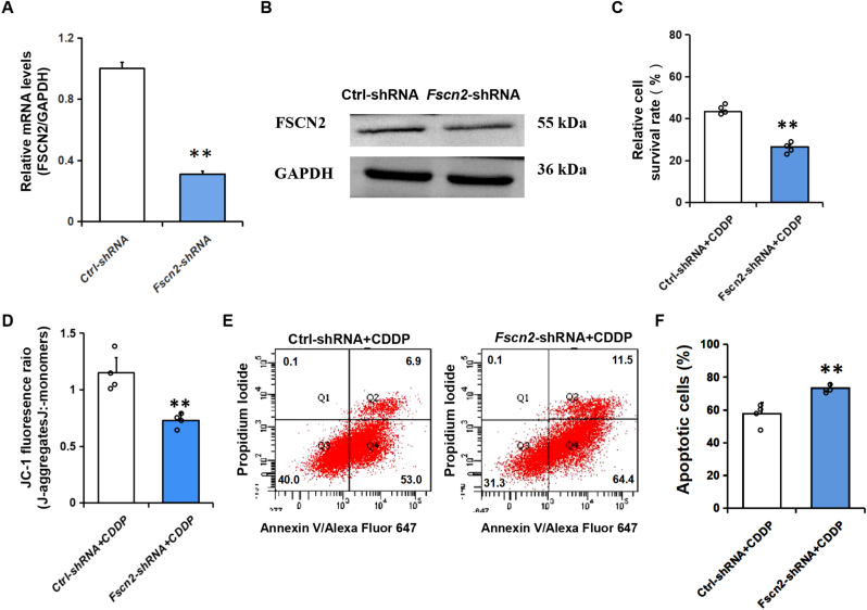
Objectives: Deletion of Fscn2 gene in mice has been linked to progressive hearing loss and degeneration of cochlear cells. Cisplatin, an antitumor drug, can cause various side effects, including ototoxicity. The aim of this study was to investigate the effects of Fscn2 on cisplatin-induced hearing impairment in mice and to explore the possible mechanism.

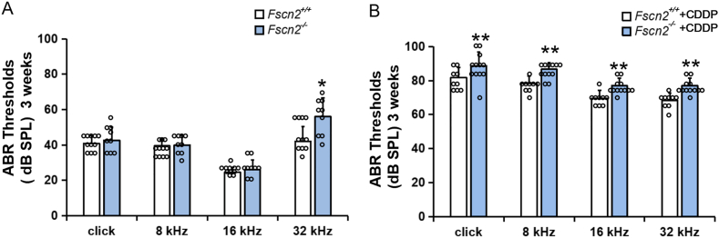
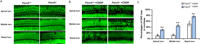
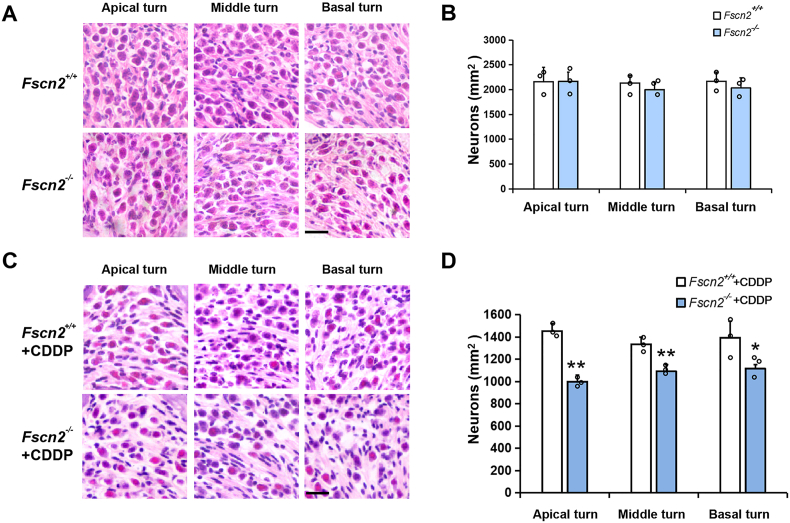
Methods: Two-week-old Fscn2 +/+ mice and Fscn2 -/- mice were treated with two doses of cisplatin, with a 3-day recovery period in between. ABR (auditory evoked brain stem response) thresholds were measured and cochlear pathology was observed at 3 weeks of age.

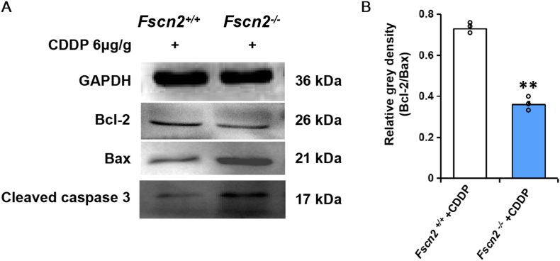
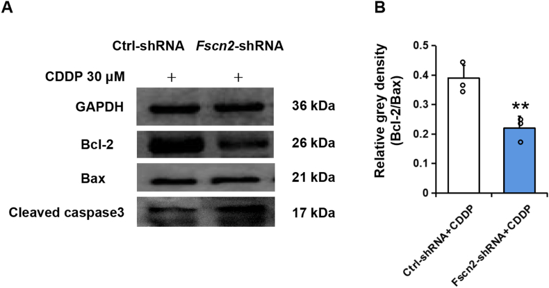
Results: Both Fscn2 +/+ and Fscn2 -/- mice showed hearing loss under the effect of cisplatin, but the impairment was more severe in Fscn2 -/- mice. Further experiments showed that the percentages of outer hair cell (OHC) and spiral ganglion neuron (SGN) loss were significantly higher in cisplatin-treated Fscn2 -/- mice compared to Fscn2 +/+ mice. Additionally, knockdown of Fscn2 in HEI-OC1 cells worsened cisplatin-induced cell apoptosis.

Conclusion: FSCN2 mediates reduction of CDDP induced ototoxicity by inhibiting cell apoptosis.

Keywords: Apoptosis; Cisplatin; Fascin2; Mouse; Ototoxicity.


Conflict of interest statement

The authors report no conflicts of interest. The authors alone are responsible for the content and writing of the paper.

Figures

Fig. 1
Fig. 1
ABR threshold tests in the 4 mouse groups. Each point represents the mean ABR threshold for one group, with an error bar indicating SD from the mean. The values of the number of individual animals' were overplotted. (n = 11 and 9 for untreated Fscn2+/+ and Fscn2−/− mice; n = 9 and 12 for treated Fscn2+/+ and Fscn2−/− mice, respectively). (A) The results show that at 3 weeks of age, there was no significant difference in the ABR thresholds between the two untreated mouse groups except for 32 kHz. (B) For the CDDP treated mice, ABR thresholds of Fscn2−/− mice were significantly higher than that of Fscn2+/+mice at the stimulation frequencies of click, 8 kHz, 16 kHz and 32 kHz. ABR; auditory-evoked brainstem response; SPL, sound pressure levels; dB, decibel. ∗P < 0.05, ∗∗P < 0.01.
Fig. 2
Fig. 2
Observation of OHCs in the mouse cochleae after the treatment with CDDP. (A) Phalloidin labeled OHC of the cochleae of untreated Fscn2+/+ mice and Fscn2−/− mice at 3 weeks of age are shown in green. (B) OHC loss was obvious and cell arrangement was irregular in the CDDP treated Fscn2+/+ and Fscn2−/− mice at 3 weeks of age, especially at the basal turns. (C) Percentages of OHC loss in the 3 turns of cochleae of Fscn2+/+ and Fscn2−/− mice. Scale bar = 10 μm. n = 5 for each group. ∗∗P < 0.01.
Fig. 3
Fig. 3
Evaluation of SGN density by HE staining. (A) SGNs were observed in the cochleae of untreated Fscn2+/+and Fscn2−/− mice at 3 weeks of age. (B) There was no significant difference in the densities of SGNs between the two untreated mouse groups. (C) SGNs were observed in the cochleae of Fscn2+/+and Fscn2−/− mice after the treatment of CDDP. (D) Densities of SGNs in the CDDP treated mice. Fscn2−/− mice had fewer SGNs than Fscn2+/+ mice in the apical, middle and basal turns. Scale bar = 20 μm.n = 3 for each group. ∗P < 0.05; ∗∗P < 0.01.
Fig. 4
Fig. 4
Expression of Bax, Bcl-2 and cleaved caspase3 in the inner ears of Fscn2−/−mice after the treatment with CDDP. (A) Levels of Bax, Bcl-2 and cleaved caspase3 were detected in the inner ears of Fscn2+/+ and Fscn2−/− mice by Western blot. (B) Cell apoptosis was evaluated by calculating the ratio of relative gray densities of Bcl2/Bax. n = 3 for each group. ∗P < 0.01.
Fig. 5
Fig. 5
Proliferation rate and apoptotic ratio of HEI-OC1 cells with low Fscn2 expression. (A–B) Evaluation of mRNA levels with RT-PCR and FSCN2 levels by Western blot in Ctrl-shRNA infected cells (Ctrl-shRNA) and Fscn2-shRNA infected cells (Fscn2-shRNA). (C) Cell proliferation rate was measured by CCK-8 assay after the treatment with 30 μM CDDP for 24 h. (D) Cell apoptosis was evaluated by JC-1 assay. A decrement in the ratio of J-aggregates to J-monomers is an indicator of early cell apoptosis. (E–F) Flow cytometry was used to detect apoptotic cells exposed to 30 μM CDDP for 24 h n = 4 for each group. ∗P < 0.05; ∗∗P < 0.01.
Fig. 6
Fig. 6
Expression of apoptosis-related proteins in Fscn2-shRNA infected cells after the treatment with CDDP. (A) Levels of Bcl-2, Bax and cleaved caspase3 were detected by Western blot. (B) The ratio of Bcl-2 to Bax represents the relative gray density of the two proteins in Western blot in Ctrl-shRNA infected cells (Ctrl-shRNA) or in Fscn2-shRNA infected cells (Fscn2-shRNA). n = 3 for each group. ∗P < 0.01.

Similar articles

References

    1. Abercrombie M., Johnson M.L. Quantitative histology of Wallerian degeneration: I. Nuclear population in rabbit sciatic nerve. J. Anat. 1946;80:37–50. - PubMed
    1. Angeli S., Lin X., Liu X.Z. Genetics of hearing and deafness. Anat. Rec. 2012;295:1812–1829. - PMC - PubMed
    1. Gao L., Ge R., Xie G., Yao D., Li P., Wang O., Ma X., Han F. Hearing improvement in A/J mice via the mouse nerve growth factor. Clin Exp Otorhinolaryngol. 2017;10:303–308. - PMC - PubMed
    1. Guillery R.W. On counting and counting errors. J. Comp. Neurol. 2002;447:1–7. - PubMed
    1. Hashimoto Y., Kim D.J., Adams J.C. The roles of fascins in health and disease. J. Pathol. 2011;224:289–300. - PubMed

LinkOut - more resources