. 2024 Dec 20;14(24):e5143.
doi: 10.21769/BioProtoc.5143.

Cryo-SEM Investigation of Chlorella Using Filter Paper as Substrate

Affiliations

Cryo-SEM Investigation of Chlorella Using Filter Paper as Substrate

Peng Wan et al. Bio Protoc. .

Abstract

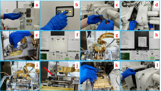
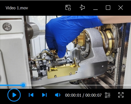
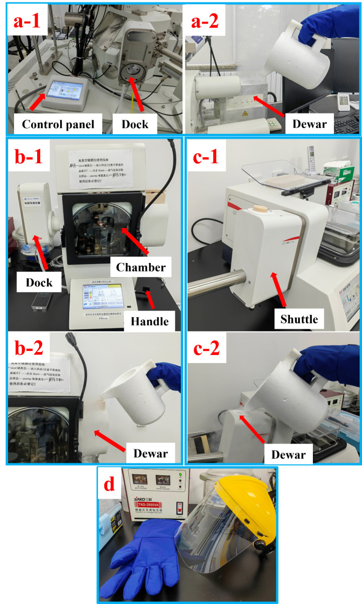
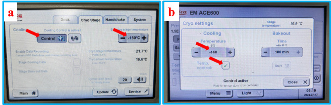
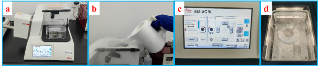
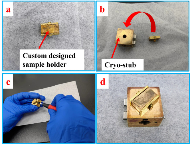
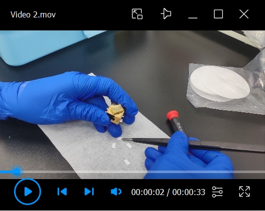
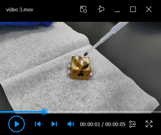
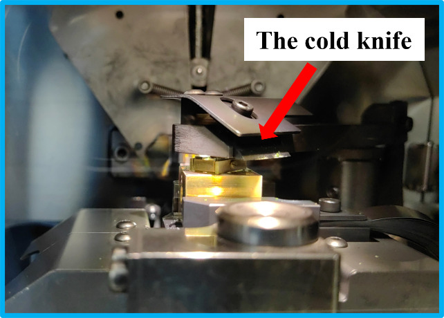
Cryo-electron microscopy (cryo-EM) is a powerful technique capable of investigating samples in a hydrated state, compared to conventional high-vacuum electron microscopy that requires samples to be completely dry. During the drying process, numerous features and details may be lost due to damage caused by dehydration. Cryo-EM circumvents these problems by cryo-fixing the samples, thereby retaining the intact and original features of hydrated samples. This protocol describes a step-by-step cryo-scanning electron microscopy (cryo-SEM) experimental procedure with Chlorella sorokiniana as the subject. By employing filter paper as the sample substrate, we propose a simple and reliable method for cryo-fixation and freeze-fracture of Chlorella sorokiniana in water suspension. The advantage of using filter paper as a substrate lies in its ability to support a thin film of sample, enabling a cold knife to make a cut effortlessly and produce a clean freeze-fractured surface for SEM investigation. By following the approach described in this protocol, both the internal structure and surface morphology of Chlorella sorokiniana can be easily resolved with high quality. This protocol is highly versatile and can be applied to samples dispersed in water or solvents, including cyanobacterial cells, algal cells, and any kind of sample that can be adsorbed onto filter paper. Key features • Introducing a reliable way for ideal freeze-fracture of a water-suspended sample using filter paper as substrate. • Detailed step-by-step descriptions of the entire experiment, covering how to operate the instruments and including some practical experimental tips. Graphical overview.

Keywords: Algae; Chlorella.; Cryo-SEM; Filter paper; Freeze fracture.


Conflict of interest statement

Competing interestsThe authors declare no competing financial interests.

Figures

Figure 1.
Figure 1.. Step-by-step procedures of the mounting of cryo-stage.
a. Loosening the spring snap of the airlock of the SEM chamber. b. Venting of the SEM chamber by pressing the “vent” button on the front panel. c. Unscrewing the bolts on the chamber door. d. Pulling open the chamber door. e. Sliding in the cryo-stage into the dovetail of the SEM stage. f. Setting the type and height of the cryo-stage. g. Mounted cryo-stage on the stage of SEM. h. Setting the coordinates of the cryo-stage. i. Cryo-stage in ready position for cryo-SEM experiment. j. Mounting the stopping piece next to the cryo-stage. k. Mounting the cold trap on top of the cryo-stage. l. Closing the chamber door.
Video 1.
Video 1.. Mounting the cryo-stage into the dovetail of the SEM stage
Figure 2.
Figure 2.. Overview of the VCT500, ACE600, and the shuttle, along with the cooling processes.
a-1. Control panel and cryo-dock of VCT500. a-2. Filling liquid nitrogen into the dewar of VCT500. b-1. ACE600 with red arrows indicating the dock, chamber, and handle of the cold knife. b-2. Filling liquid nitrogen into the dewar of ACE600. c-1. The shuttle attached to the VCM. c-2. Filling liquid nitrogen into the dewar of the shuttle. d. Mask and cryo-gloves for safety when handling liquid nitrogen.
Figure 3.
Figure 3.. Setting the temperatures and the activation of cooling on the control panel of VCT500 (a) and ACE600 (b)
Figure 4.
Figure 4.. Overview of the VCM and preparations for cryo-fixation.
a. Picture of VCM with the shuttle attached. b. Filling liquid nitrogen into the reservoir of VCM. c. Control panel of VCM. d. Liquid nitrogen bath ready for cryo-fixation.
Figure 5.
Figure 5.. Setup of the custom-designed sample holder on the cryo-stub
Video 2.
Video 2.. Fixation of the filter paper strips onto the sample holder
Video 3.
Video 3.. Appling a droplet of Chlorella sorokiniana onto the filter paper substrate
Video 4.
Video 4.. Cryo-fixation of the sample in the liquid nitrogen bath
Video 5.
Video 5.. Retrieving the cryo-stub from the VCM to the shuttle
Video 6.
Video 6.. Removal of the shuttle from the VCM and its attachment to the ACE600
Video 7.
Video 7.. Transfer of the cryo-stub onto the cryo-stage of the ACE600
Figure 6.
Figure 6.. Setting the height of the cold knife.
a. Selecting the freeze-fracture module from the main menu. b. Setting the height of the cold knife.
Video 8.
Video 8.. Freeze-fracture of the samples with cold knife
Figure 7.
Figure 7.. Bent filter paper substrate showing both the cross-section and the surface
Video 9.
Video 9.. Clearing of the debris after freeze-fracture
Figure 8.
Figure 8.. Setting up the freeze-etching parameters.
a. Selection of the freeze-etching module from the main menu. b. Setting up the temperature of etching, the holding time, and the ramp. c. Interface of freeze-etching.
Figure 9.
Figure 9.. Cold knife right on top of the sample during freeze-etching
Figure 10.
Figure 10.. Sputter coating.
a. Setting up the parameters for sputter coating. b. Interface of sputter coating.
Video 10.
Video 10.. Retrieving the cryo-stub from ACE600 back to the shuttle
Video 11.
Video 11.. Transferring the cryo-stub from the ACE600 to the cryo-stage of SEM
Video 12.
Video 12.. SEM operation
Video 13.
Video 13.. Retrieving the cryo-stub from the SEM back to the shuttle
Figure 11.
Figure 11.. Baking out all the equipment.
a. VCT500. b. ACE600. c. VCM and the shuttle.
Figure 12.
Figure 12.. SEM images of freeze-fractured samples showing the surface and internal structure.
a. Fractured sample of Chlorella sorokiniana without etching. b. After etching.

Similar articles

References

    1. Fujikawa S. and Endoh K.(2020). Cryo-Scanning Electron Microscopy to Study the Freezing Behavior of Plant Tissues. Methods Mol Biol. 2156: 99-117. - PubMed
    1. Gosden R. G., Yin H., Bodine R. J. and Morris G. J.(2010). Character, distribution and biological implications of ice crystallization in cryopreserved rabbit ovarian tissue revealed by cryo-scanning electron microscopy. Hum Reprod. 25(2): 470-478. - PubMed
    1. Nakatomi R., Hayashida T., Fujimoto K., Tohyama K. and Hashikawa T.(2005).Cryo-SEM and subsequent TEM examinations of identical neural tissue specimen. Brain Res Protoc. 14(2): 100-106. - PubMed
    1. Read N. D. and Jeffree C. E.(1991). Low‐temperature scanning electron microscopy in biology. J Microsc. 161(1): 59-72. - PubMed
    1. Yan H., Wang Y., Zhang J., Cui X., Wu J., Zhou J., Chen Y., Lu J., Guo R., Ou M., et al. .(2021). Rice Root Hair Phenotypes Imaged by Cryo-SEM. Bio Protoc. 11(11): e4037. - PMC - PubMed

LinkOut - more resources