. 2024 Nov 11;10(12):2213-2228.
doi: 10.1021/acscentsci.4c01377. eCollection 2024 Dec 25.

Examining the Relationship between Aptamer Complexity and Molecular Discrimination of a Low-Epitope Target

Affiliations

Examining the Relationship between Aptamer Complexity and Molecular Discrimination of a Low-Epitope Target

Linlin Wang et al. ACS Cent Sci. .

Abstract

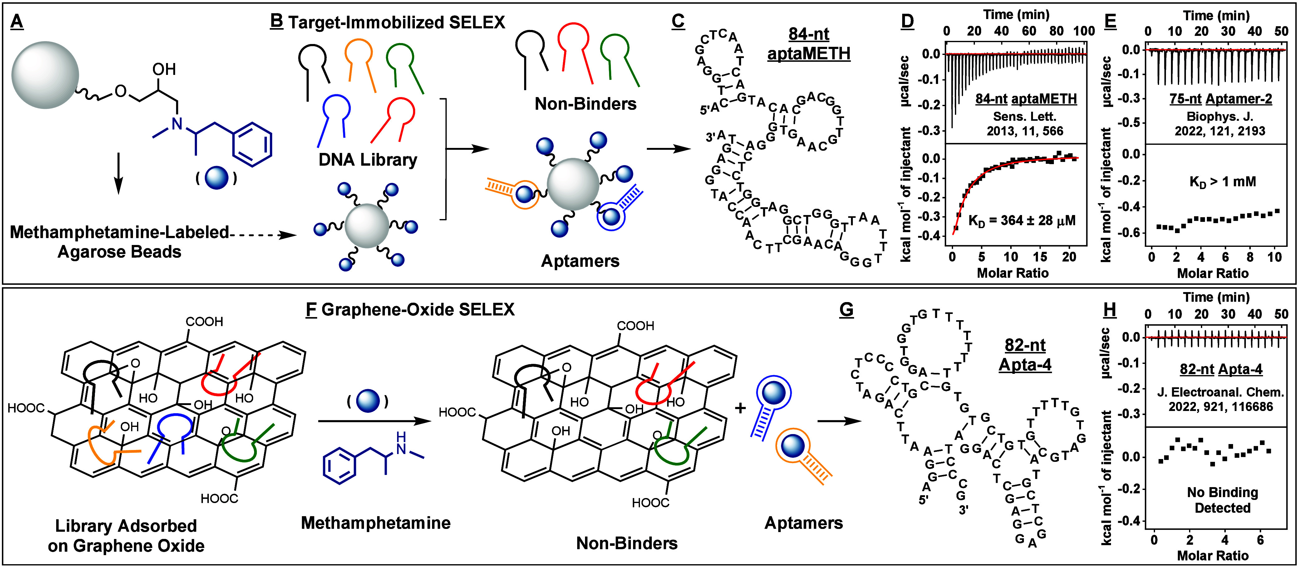
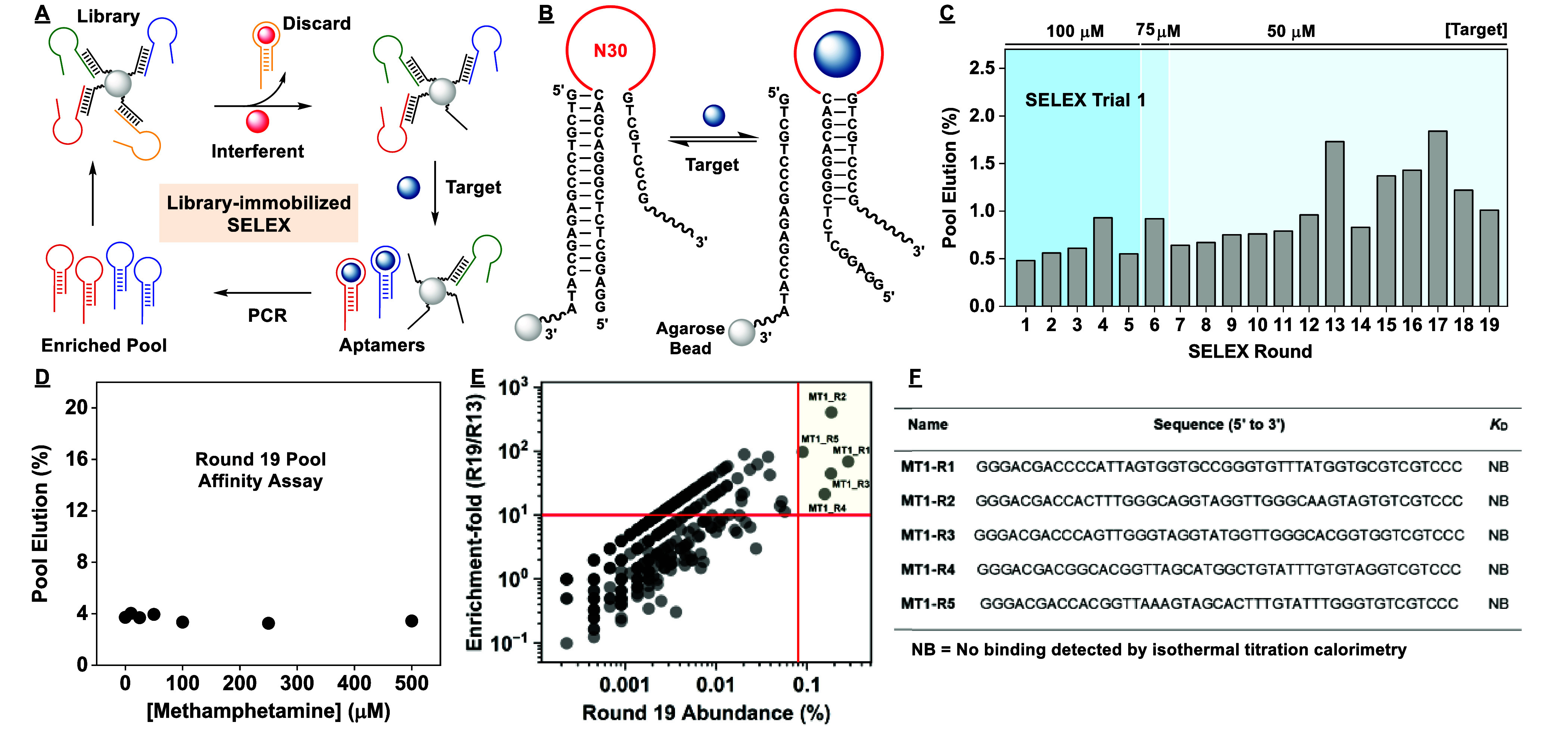
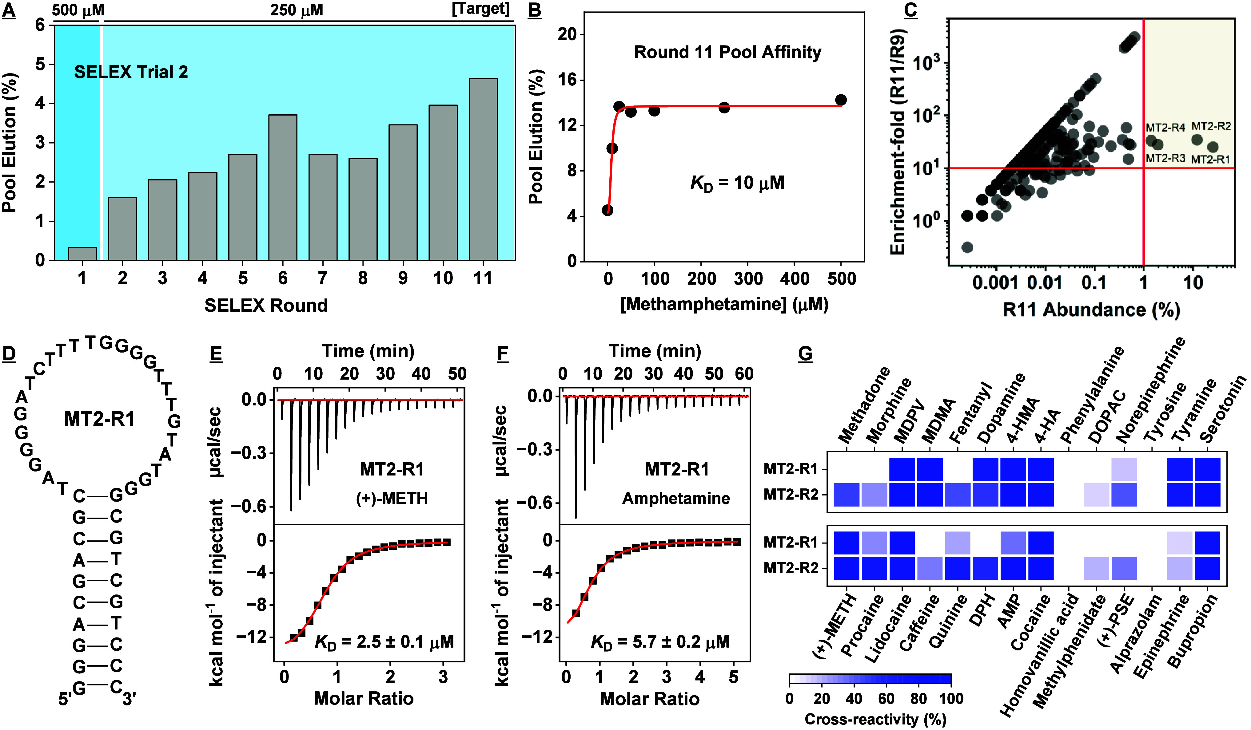
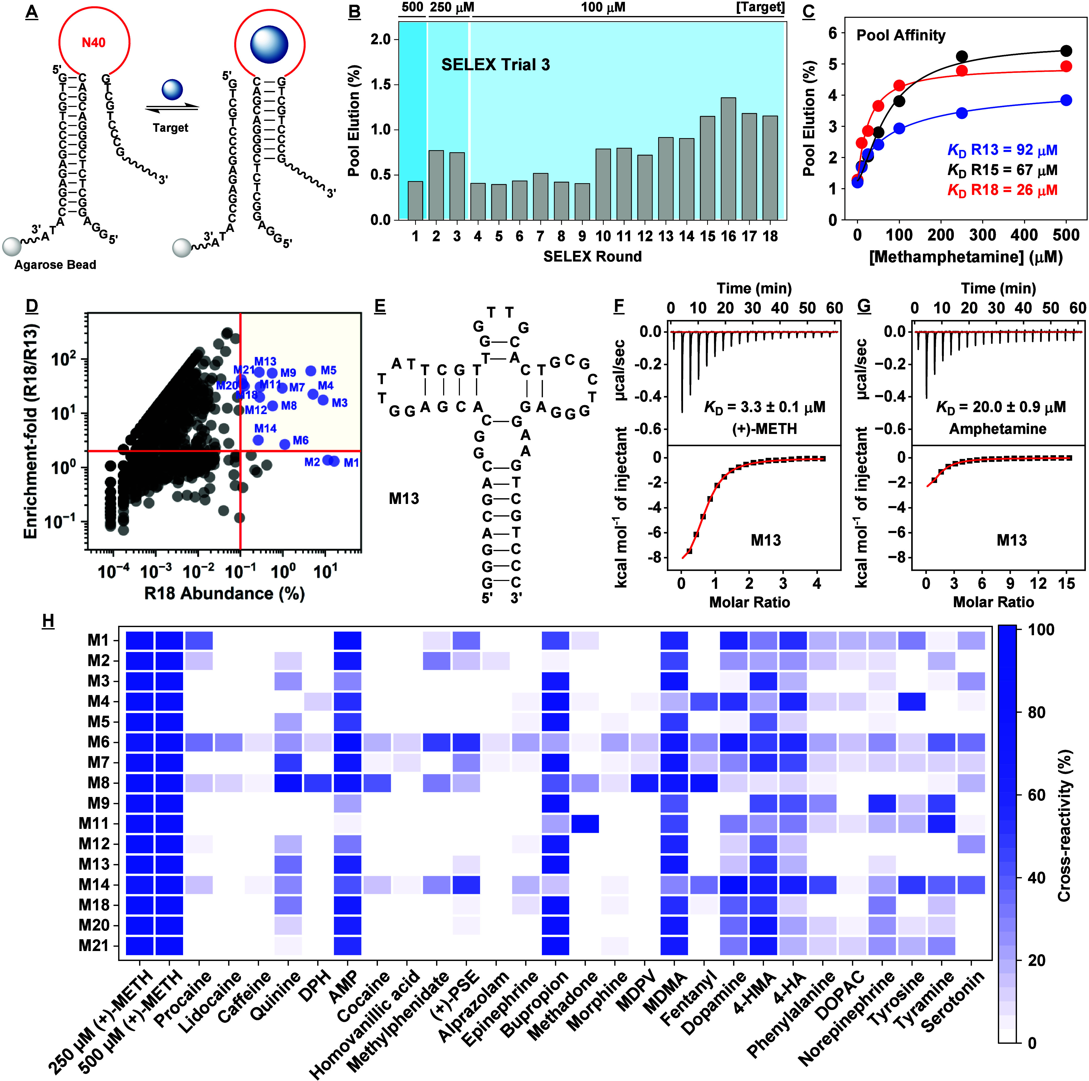
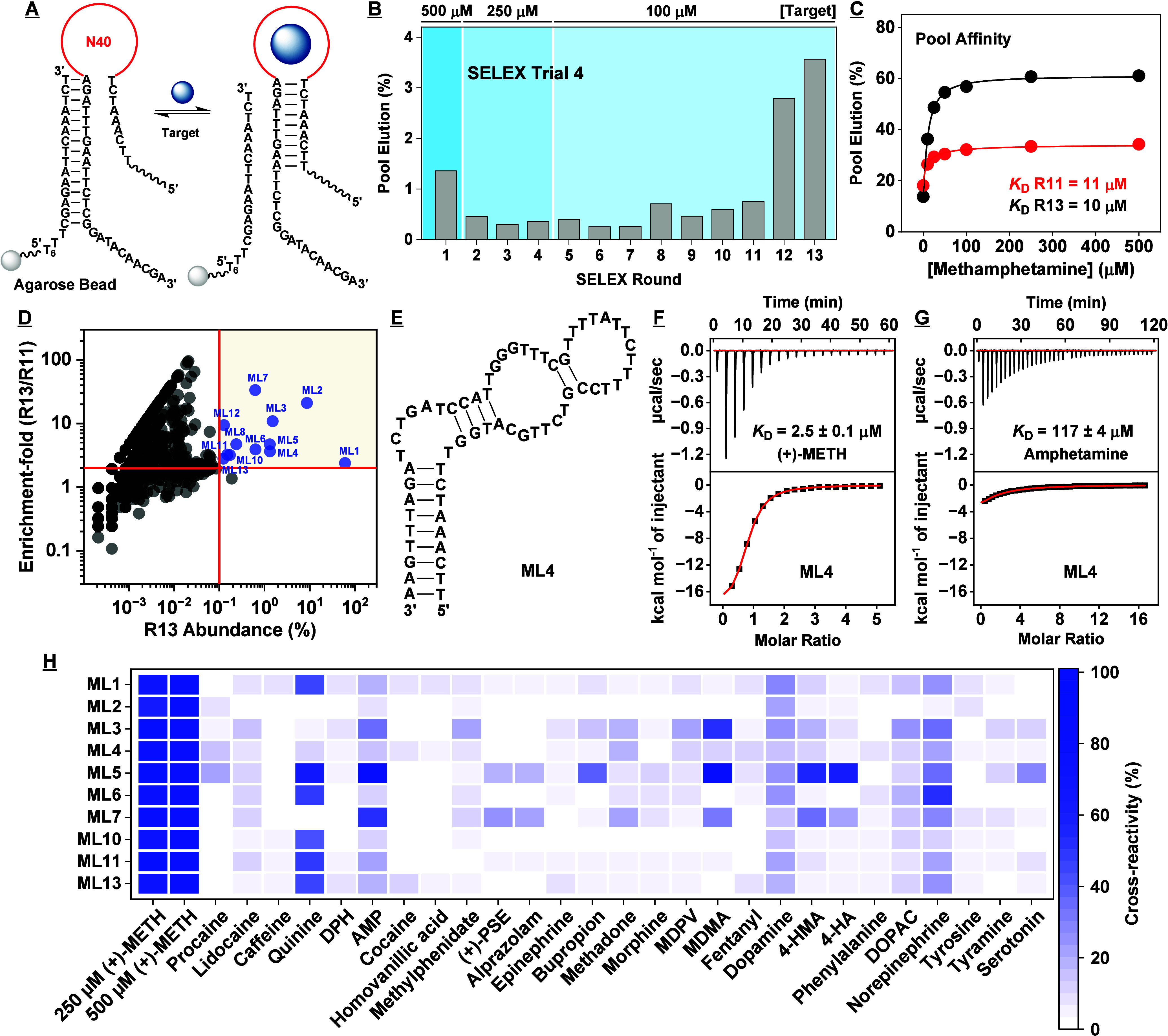
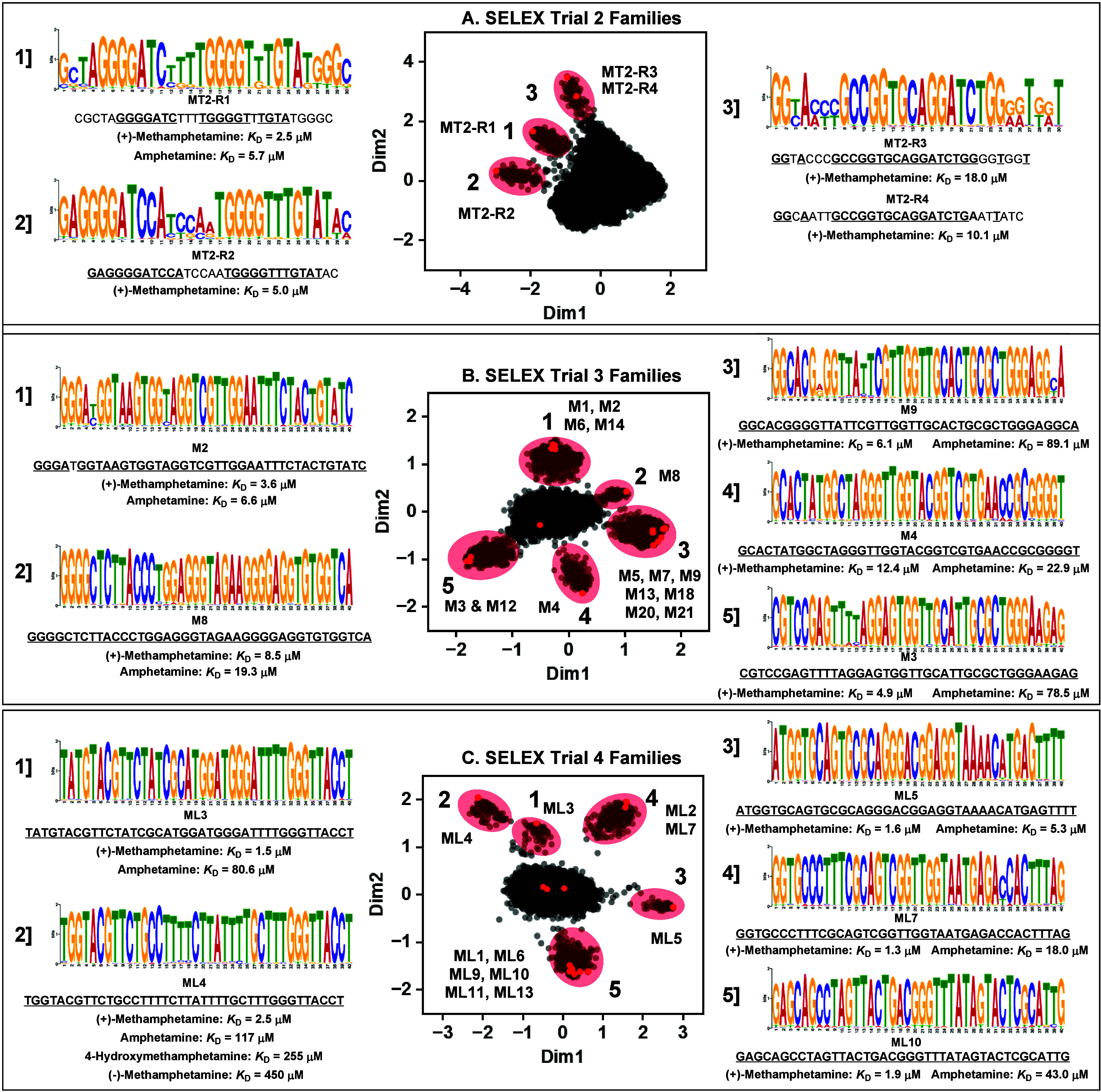
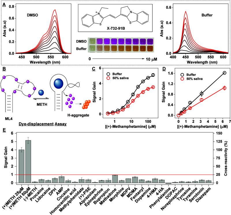
Aptamers are oligonucleotide-based affinity reagents that are increasingly being used in various applications. Systematic evolution of ligands by exponential enrichment (SELEX) has been widely used to isolate aptamers for small-molecule targets, but it remains challenging to generate aptamers with high affinity and specificity for targets with few functional groups. To address this challenge, we have systematically evaluated strategies for optimizing the isolation of aptamers for (+)-methamphetamine, a target for which previously reported aptamers have weak or no binding affinity. We perform four trials of library-immobilized SELEX against (+)-methamphetamine and demonstrate that N30 libraries do not yield high-quality aptamers. However, by using a more complex N40 library design, stringent counter-SELEX, and fine-tuned selection conditions, we identify aptamers with high affinity for (+)-methamphetamine and better selectivity relative to existing antibodies. Bioinformatic analysis from our selections reveals that high-quality aptamers contain long conserved motifs and are more informationally dense. Finally, we demonstrate that our best aptamer can rapidly detect (+)-methamphetamine at toxicologically relevant concentrations in saliva in a colorimetric dye-displacement assay. The insights provided here demonstrate the challenges in generating high-quality aptamers for low complexity small-molecule targets and will help guide the design of more efficient future selection efforts.


Conflict of interest statement

The authors declare no competing financial interest.

Figures

Figure 1
Figure 1
Affinity characterization of aptamers previously isolated for methamphetamine by other groups. Aptamers have been previously isolated using target-immobilized SELEX, in which (A) methamphetamine is conjugated to agarose beads via its amino group. (B) These beads are incubated with the DNA library, and binders are physically partitioned from binding-incompetent sequences. (C) Ebrahimi et al. identified 84-nt aptaMETH in this way and reported that this aptamer binds (+)-methamphetamine with a KD of 100 nM. (D) However, our isothermal calorimetry (ITC) results showed a far higher KD of 364 μM in their selection buffer. (E) Sester et al. used a similar approach to isolate 75-nt Aptamer-2, with a reported KD of 244 nM, but our ITC results again indicated a higher KD of >1 mM for (+)-methamphetamine in their reported selection buffer. (F) Bor et al. utilized graphene-oxide SELEX to isolate DNA aptamers for methamphetamine, based on binding-induced desorption of target-specific aptamers from graphene oxide. (G) The resulting 82-nt Apta-4 aptamer reportedly bound methamphetamine with a KD of 1.3 μM. (H) In contrast, our ITC results indicate no binding at all in their reported selection buffer.
Figure 2
Figure 2
Results of the first SELEX trial. (A) Simplified scheme of library-immobilized SELEX. (B) The aptamer is initially hybridized to a biotinylated cDNA strand immobilized on agarose microbeads. Aptamer-target binding displaces the aptamer from the cDNA, releasing the aptamer into solution. (C) Pool elution in each round of the first SELEX trial. (D) Binding affinity of the Round 19 pool to (+)-methamphetamine was determined using a gel-elution assay. The pool displayed no apparent affinity for the target. (E) Round 13 and 19 pools were subjected to high-throughput sequencing (HTS). Enrichment-fold between Rounds 13 and 19 was plotted as a function of Round 19 abundance. Five top-ranked sequences (upper right) had abundance >0.08% and enrichment-fold >10. (F) None of these sequences displayed measurable affinity for (+)-methamphetamine based on ITC.
Figure 3
Figure 3
Results of the second SELEX trial. (A) Pool elution by target in each round of the second SELEX trial. (B) Binding affinity of the Round 11 pool to (+)-methamphetamine as determined using a gel-elution assay. (C) Enrichment-fold between Rounds 9 and 11 plotted as a function of Round 11 abundance. (D) Secondary structure of the most abundant aptamer discovered in this trial, MT2-R1. The binding affinity of MT2-R1 to (E) (+)-methamphetamine and (F) amphetamine was determined using ITC. (G) We assessed the specificity of MT2-R1 and MT2-R2 toward several interferents using the exonuclease digestion assay. Heat-map indicates cross-reactivity relative to (+)-METH. The concentration of target and interferent was 250 μM, except for alprazolam, which was 50 μM due to solubility limits.
Figure 4
Figure 4
Results for the third SELEX trial. (A) In this trial, we employed an N40 library for library-immobilized SELEX. (B) Pool elution by (+)-methamphetamine for each round of SELEX. (C) Binding affinity for (+)-methamphetamine as determined using the gel-elution assay for the Round 13, 15, and 18 pools. (D) Enrichment-fold of sequences between Rounds 13 and 18 plotted as a function of Round 18 abundance. Sequences with abundance > 0.1% and enrichment-fold > 2 are named and marked in blue. (E) Secondary structure of one of the highly enriched aptamers, M13, as predicted by NUPACK. Binding affinity of M13 to (F) (+)-methamphetamine and (G) amphetamine as determined using ITC. (H) Specificity of aptamers discovered in this trial to a panel of interferents as assessed via exonuclease digestion assay. Heat-map indicates cross-reactivity relative to 250 μM (+)-METH. The concentration of interferent employed was 250 μM, but 100 μM for quinine and 50 μM for alprazolam due to solubility limitations.
Figure 5
Figure 5
Results for the fourth SELEX trial. (A) In this trial, we employed an N40 library for library-immobilized SELEX. (B) Pool elution by the target for each round of SELEX. (C) Binding affinity for (+)-methamphetamine based on a gel-elution assay for the Round 11 and 13 pools. (D) Enrichment-fold of sequences between Rounds 11 and 13 plotted as a function of Round 13 abundance. Sequences with abundance >0.1% and enrichment-fold >2 are named and marked in blue. (E) Secondary structure of aptamer ML4 as predicted by NUPACK. Binding affinity of ML4 for (F) (+)-methamphetamine and (G) amphetamine as determined using ITC. (H) Specificity of aptamers discovered in this trial to a panel of interferents as assessed via exonuclease digestion assay. Heat-map indicates cross-reactivity relative to 500 μM (+)-methamphetamine. The concentration of interferent employed was 250 μM; alprazolam was 50 μM.
Figure 6
Figure 6
Identification of aptamer families and conserved sequence motifs from each trial of SELEX for (+)-methamphetamine via bioinformatic analysis. Families and motifs discovered in the final pool of the (A) second, (B) third, and (C) fourth SELEX trials. Plots in the middle represent the sequence space produced by Raptgen, with each dot representing a unique sequence. Aptamers close to each other in space are related to each other in sequence. Aptamer families in these plots are highlighted in red, and representative members are named. The primary motif in each family was determined using GLAM2, and a representative aptamer of that family is listed below the sequence logo along with its target-binding affinity and affinity for structurally related analogs of (+)-methamphetamine.
Figure 7
Figure 7
Colorimetric detection of (+)-methamphetamine with an aptamer-based dye-displacement assay. (A) Absorbance spectra of the cyanine dye X-732–91B dissolved in DMSO (left) and aqueous buffer (right) at concentrations of 0–10 μM. The structure of the dye is shown at the top center, and a photograph of solutions containing various concentrations of dye is shown at bottom center. (B) Scheme of the dye-displacement assay using aptamer ML4 and X-732–91B. Target binding displaces the dye from the aptamer into solution, causing the dye to aggregate and inducing a concomitant color change. (C) Calibration curve of this assay in both buffer (black) and 50% saliva (red). (D) Response of the assay to 0–6.4 μM (+)-methamphetamine. (E) Assay cross-reactivity to 50 μM interferents. The red line demarcates 25% cross-reactivity relative to 25 μM (+)-methamphetamine.

Similar articles

Cited by

References

    1. Ellington A. D.; Szostak J. W. In Vitro Selection of RNA Molecules That Bind Specific Ligands. Nature 1990, 346 (6287), 818–822. 10.1038/346818a0. - DOI - PubMed
    1. Tuerk C.; Gold L. Systematic Evolution of Ligands by Exponential Enrichment: RNA Ligands to Bacteriophage T4 DNA Polymerase. Science 1990, 249 (4968), 505–510. 10.1126/science.2200121. - DOI - PubMed
    1. Dunn M. R.; Jimenez R. M.; Chaput J. C. Analysis of Aptamer Discovery and Technology. Nat. Rev. Chem. 2017, 1 (10), 0076.10.1038/s41570-017-0076. - DOI
    1. Yu H.; Alkhamis O.; Canoura J.; Liu Y.; Xiao Y. Advances and Challenges in Small-Molecule DNA Aptamer Isolation, Characterization, and Sensor Development. Angew. Chem., Int. Ed. 2021, 60 (31), 16800–16823. 10.1002/anie.202008663. - DOI - PMC - PubMed
    1. Liu J.; Cao Z.; Lu Y. Functional Nucleic Acid Sensors. Chem. Rev. 2009, 109 (5), 1948–1998. 10.1021/cr030183i. - DOI - PMC - PubMed

LinkOut - more resources