Review
. 2024 Dec 24:19:13901-13923.
doi: 10.2147/IJN.S486858. eCollection 2024.

The Genetic and Epigenetic Toxicity of Silica Nanoparticles: An Updated Review

Affiliations
Review

The Genetic and Epigenetic Toxicity of Silica Nanoparticles: An Updated Review

Manjia Zheng et al. Int J Nanomedicine. .

Abstract

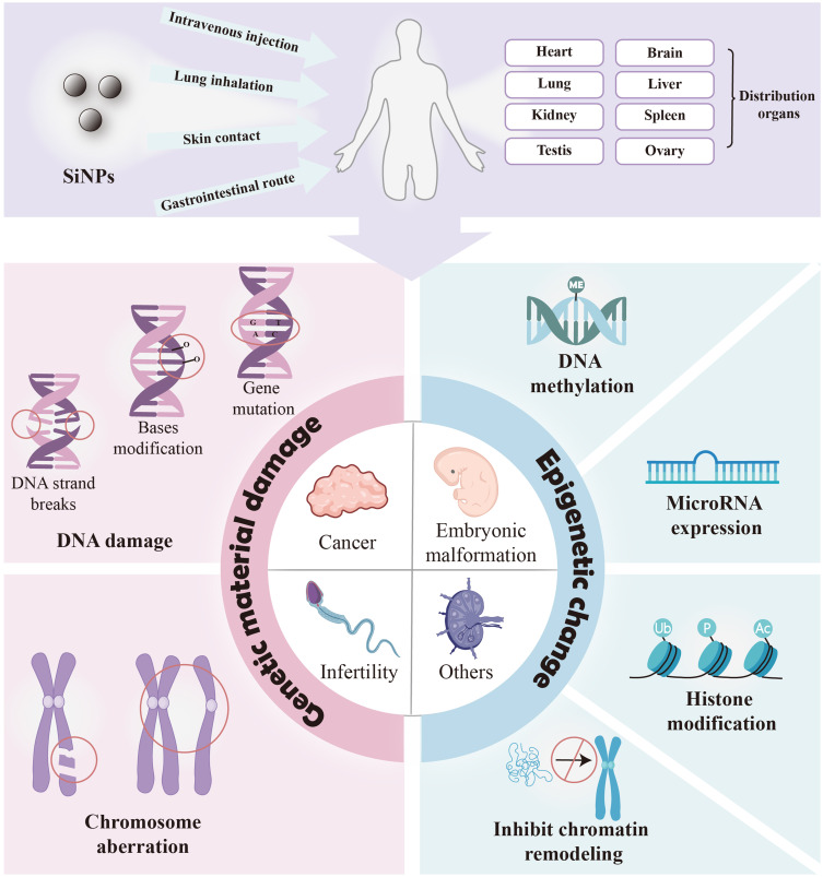
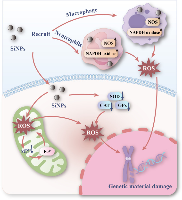
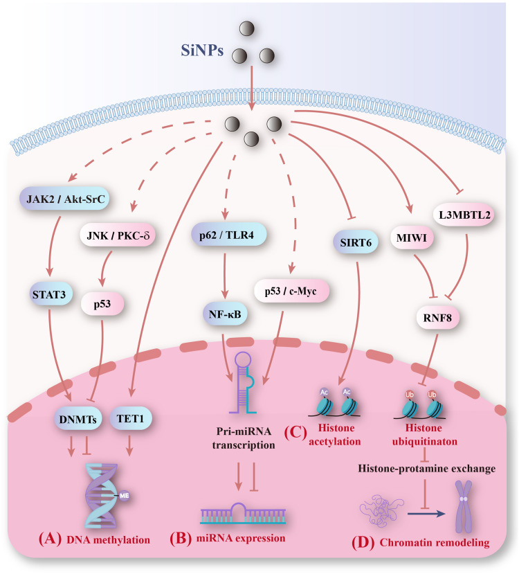
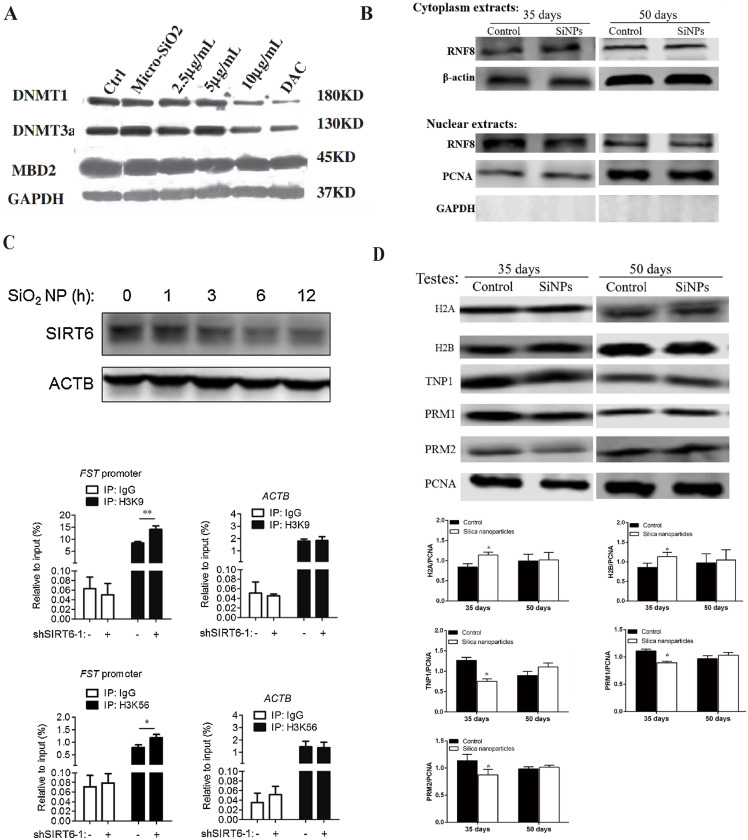
Silica nanoparticles (SiNPs) are widely used in biomedical fields, such as drug delivery, disease diagnosis, and molecular imaging. An increasing number of consumer products containing SiNPs are being used without supervision, and the toxicity of SiNPs to the human body is becoming a major problem. SiNPs contact the human body in various ways and cause damage to the structure and function of genetic material, potentially leading to carcinogenesis, teratogenicity and infertility. This review summarizes SiNPs-induced genetic and epigenetic toxicity, especially to germ cells, and explore their potential mechanisms. SiNPs cause genetic material damage mainly by inducing oxidative stress. Furtherly, the molecular mechanisms of epigenetic toxicity are discussed in detail for the first time. SiNPs alter DNA methylation, miRNA expression, histone modification and inhibit chromatin remodeling by regulating epigenetic-related enzymes and transcription factors. This review is beneficial for investigating potential solutions to avoid toxicity and provide guidance for better application of SiNPs in the biomedical field.

Keywords: DNA damage; epigenetic; genotoxicity; germ cells; silica nanoparticles.


Conflict of interest statement

The authors report no conflicts of interest in this work.

Figures

Figure 1
Figure 1
Genetic and epigenetic toxicity and potential risks of silica nanoparticles. SiNPs enter the human body and induce genetic material damage and abnormal epigenetic changes, causing potential risks such as carcinogenesis, teratogenesis and infertility.
Figure 2
Figure 2
Genetic material damage induced by SiNPs. (A) Mutant frequencies in MEF-LacZ cells exposed to (a) 10-nm and (b) 30-nm spherical A-SiNPs. The 30-nm SiNPs increased the mutation frequency in a dose-dependent manner. *P < 0.05. Source: Park MVDZ, Verharen HW, Zwart E, et al. Genotoxicity evaluation of amorphous silica nanoparticles of different sizes using the micronucleus and the plasmid lacZ gene mutation assay. Nanotoxicology. 2011;5(2):168–181. reprinted by permission of the publisher (Taylor&FrancisLtd, http://www.tandfonline.com). (B) Effect of SiNPs on the expression of γ-H2AX in GC-2 cells after 24 h of exposure. γ-H2AX (marked with red arrows) is a biomarker of DNA strand breaks. Obvious γ-H2AX-positive staining was observed in the 25 and 50 μg/mL SiNPs groups. Source: Reprinted from Zhang J, Liu J, Ren L, et al. Silica nanoparticles induce abnormal mitosis and apoptosis via PKC-delta mediated negative signaling pathway in GC-2 cells of mice. Chemosphere. 2018;208:942–950. With permission from Elsevier. (C) Western blot analyses of γ-H2AX proteins in the ovaries of Balb/c female mice after exposure to SiNPs by intratracheal instillation. On the 15th day after the first dose, the expression of γ-H2AX significantly increased. On the 30th day after the first dose, the γ-H2AX level did not obviously differ among the groups. C, control group; L, 7 mg/kg SiNPs group; M, 21 mg/kg SiNPs group; H, 35 mg/kg SiNPs group. *P < 0.05. Source: Reprinted with permission from Liu J, Yang M, Jing L, et al. Silica nanoparticle exposure inducing granulosa cell apoptosis and follicular atresia in female Balb/c mice. Environ Sci Pollut Res Int. 2018;25(4):3423–3434. Springer Nature. (D) Changes in the nuclear shape of HUVECs, as determined by the alkaline comet assay. The typical “comet” shapes of the cell nuclei were observed in the A-SiNPs group (exposed for 4 h). DNA damage was interpreted by the OTM. The OTM values revealed the dose- and size-dependent effects on DNA damage induced by SiNPs. *P < 0.05, **P < 0.01. Source: Reprinted with permission from Zhou F, Liao F, Chen L, Liu Y, Wang W, Feng S. The size-dependent genotoxicity and oxidative stress of silica nanoparticles on endothelial cells. Environ Sci Pollut Res Int. 2019;26(2):1911–1920. Springer Nature. (E) Chromosomal damage in HUVECs was detected via a cytokinesis-block MN assay. MNs in binucleated cells (marked with white arrows) exposed to A-SiNPs. A-SiNPs (exposed for 24 h) had dose-dependent effects on MN%. The results are presented as the percentage of micronucleated cells (MN%) per 1000 binucleated cells. *P < 0.05, **P < 0.01. Source: Reprinted with permission from Zhou F, Liao F, Chen L, Liu Y, Wang W, Feng S. The size-dependent genotoxicity and oxidative stress of silica nanoparticles on endothelial cells. Environ Sci Pollut Res Int. 2019;26(2):1911–1920. Springer Nature. MEF-LacZ, containing lacZ as a reporter gene; A-SiNPs, amorphous silica nanoparticles; γ-H2AX, histone family member X phosphorylation; OTM, olive tail moment; GC-2 cells, spermatocyte lines; MN, micronucleus.
Figure 3
Figure 3
Food-grade SiNPs (E 551) induce genome-wide DNA methylation changes in mothers and fetuses. (A) E 551 accumulated in maternal and fetal liver tissues, causing genome-wide DNA methylation changes in liver tissues after high-dose prenatal exposure. The methylation and altered expression of genes are related mainly to glycolipid metabolism, which impairs glucose tolerance in pregnant mice. E 551 has a risk of inducing metabolic disorders in both the maternal and fetal liver, leading to fetal resorption. (B) (a) Representative uterine morphology after E 551 exposure at GD19. The red arrows indicate that low-dose and high-dose E551 resulted in fetal resorption. (b) Normal fetal rates after E551 exposure. High-dose exposure decreased normal fetal rates. (c) Representative fetal and placental morphology at GD19. (d and e) Weights (d) and body lengths (e) of all fetuses in the control group (n = 14), low-dose E 551 group (n = 7), and high-dose E 551 group (n = 7). (C) Genomic 5-mC and 5-hmC levels in the maternal liver and fetal liver, n=7 for each group. 5-mC levels increased in high-dose E 551-exposed livers. The 5-hmC levels in the maternal liver increased only in the high-dose E 551 group. *P < 0.05, ***P < 0.001, compared with the control group. Source: Reprinted from Zhan Y, Lou H, Shou R, et al. Maternal exposure to E 551 during pregnancy leads to genome-wide DNA methylation changes and metabolic disorders in the livers of pregnant mice and their fetuses. J Hazard Mater. 2024;465:133233. Copyright 2024, with permissions from Elsevier. GD19, gestational day 19; 5-methylcytosine; 5-hmC, 5-hydroxymethylcytosine.
Figure 4
Figure 4
SiNPs-induced changes in the miRNA expression profile of GC-2 spd cells. (A) The expression levels of 15 miRNAs changed in GC-2 spd cells after SiNPs (5 μg/mL, 24 h) exposure. The relative up- and downregulation of miRNAs are indicated by yellow and blue, respectively. (B) Percentages of differentially expressed miRNAs in GC-2 spd cells. Fifteen miRNAs (0.08%) were differentially expressed. Among them, 5 were upregulated (33.3%), and 10 were downregulated (66.7%). (C) Pathways associated with significantly up- and downregulated miRNAs according to the GO enrichment database. Top 30 significant GO terms for the 15 miRNAs. (a) Biological processes of upregulated miRNA target genes. Biological processes primarily involve small-molecule metabolic processes. (b) Cellular components of upregulated miRNA target genes. Cellular components primarily include intracellular components. (c) Molecular functions of upregulated miRNA target genes, which involve mainly nucleoside phosphate binding. (d) Biological processes of downregulated miRNA target genes, which primarily involve the regulation of tyrosine phosphorylation of the Stat1 protein. (e) Cellular components of downregulated miRNA target genes, which primarily involve the membrane. (f) Molecular functions of downregulated miRNA target genes, which involve mainly core promoter sequence-specific DNA binding. X-axis, negative logarithm of the P value (-LgP); the larger the number is, the smaller the P value. Source: Reprinted from Zhou G, Ren L, Yin H, et al. The alterations of miRNA and mRNA expression profile and their integration analysis induced by silica nanoparticles in spermatocyte cells. NanoImpact. 2021;23:100348. Copyright 2021, with permission from Elsevier. GC-2 spd cells, spermatocyte lines.
Figure 5
Figure 5
SiNPs inhibit histone ubiquitination and chromatin remodeling. (A) SiNPs (20 mg/kg.bw) inhibited ubH2A/ubH2B protein expression in nuclear extracts of elongating spermatids from male ICR mice after intratracheal instillation for 35 days. After the 15-day withdrawal period, the ubH2A/ubH2B levels recovered. Source: Reprinted from Liu J, Li X, Zhou G, et al. Silica nanoparticles inhibiting the differentiation of round spermatid and chromatin remodeling of haploid period via MIWI in mice. Environ Pollut. 2021;284:117446. Copyright 2021, with permission from Elsevier. (B) Western blot analyses of ubH2A/ubH2B in the nuclear extracts of germ cells from male ICR mice after exposure to SiNPs by intratracheal instillation every 3 days for 15 days. SiNPs inhibited the expression of ubH2A/ubH2B in a dose-dependent manner. The internal control protein was PCNA. Source: Reprinted from Liu J, Li X, Zhou G, et al. Silica nanoparticles induce spermatogenesis disorders via L3MBTL2-DNA damage-p53 apoptosis and RNF8-ubH2A/ubH2B pathway in mice. Environ Pollut. 2020;265(Pt A):114974. Copyright 2020, with permission from Elsevier. (C) TEM images showing a defect in DNA condensation in sperm heads after exposure to SiNPs (20 mg/kg.bw) for 35 days. After the 15-day withdrawal period, there was no significant difference in the amount of sperm nuclear chromatin. (a) Control group after 35 days. (b) SiNPs group after 35 days. (c) Control group after the 15-day withdrawal period. (d) SiNPs group after the 15-day withdrawal period. The black thick arrow indicates the less condensed chromatin in the sperm. Source: Reprinted from Liu J, Li X, Zhou G, et al. Silica nanoparticles inhibiting the differentiation of round spermatid and chromatin remodeling of haploid period via MIWI in mice. Environ Pollut. 2021;284:117446. Copyright 2021, with permission from Elsevier. (D) Effects of SiNPs on sperm quality. (a) Epididymal sperm morphology detected via sperm smears. The black arrows point to sperm folded at the neck, and the white arrow points to sperm with its head falling off. More sperm with abnormal morphology, including neck folding and head shedding, were observed in the 35 days SiNPs group than in the 35 days SiNPs +15 days recovery group. (b) Electron microscope image of the structure of each segment of the sperm flagella. The cross section of the middle piece includes the CP, OD, ODF and MS. The cross section of the principal piece contains the CP, OD, ODFs, and FS. The solid arrows represent abnormal structures. The sperm flagella were significantly damaged in the 35 days SiNPs group. Source: Reprinted with permission from Sang Y, Liu J, Dong X, et al. Silica nanoparticles induce male reproductive toxicity via Crem hypermethylation mediated spermatocyte apoptosis and sperm flagella damage. Environ Sci Pollut Res. 2024;31(9):13856–13866. Springer Nature. ubH2A, ubiquitinated H2A; ubH2B, ubiquitinated H2A; PCNA, proliferating cell nuclear antigen; bw, body weight; ICR mice, Institute of Cancer Research mice; CP, central pair; OD, outer doublet microtubules; ODF, outer dense fiber; MS, mitochondrial sheath; FS, fibrous sheath.
Figure 6
Figure 6
SiNPs induce ROS production and disrupt mitochondrial structure. (A) ROS assay of hCECs exposed to 100 μg/mL MSiNPs or MSiNP-Ag+ for 24 h. Immunofluorescence images of specific markers in hCECs, including intracellular ROS (red), DAPI (blue) and FITC (green), are shown. The ROS levels of hCECs increased in the MSiNP and MSiNP-Ag+ groups. Source: Reprinted with permission from Royal Society of Chemistry, Chen X, Zhu S, Hu X, et al. Toxicity and mechanism of mesoporous silica nanoparticles in eyes. Nanoscale. 2020;12(25):13637–13653. permission conveyed through Copyright Clearance Center, Inc. (B) SiNPs decreased the activities of SOD and GSH-Px in a dose-dependent manner in the aortic arch of Sprague‒Dawley rats after 30 days of exposure via intratracheal instillation. **P < 0.01. Source: Reprinted with permission from Feng L, Yang X, Liang S, et al. Silica nanoparticles trigger the vascular endothelial dysfunction and prethrombotic state via miR-451 directly regulating the IL6R signaling pathway. Part Fibre Toxicol. 2019;16(1):16. (http://creativecommons.org/licenses/by/4.0/). (C) TEM image of mitochondrial morphology after exposure to SiNPs (50 μg/mL, 24 h). More aberrantly shaped mitochondria were observed (red arrow) in SiNPs group. The mitochondria in the SiNPs group were mainly short rod-shaped. Scale bars: 2 μm or 500 nm. Source: Reprinted with permission from Royal Society of Chemistry, Qi Y, Ma R, Li X, et al. Disturbed mitochondrial quality control involved in hepatocytotoxicity induced by silica nanoparticles. Nanoscale. 2020;12(24):13034–13045. permission conveyed through Copyright Clearance Center, Inc. MSiNPs, mesoporous SiNPs; MSiNPs-Ag+, silver ion-adsorbed mesoporous SiNPs; SOD, superoxide dismutase; GSH-Px, glutathione peroxidase; hCECs, primary human corneal epithelial cells; Mt, mitochondrion; ER, endoplasmic reticulum; Av, autophagic vacuole.
Figure 7
Figure 7
Mechanisms of genetic material damage induced by SiNPs. SiNPs damage genetic material through oxidative stress. On the one hand, SiNPs enter the cell, cause the mitochondria to produce ROS, and lead to the depletion of the antioxidant defense system in the cell, causing damage to the genetic material. On the other hand, SiNPs recruit immune cells such as macrophages and neutrophils to produce ROS, and the release of large amounts of ROS causes damage to genetic material.
Figure 8
Figure 8
Mechanisms of epigenetic changes induced by SiNPs. (A) SiNPs alter DNA methylation levels by regulating DNMT expression. (B) SiNPs affect the transcription of miRNA genes by regulating transcription factors and subsequently altering miRNA expression. (C) SiNPs promote histone acetylation by decreasing SIRT6 and inhibit histone ubiquitination by reducing RNF8. (D) SiNPs inhibit chromatin remodeling by inhibiting histone ubiquitination.
Figure 9
Figure 9
SiNPs induce epigenetic changes by regulating enzymes. (A) 15-nm SiNPs (10μg/mL, 24 h) exposure decreased DNMT1 and DNMT3a protein levels in HaCaT cells. Source: Reprinted from Gong C, Tao G, Yang L, Liu J, Liu Q, Zhuang Z. SiO(2) nanoparticles induce global genomic hypomethylation in HaCaT cells. Biochem Biophys Res Commun. 2010;397(3):397–400. Copyright 2010, with permission from Elsevier. (B) SiNPs (20 mg/kg.bw) exposure for 35 days inhibited RNF8 expression in nuclear extracts of testes. After the 15-day withdrawal period, the RNF8 levels recovered. Source: Reprinted from Liu J, Li X, Zhou G, et al. Silica nanoparticles inhibiting the differentiation of round spermatid and chromatin remodeling of haploid period via MIWI in mice. Environ Pollut. 2021;284:117446. Copyright 2021, with permission from Elsevier. (C) SiNPs (50 μg/mL) decreased SIRT6 protein expression in A549 cells. A549 cells were infected with a virus expressing shRNA targeting SIRT6 or control shRNA. ChIP analysis was performed with antibodies against Ac-H3K9, Ac-H3K56 or control IgG and analyzed by qPCR. SIRT6 reduced Ac-H3K9 and Ac-H3K56 levels at the FST promoter region. The ACTB gene was used as a control. *P < 0.05, **P < 0.01. Source: Reprinted from Zhang L, Han B, Xiang J, Liu K, Dong H, Gao X. Silica nanoparticle releases SIRT6-induced epigenetic silencing of follistatin. Int J Biochem Cell Biol. 2018;95:27–34. Copyright 2018, with permission from Elsevier. (D) SiNPs (20 mg/kg.bw) altered the levels of histones and protamine. On day 35 after the first dose, histones (H2A, H2B, H3, H4) were dramatically upregulated, and TNP1, PRM1 and PRM2 were downregulated in the SiNP group. After the 15-day withdrawal period, the injury was reversed. n=5 for each group. *P< 0.05. Source: Reprinted from Liu J, Li X, Zhou G, et al. Silica nanoparticles inhibiting the differentiation of round spermatid and chromatin remodeling of haploid period via MIWI in mice. Environ Pollut. 2021;284:117446. Copyright 2021, with permission from Elsevier. Micro-SiO2, microsized SiNPs; DAC, 5-aza-deoxycytidine, a DNA methyltransferase inhibitor; DNMT, DNA methyltransferase; MBD2, methyl-CpG binding protein 2; RNF8, ring finger protein 8; GAPDH, glyceraldehyde-3-phosphate dehydrogenase; SIRT6, sirtuin 6; ACTB, beta-actin; FST, follistatin; A549 cells, lung epithelial cells; ChIP, chromatin immunoprecipitation; Ac-H3K9, acetylated H3K9; Ac-H3K56, acetylated H3K56.

Similar articles

References

    1. Li Z, Mu Y, Peng C, Lavin MF, Shao H, Du Z. Understanding the mechanisms of silica nanoparticles for nanomedicine. Wiley Interdiscip Rev Nanomed Nanobiotechnol. 2021;13(1):e1658. doi:10.1002/wnan.1658 - DOI - PMC - PubMed
    1. Ghaferi M, Koohi Moftakhari Esfahani M, Raza A, Al Harthi S, Ebrahimi Shahmabadi H, Alavi SE. Mesoporous silica nanoparticles: synthesis methods and their therapeutic use-recent advances. J Drug Target. 2021;29(2):131–154. doi:10.1080/1061186x.2020.1812614 - DOI - PubMed
    1. Huang Y, Li P, Zhao R, et al. Silica nanoparticles: biomedical applications and toxicity. Biomed Pharmacother. 2022;151:113053. doi:10.1016/j.biopha.2022.113053 - DOI - PubMed
    1. Ding R, Li Y, Yu Y, Sun Z, Duan J. Prospects and hazards of silica nanoparticles: biological impacts and implicated mechanisms. Biotechnol Adv. 2023;69:108277. doi:10.1016/j.biotechadv.2023.108277 - DOI - PubMed
    1. Inoue M, Sakamoto K, Suzuki A, et al. Size and surface modification of silica nanoparticles affect the severity of lung toxicity by modulating endosomal ROS generation in macrophages. Part Fibre Toxicol. 2021;18(1):21. doi:10.1186/s12989-021-00415-0 - DOI - PMC - PubMed

LinkOut - more resources