. 2024 Dec 24:19:13839-13855.
doi: 10.2147/IJN.S498950. eCollection 2024.

Injectable Polyhydroxyalkanoate-Nano-Clay Microcarriers Loaded with r-BMSCs Enhance the Repair of Cranial Defects in Rats

Affiliations

Injectable Polyhydroxyalkanoate-Nano-Clay Microcarriers Loaded with r-BMSCs Enhance the Repair of Cranial Defects in Rats

Hai Ci et al. Int J Nanomedicine. .

Abstract

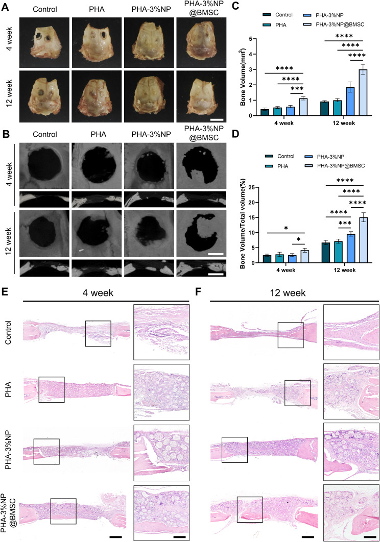
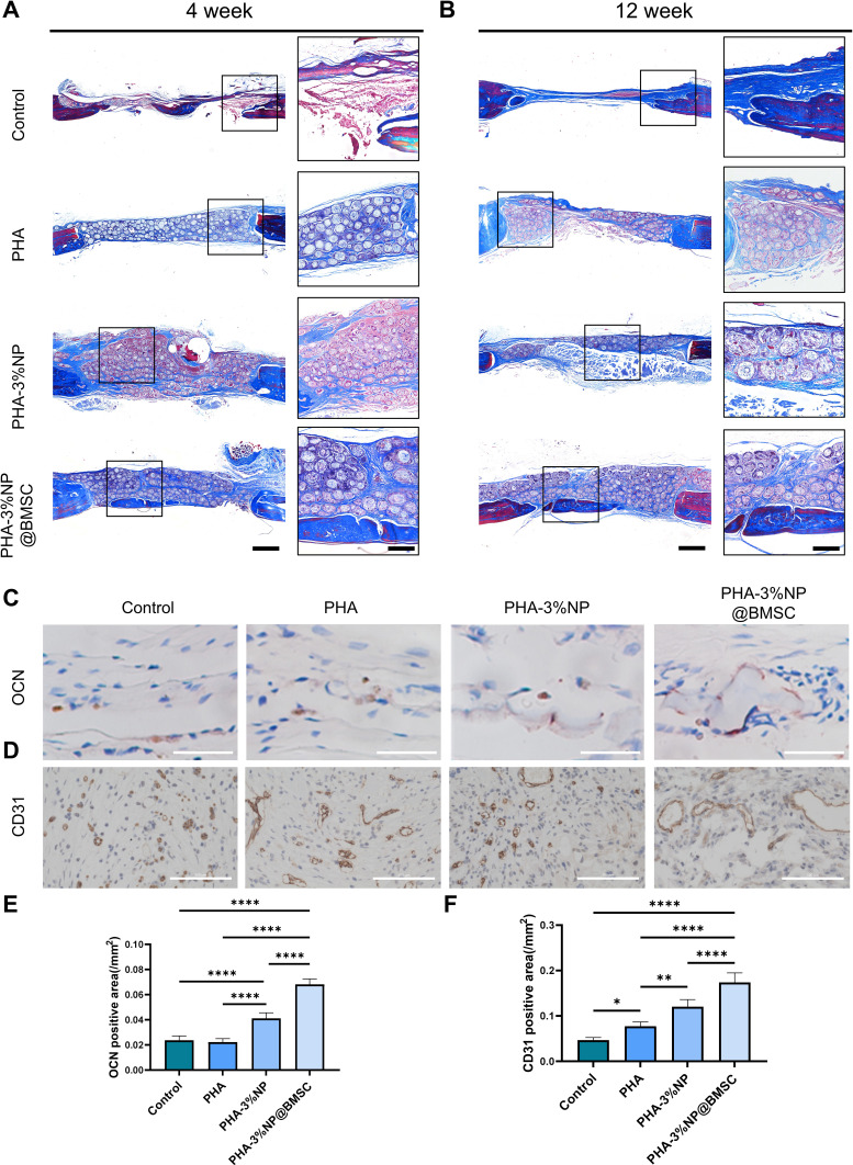
Purpose: Successful regeneration of cranial defects necessitates the use of porous bone fillers to facilitate cell proliferation and nutrient diffusion. Open porous microspheres, characterized by their high specific surface area and osteo-inductive properties, offer an optimal microenvironment for cell ingrowth and efficient ossification, potentially accelerating bone regeneration.

Materials and methods: An in vitro investigation was conducted to assess the physicochemical properties, porosity, and biocompatibility of PHA-nano-clay open porous microspheres. Subsequently, PHA-nano-clay microspheres loaded with rat bone marrow mesenchymal stem cells were implanted into 5 mm cranial defects in rats for a duration of 12 weeks and were evaluated through histological and immunohistochemical analyses.

Results: The incorporation of nano-clay into PHA resulted in improved mechanical properties of the porous scaffolds. Furthermore, cell adhesion, viability, and morphology on the scaffolds were maintained. The PHA-3% nano-clay open porous microspheres effectively enhanced the repair of cranial defects compared to the control group, without recurrence or complications.

Conclusion: Porous PHA-nano-clay microspheres, with their high specific surface area, biodegradability, and osteo-inductive properties, can be utilized as a bone-filling material for improved bone defect repair through cell delivery. In particular, PHA-3% nano-clay open porous microspheres exhibit promising therapeutic potential in the repair of cranial defects.

Keywords: P34HB; bone-filling biomaterial; cranial defects; open porous microspheres; osteo-inductivity.


Conflict of interest statement

The authors report no conflicts of interest in this work.

Figures

None
Graphical abstract
Figure 1
Figure 1
Preparation, morphology, and biocompatibility of PLA, PCL, and PHA OPMs and SMs. (A) Morphology, Nile red staining and live and dead r-BMSCs of PLA, PCL, and PHA SMs. (B) Morphology, Nile red staining and live and dead r-BMSCs of PLA, PCL, and PHA OPMs. (C) Quantification of average cell number on PLA, PCL, and PHA SMs from days 1 to 10. (D) Quantification of average cell number on PLA, PCL, and PHA OPMs from days 1 to 10. (E) CLSM images of r-BMSCs cultured on the PLA, PCL, and PHA OPMs and SMs with phalloidin on days 10 (red: β-actin, blue: 4’,6-diamidino-2-phenylindole (DAPI)). (F) Quantification of r-BMSCs cytoskeleton area/microsphere area of PLA, PCL, and PHA OPMs and SMs on days 10. The bars are 100 µm. (*p < 0.05; ****p < 0.0001).
Figure 2
Figure 2
Fabrication and optimization of PHA OPMs and PHA-NP OPMs. (A) Morphology and Nile red staining of PHA OPMs and PHA-NP OPMs. (B) Porosity rate of PHA OPMs and PHA-NP OPMs. (C) Morphology and Nile red staining of PHA-3%NP OPMs with different homogenizer speeds during primary emulsification. (D) Porosity rate of PHA-3%NP OPMs with different homogenizer speeds during primary emulsification. (E) Morphology and Nile red staining of PHA-3%NP OPMs with different homogenizer speeds during secondary emulsification. (F) Porosity rate of PHA-3%NP OPMs with different homogenizer speeds during secondary emulsification. (G) Morphology and Nile red staining of PHA-3%NP OPMs with different size. (H)Porosity rate of PHA-3%NP OPMs with different size. (I) Phalloidin CLSM images of r-BMSCs cultured on PHA-3%NP OPMs with different size from days 4, 7, and 14 (red: β-actin, blue: DAPI). (J) Quantification of r-BMSCs cytoskeleton area/microsphere area on PHA-3%NP OPMs with different size on days 4, 7, and 14. Data for each sample were obtained from > 200 parallels randomly selected from 20 microspheres. The bars are 100 µm. (*p < 0.05; **p < 0.01; ****p < 0.0001).
Figure 3
Figure 3
Proliferation of r-BMSCs on/in PHA OPMs and PHA-NP OPMs. (A) Bright field images of PHA OPMs and PHA-NP OPMs with 300μm size. The bars are 1mm. (B) CLSM images of r-BMSCs cultured on the microspheres with live/dead cell staining on days 1, 4, 7, and 10 (green: live cells, red: dead cells, blue: DAPI). The bars are 100 µm. (C) Quantitative analysis revealed cell viability on the OPMs on days 1, 4, 7, and 14. (D) CLSM images of r-BMSCs cultured on the microspheres with phalloidin on days 1, 4, 7, and 14 (red: β-actin, blue: DAPI). The bars are 100 µm. (E) Quantitative analysis revealed the cytoskeleton area/microsphere area on the OPMs on days 1, 4, 7, and 14. (****p < 0.0001).
Figure 4
Figure 4
Morphology and in vitro differentiation of r-BMSCs on/in OPMs (A) SEM images of r-BMSCs grown on the PHA OPMs and PHA-3%NP OPMs. (B) Bright field images of section of PHA OPMs and PHA-3%NP OPMs. (C) CCK-8 analysis of r-BMSCs on/in PHA OPMs, PHA-3%NP OPMs on days 1, 4, 7, and 10. (D) Pico Green dsDNA assay of r-BMSCs on/in PHA OPMs, PHA-3%NP OPMs on days 1, 4, 7, and 10. (E-H) Quantitative real-time polymerization chain reaction of bone-related gene markers expression of r-BMSCs during osteo-inductive differentiation for 14 and 21 days on/in PHA OPMs versus PHA-3%NP OPMs. (I) Alizarin red staining of calcium (Ca2+) deposition of r-BMSCs grown on/in microspheres. The bars are 100 µm. (J) Quantitative analysis of Ca2+ deposition of r-BMSCs grown on/in microspheres. (**p < 0.01; ***p < 0.001; ****p < 0.0001).
Figure 5
Figure 5
In vivo evaluation of new bone formation. (A)Macroscopic observation of implantation samples 4 and 12 weeks after surgery. The bars are 1cm. (B) Micro-CT 3D reconstruction images showing bone defects at 4 and 12 weeks after implantation. The bars are 2.5 mm. (C)Quantitative analysis of the micro-CT data of Bone volume and (D) bone volume/total volume ratio of porous microspheres. (E) Images of H&E staining of cranial defects recovery at 4 weeks after implantation. (F) Images of H&E staining of cranial defects recovery at 12 weeks after implantation. The bars are 1mm. (*p < 0.05; ***p < 0.001; ****p < 0.0001).
Figure 6
Figure 6
Histological and immunohistochemical analysis following scaffolds implantation. (A) Images of Masson’s trichrome staining of cranial defects at 4 weeks after implantation. (B) Images of Masson’s trichrome staining of cranial defects at 12 weeks after implantation. The bars are 1mm. (C) IHC analysis of OCN at 12 weeks following scaffold implantation. The bars are 100µm. (D)IHC analysis of CD31 at 12 weeks following scaffold implantation. The bars are 100µm. (E)Quantitative analysis of OCN at 12 weeks following scaffold implantation. (F) Quantitative analysis of CD31 at 12 weeks following scaffold implantation. (*p < 0.05; **p < 0.01; ****p < 0.0001).

Similar articles

Cited by

References

    1. Hassanein AH, Arany PR, Couto RA, et al. Cranial particulate bone graft ossifies calvarial defects by osteogenesis. Plast Reconstr Surg. 2012;129(5):796–802. doi:10.1097/PRS.0b013e31824a2bdd - DOI - PubMed
    1. Li Y, Zha Y, Hu W, et al. Monoporous microsphere as a dynamically movable drug carrier for osteoporotic bone remodeling. Adv Healthc Mater. 2023;12(16):1–12. doi:10.1002/adhm.202201242 - DOI - PubMed
    1. Li Y, Zhang X, Dai C, et al. Bioactive three-dimensional graphene oxide foam/polydimethylsiloxane/zinc silicate Scaffolds with enhanced osteoinductivity for bone regeneration. ACS Biomater Sci Eng. 2020;6(5):3015–3025. doi:10.1021/acsbiomaterials.9b01931 - DOI - PubMed
    1. Dai C, Li Y, Pan W, et al. Three-dimensional high-porosity Chitosan/Honeycomb Porous Carbon/Hydroxyapatite Scaffold with enhanced osteoinductivity for bone regeneration. ACS Biomater Sci Eng. 2020;6(1):575–586. doi:10.1021/acsbiomaterials.9b01381 - DOI - PubMed
    1. Guo L, Chen H, Li Y, Zhou J, Chen J. Biocompatible scaffolds constructed by chondroitin sulfate microspheres conjugated 3D-printed frameworks for bone repair. Carbohydr Polym. 2023;299(September 2022):120188. doi:10.1016/j.carbpol.2022.120188 - DOI - PubMed

MeSH terms

Substances

LinkOut - more resources