Review
. 2024 Dec 25:19:13925-13946.
doi: 10.2147/IJN.S497590. eCollection 2024.

Application of Nanomaterials Targeting Immune Cells in the Treatment of Chronic Inflammation

Affiliations
Review

Application of Nanomaterials Targeting Immune Cells in the Treatment of Chronic Inflammation

Zhen Ci et al. Int J Nanomedicine. .

Abstract

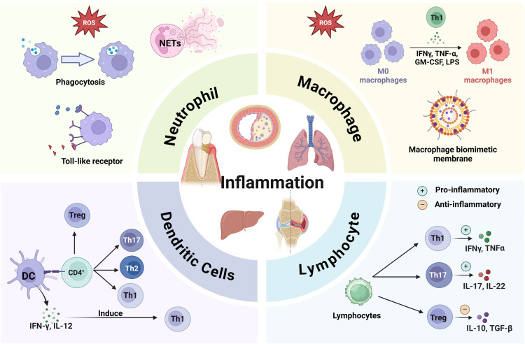
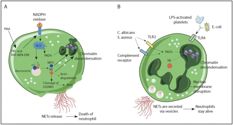
Chronic inflammation is a common characteristic of all kinds of diseases, including autoimmune diseases, metabolic diseases, and tumors. It is distinguished by the presence of low concentrations of inflammatory factors stimulating the body for an extended period, resulting in a persistent state of infection. This condition is manifested by the aggregation and infiltration of mononuclear cells, lymphocytes, and other immune cells, leading to tissue hyperplasia and lesions. Although various anti-inflammatory medications, including glucocorticoids and non-steroidal anti-inflammatory drugs (NSAIDs), have shown strong therapeutic effects, they lack specificity and targeting ability, and require high dosages, which can lead to severe adverse reactions. Nanoparticle drug delivery mechanisms possess the capacity to minimize the effect on healthy cells or tissues due to their targeting capabilities and sustained drug release properties. However, most nanosystems can only target the inflammatory sites rather than specific types of immune cells, leaving room for further improvement in the therapeutic effects of nanomaterials. This article reviews the current research progress on the role of diverse immune cells in inflammation, focusing on the functions of neutrophils and macrophages during inflammation. It provides an overview of the domestic and international applications of nanomaterials in targeted therapy for inflammation, aiming to establish a conceptual foundation for the utilization of nanomaterials targeting immune cells in the treatment of chronic inflammation and offer new perspectives for the avoidance and management of inflammation.

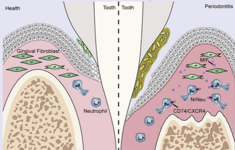
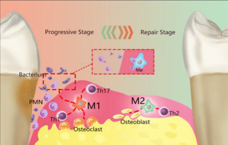
Keywords: chronic inflammation; macrophage; nanomaterials; neutrophil; periodontitis; target therapy.


Conflict of interest statement

All authors declared no conflicts of interest in this work.

Figures

None
Graphical abstract
Figure 1
Figure 1
Mechanisms of NET formation. (A) PMA and other stimuli induce lytic-NET formation. Neutrophils are stimulated with PMA, resulting in the activation of NADPH oxidase via PKC and Raf-MEK-ERK signaling pathways, consequently generating ROS. Subsequently, PAD4 is activated and citrullinates arginine on histones, causing chromatin decondensation. MPO and NE are discharged from cytoplasmic azurophilic granules and then translocated to the nucleus, contributing to the unfolding of chromatin. The nuclear envelope subsequently disintegrates, discharging the chromatin into the cytosol, where it blends with cytosolic proteins. NE also cleaves GSDMD in the cytosol to its active form (GSDMD-NT), which, besides forming pores in the plasma membrane, also mediates pore formation in nuclear and granule membranes, enhancing the release of NE and other granular content. Finally, NETs are released, and the neutrophil undergoes cell death. (B) Nonlytic NET formation is induced by the recognition of stimuli via Toll-like receptor 2 (TLR2), TLR4, or complement receptors, independent of NAPDH oxidase activation. S. aureus and C. albicans activate TLR2 and complement receptors, respectively, while E. coli or LPS-activated platelets activate TLR4. Along with PAD4 activation and NE translocation to the nucleus, chromatin decondensation proceeds, and protein-decorated chromatin is expelled via vesicles without plasma membrane disruption. After the release of NETs, neutrophils remain alive for further functions. Reprinted from Blood, Vol 133/Edition 20, Castanheira FVS, Kubes P. Neutrophils and NETs in modulating acute and chronic inflammation, Page numbers 2178–2185, Copyright 2019, with permission from Elsevier.
Figure 2
Figure 2
In inflamed gingival tissue, fibroblasts decreased while neutrophils increased; Fibroblasts induce excessive formation of NETS through MIFCD74/CXCR4 ligand receptor axis, thereby promoting the progression of periodontitis). Reprinted from Journal of Advanced Research, Qiu W, Guo R, Yu H, et al. Single-cell atlas of human gingiva unveils a NETs-related neutrophil subpopulation regulating periodontal immunity, Copyright 2024, with permission from Elsevier.
Figure 3
Figure 3
Polarized macrophages play a crucial role in the initiation and progression of Parkinson’s disease (PD). In PD, resident macrophages polarize into two primary phenotypes, M1 and M2, which respectively govern the inflammatory development and resolution phases. M1 macrophages are primarily proinflammatory and produce a series of proinflammatory factors, working in conjunction with Th1 cells, Th2 cells, and other cells. By collaborating with Th1-type immune cells, M1 macrophages can remove periodontal pathogenic microorganisms through the recruitment of PMNs. Simultaneously, M1 macrophages activate osteoclasts, leading to the absorption of the alveolar ridge. M2 macrophages primarily play an anti-inflammatory role and are mainly involved in immune interactions with Th2 cells. M2 macrophages terminate inflammatory development via accelerating the apoptosis for M1 macrophages and PMNs, perform tissue repair through various anti-inflammatory factors, and could activate osteoblasts to recover bone tissue. Reproduced from Sun X, Gao J, Meng X, Lu X, Zhang L, Chen R. Polarized Macrophages in Periodontitis: characteristics, Function, and Molecular Signaling. Front Immunol. 2021;12:763334.
Figure 4
Figure 4
The preparation of LP, RLP and Effero‐RLP. The schematic of apoptotic RBC membrane preparation and the fusion with liposome particles. Reproduced from Han R, Ren Z, Wang Q, et al. Synthetic Biomimetic Liposomes Harness Efferocytosis Machinery for Highly Efficient Macrophages-Targeted Drug Delivery to Alleviate Inflammation. Adv Sci. 2024;11(29):e2308325.
Figure 5
Figure 5
Schematic illustration of Motor@M2M@SAM preparation and its mechanism for UC treatment. (A) Scheme depicting the fabrication process of Motor@M2M. (B) Scheme illustrating the fabrication process of Motor@M2M@SAM using microfluidic technology. (C) Mechanism for UC Treatment: Upon oral administration, SAM is disrupted as it enters the colon. Subsequently, Motor@M2M is released from the hydrogel into the colonic lumen. The propelling force of O2 bubbles, generated by the decomposition of local H2O2 in the inflammatory microenvironment, facilitates the penetration of Motor@M2M through the mucus layer. These nanomotors then target inflammatory colon cells through a macrophage-like function. They specifically interact with colon epithelial cells. Acting as decoys, Motor@M2M neutralizes inflammatory cytokines through receptor-ligand interactions and absorption. Ultimately, Motor@M2M exerts therapeutic effects against UC by scavenging ROS, reducing inflammation, reprogramming macrophages, repairing the epithelial barrier, and rebalancing the microbiota. Reproduced from Luo R, Liu J, Cheng Q, Shionoya M, Gao C, Wang R. Oral microsphere formulation of M2 macrophage-mimetic Janus nanomotor for targeted therapy of ulcerative colitis. Sci Adv. 2024;10(26):eado6798.

Similar articles

References

    1. He Y, Yue Y, Zheng X, Zhang K, Chen S, Du Z. Curcumin, inflammation, and chronic diseases: how are they linked? Molecules. 2015;20(5):9183–9213. doi:10.3390/molecules20059183 - DOI - PMC - PubMed
    1. Chen L, Deng H, Cui H, et al. Inflammatory responses and inflammation-associated diseases in organs. Oncotarget. 2018;9(6):7204–7218. doi:10.18632/oncotarget.23208 - DOI - PMC - PubMed
    1. Henson PM. Dampening inflammation. Nat Immunol. 2005;6(12):1179–1181. doi:10.1038/ni1205-1179 - DOI - PubMed
    1. Nasef NA, Mehta S, Ferguson LR. Susceptibility to chronic inflammation: an update. Arch Toxicol. 2017;91(3):1131–1141. doi:10.1007/s00204-016-1914-5 - DOI - PubMed
    1. Grivennikov SI, Greten FR, Karin M. Immunity, inflammation, and cancer. Cell. 2010;140(6):883–899. doi:10.1016/j.cell.2010.01.025 - DOI - PMC - PubMed

MeSH terms

Substances

LinkOut - more resources