. 2024 Dec 27:2024:0080.
doi: 10.34133/bmef.0080. eCollection 2024.

Recent Advances and Future Directions in Sonodynamic Therapy for Cancer Treatment

Affiliations

Recent Advances and Future Directions in Sonodynamic Therapy for Cancer Treatment

Priyankan Datta et al. BME Front. .

Abstract

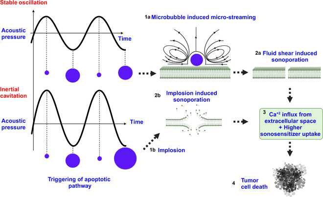
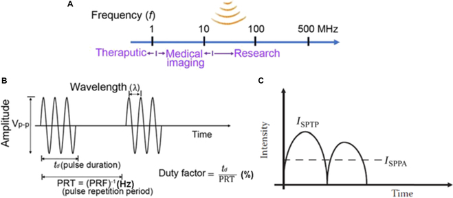
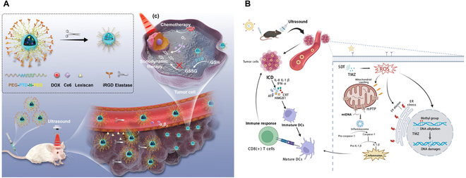
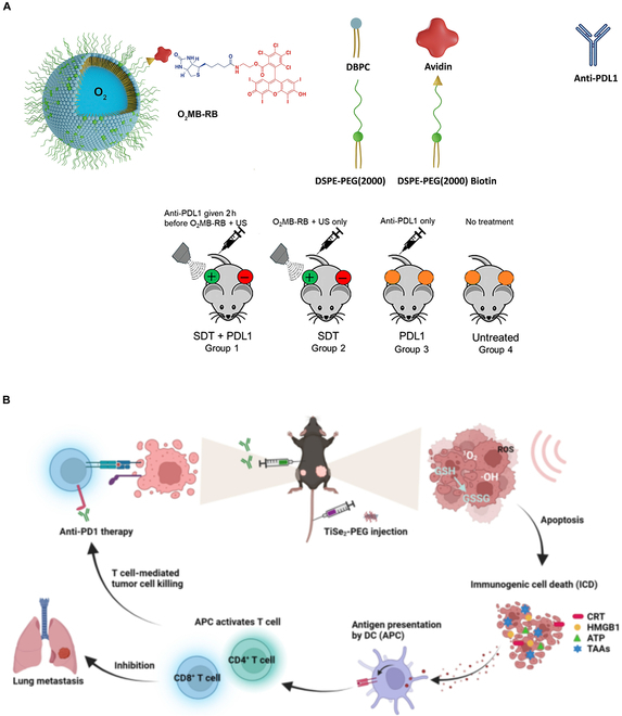
Deep-tissue solid cancer treatment has a poor prognosis, resulting in a very low 5-year patient survival rate. The primary challenges facing solid tumor therapies are accessibility, incomplete surgical removal of tumor tissue, the resistance of the hypoxic and heterogeneous tumor microenvironment to chemotherapy and radiation, and suffering caused by off-target toxicities. Here, sonodynamic therapy (SDT) is an evolving therapeutic approach that uses low-intensity ultrasound to target deep-tissue solid tumors. The ability of ultrasound to deliver energy safely and precisely into small deep-tissue (>10 cm) volumes makes SDT more effective than conventional photodynamic therapy. While SDT is currently in phase 1/2 clinical trials for glioblastoma multiforme, its use for other solid cancer treatments, such as breast, pancreatic, liver, and prostate cancer, is still in the preclinical stage, with further investigation required to improve its therapeutic efficacy. This review, therefore, focuses on recent advances in SDT cancer treatments. We describe the interaction between ultrasound and sonosensitizer molecules and the associated energy transfer mechanism to malignant cells, which plays a central role in SDT-mediated cell death. Different sensitizers used in clinical and preclinical trials of various cancer treatments are listed, and the critical ultrasound parameters for SDT are reviewed. We also discuss approaches to improve the efficacies of these sonosensitizers, the role of the 3-dimensional spheroid in vitro investigations, ultrasound-controlled CAR-T cell and SDT-based multimodal therapy, and machine learning for sonosensitizer optimization, which could facilitate clinical translation of SDT.


Conflict of interest statement

Competing interests: The authors declare that they have no competing interests.

Figures

Fig. 1.
Fig. 1.
Schematic of a photosensitizer activated by light followed by photodynamic reaction-mediated tumor cell death. This image is reproduced with permission from [11]. https://creativecommons.org/licenses/by/4.0/ PpIX is a photosensitizer that absorbs light and becomes excited (steps 1 and 2). Deexcitation (steps 3 and 4) releases fluorescence energy at a higher wavelength than of the incident light. The released energy produces singlet oxygen (steps 5 and 6), which produces ROS and triggers the cell death pathway.
Fig. 2.
Fig. 2.
Schematic illustration of sonoluminescence onset during single microbubble compression when the microbubble is subjected to ultrasound. The microbubble starts to grow during the negative cycle of the applied ultrasound and reaches its maximum. The bubble size reduces as the ultrasound field reverses toward its minima. At its minimum size, the temperature rise inside the bubble is sufficient to cause weak gas ionization, producing plasma consisting of electrons and ions. The deceleration of electrons near (I) positive ions, (II) atoms, or (III) their recombination produces electromagnetic radiation, known as “Bremsstrahlung”. This electromagnetic radiation in the visible range is called sonoluminescence.
Fig. 3.
Fig. 3.
Schematic illustration of sonosensitizer (SS) activation upon receiving energy from the sonoluminescence of a microbubble. SS is excited to its singlet excited state (SS1) from a ground state (SS0). SS1 deexcites either directly to SS0 or through SS3 to SS0. During deexcitation, either one or both type I and type II reactions occur. During a type I reaction, free radicals are generated either from a singlet or triplet excited state of the sonosensitizer. For a type II reaction, only a triplet excited state of the sonosensitizer reacts with molecular oxygen, producing singlet oxygen. Free radicals or singlet oxygen produces a cytotoxic effect on tumor cells, resulting in cell death.
Fig. 4.
Fig. 4.
Schematic representation of the 2 types of reaction paths after sonosensitizer activation [37]. The type I reaction involves either a singlet or triplet excited state of the sonosensitizer, where electron or hydrogen atom transfer produces intermediate, short-lived free radicals (or ROS). A type II reaction is a direct energy transfer process where the triplet excited state of the sonosensitizer reacts with the ground state molecular oxygen, producing highly reactive, cytotoxic singlet oxygen (1O2), which participates in different cell damage pathways.
Fig. 5.
Fig. 5.
(A) Schematic representation of the sonochemical process during the acoustic cavitation of a microbubble. Region I: Maximum temperature rise zone due to gas compression inside the bubble during the positive cycle of the ultrasound. Region II: Pyrolysis of water vapor molecules forms highly reactive free radicals (OH·, H·). Region III: Pyrolysis of hydrophilic and hydrophobic substances. Region IV: Bulk liquid zone, where free radicals escape and react with surrounding organic molecules. (B) Different water-soluble azocompounds can generate free radicals through pyrolysis and can serve as sensitizers in SDT. AAPH, 2,2′-azobis(2-methylpropionamidine) dihydrochloride; ADMF, 1,1′-azobis (N,N′-dimethylformamide); VA-044, 2,2′-azobis (N,N′-dimethyleneisobutyramidine) dihydrochloride (VA-044); V-30, 2-(carbamoylazo)-isobutyronitrile. This image is reproduced with permission from [12], Copyright 2006, John Wiley and Sons. (C) Schematic representation of the generalized pyrolysis reaction of an azo compound-based sensitizer and generation of peroxyl radical (ROO∙) in the presence of oxygen.
Fig. 6.
Fig. 6.
Mechanical pathway for ROS generation during SDT. (1) Application of ultrasound. (2) Mechanical force activates the cation channel (Piezo 1). (3) Ca+2 ions enter into the cytosol from extracellular space. (4) Endoplasmic reticulum (ER) also releases Ca+2 ions into the cytosol. (5) Increase in Ca+2 concentration leads to calcium overload. (6) Calcium overload opens the mPTP in the IMM. (7) This leads to the release of cytochrome c (Cyt c). (8a) The process results in disruption of the electron transport chain or (8b) activates enzymes such as apoptosomes. (9) Disruption of the electron transport chain increases electron leakage, and the ROS concentration consequently increases. (10 and 11) Caspase 9 is released, which induces programmed cell death or apoptosis.
Fig. 7.
Fig. 7.
Schematic of sonomechanical pathway in the presence of a microbubble during SDT. At a lower ultrasound pressure, the microbubble undergoes SO, whereas at a higher ultrasound pressure, IC occurs. (1a) SO mediated microstreaming. (1b) Bubble implosion. (2a) Microstreaming-induced fluid shear leads to the formation of transient pores at the plasma membrane. (2b) Implosion-induced sonoporation at the plasma membrane. (3) Sonoporation allows both Ca+2 influx from the extracellular space into the cytosol and higher sonosensitizer uptake by the tumor cells. (4) Triggering of cytotoxic pathways via Ca+2 overloading and sonosensitizer activation.
Fig. 8.
Fig. 8.
(A) Schematic representation of sound waves at different frequencies and corresponding application areas. (B) Critical ultrasound parameters, which are commonly used in SDT and for other application areas. This image is reproduced with permission from [65], Copyright 2021, SAGE Publications. (C) Schematic diagram of ISPPA, adapted from [62].
Fig. 9.
Fig. 9.
Schematic representation of the current SDT-based multimodal therapeutic approach for GBM. (A) Chlorin e6-assisted SDT combined with Lexiscan-loaded poly (2,2″-thiodiethylene 3,3″-dithiodipropionate) nanoparticles for doxorubicin delivery in brain tumors in vivo. The SDT-chemotherapy combined therapy shows an improvement in the efficacy via depletion of GSH. This image is reproduced with permission from [67]. https://creativecommons.org/licenses/by/4.0/ (B) TMZ is used as a sonosensitizer, which causes mitochondrial membrane permeabilization and ER stress. This induces mtDNA release within the cytoplasm and triggers the immunogenic signal [e.g., interleukin-1β (IL-1β)] that facilitates dendritic cell activation followed by glioma cell death. This image is reproduced with permission from [72], Copyright 2023 Elsevier Inc.
Fig. 10.
Fig. 10.
Recent advances of sonosensitizers in SDT-mediated BC therapy. (A) For the first time, 4 BODIPY derivative sonosensitizers are used for treating breast tumors (4T1 cell line). The sensitizer with the lowest singlet to triplet state transition energy gap (1.1243 eV) exhibits the highest antitumor effect in vivo. This image is reproduced with permission from [74], Copyright 2023 Elsevier Masson SAS. (B) [Ru(bpy)3]2+ is used as a novel sonosensitizer for treating 4T1 tumor-bearing mice. [Ru(bpy)3]2+ has a very low energy gap (0.1239 eV) between LUMO and HOMO, which leads to its activation by ultrasound stimulation (0.1 to 0.3 W/cm2, 3 MHz, 20-min stimulation) and, thereby, generates cytotoxic singlet oxygen. Further, during SDT, NADPH to NAD+ oxidation causes redox imbalance in the TME, which results in an arrest of tumor growth. This image is reproduced with permission from [75]. https://creativecommons.org/licenses/by/4.0/ (C) FeOOH-MnO2 nanocomposite exhibits dual features. It inhibits the electron-hole pair recombination and, hence, increases ROS generation (singlet oxygen and hydroxyl radicals). Additionally, due to its catalytic activity, H2O2 to O2 decomposition causes hypoxia alleviation in the TME and GSH depletion. This collectively enhances ROS generation rate and SDT efficacy in mice bearing BC. This image is reproduced from [77]. https://creativecommons.org/licenses/by/4.0/ (D) α-Fe2O3 with Pt nanocrystal-based photosensitizer, which reduces the energy gap and enhances ROS generation, followed by an increase in SDT efficacy in 4T1 tumor-bearing mice. This image is reproduced from [78]. https://creativecommons.org/licenses/by/4.0/
Fig. 11.
Fig. 11.
SDT-based multimodal therapy for treating pancreatic cancer. (A) O2-microbubble loaded with Rose Bengal sensitizer is used in combination with anti-PDL1. The combined therapy showed the maximum antitumor effect on pancreatic cancer tumor-bearing mouse models compared to SDT as well as immune checkpoint inhibitor alone. This image is reproduced with permission from [24], Copyright 2021 Elsevier B.V. (B) SDT is combined with anti-PD1 with TeS2 nanosheets as a sensitizer. SDT induces an immunogenic signal that enhances tumor-associated antigen and cytokine release, followed by dendritic cell activation. This ultimately enhances CD8+ infiltration and therapeutic efficacy. This image is reproduced with permission from [82]. https://creativecommons.org/licenses/by/4.0/
Fig. 12.
Fig. 12.
Multimodal SDT for HCC treatment. (A) HMME sensitizer is combined with doxorubicin for SDT-chemotherapy. Redrawn from [86] using Biorender. (B) Combination of Cas9/single-guide RNA (sgRNA) with HMME shows a promising antitumor effect due to the knockdown of the NFE2L2 transcription factor, which plays an important role in the reduction of oxidative stress and prevents cell damage. This image is reproduced with permission from [87]. https://creativecommons.org/licenses/by-nc-nd/4.0/ (C) The application of IR820 sensitizer dye within the NB in combination with PI-103 shows a synergistic effect due to its inhibitory nature in the PI3K/ mTOR signaling pathway. This image is reproduced with permission from [88]. http://creativecommons.org/licenses/by-nc/3.0/. (D) si-NUDT1 is used with polythiophene, which shows a substantial antitumor effect due to the silencing of the NUDT1 gene and the singlet oxygen as well as ROS generation during ultrasound application. This image is reproduced (adapted) with permission from [89], Copyright 2024, American Chemical Society.
Fig. 13.
Fig. 13.
SDT for PC treatment. (A) Hematoporphyrin conjugated with glutamate and tyrosine via hydrophobic and ππ interactions, forming a self-assembled nanoparticle HPNP. The acidic TME, along with the presence of cathepsin B, causes the digestion of the HPNP, which reduces its size. This allows higher accumulation of the sensitizer molecule due to an increase in its diffusion within the tumor cell and promising therapeutic efficacy in terms of reduction of the tumor volume within 24 h after the dose administration in vivo. This figure is reproduced from [27]. https://creativecommons.org/licenses/by/4.0/. (B) SDT-based multimodal therapeutic approach has been adopted. Ce6, along with anti-PDL1, is encapsulated within the NB. The ultrasound stimulation enhances ROS generation, and the presence of an immune checkpoint inhibitor, anti-PDL1, enhances immune response, which finally leads to immunogenic cell death. This figure is reproduced with permission from [91]. http://creativecommons.org/licenses/by-nc/3.0/
Fig. 14.
Fig. 14.
Porphyrin sonosensitizers for SDT-based clinical trials [96].
Fig. 15.
Fig. 15.
(A) The direct excitation from the ground state (S0) to the triplet state (T1) is a spin-forbidden process. However, the use of heavy metal atoms can directly excite the sensitizer molecule to its triplet state and can enhance the cytotoxic singlet oxygen generation. This figure is reproduced with permission from [101]. https://creativecommons.org/licenses/by/3.0/ (B) HOMO inversion process: Substitution of electron donor groups causing destabilizing HOMO by MC to LC HOMO state. This enhances the absorption spectrum of the sensitizer molecule. This figure is reproduced with permission from [101]. https://creativecommons.org/licenses/by/3.0/ (C) Structure of “cMa” complexes (c: carbene; M: coinage d10 metal; a: anionic amide ligand). These recently developed complexes have high potential for use as novel sonosensitizer molecules as alternatives to MC complexes due to their low cost, low toxicity, and, most importantly, the capability of avoiding nonradiative energy transfer. This figure is reproduced (adapted) with permission from [98], Copyright 2024, American Chemical Society.
Fig. 16.
Fig. 16.
(A) Pt-Ru-based hybrid complex exhibits multienzymatic catalytic activity and hence can act as a potential sensitizer with tumor hypoxia alleviation as well as a GSH-depleting substance. This can lead to an increase in SDT efficacy. This image is reproduced from [109]. https://creativecommons.org/licenses/by-nc-nd/4.0/ (B and C) Ru (II)-based and Ir-based metal complexes. These metal complexes have a low LUMO-HOMO energy gap, oxidize NADH to NAD+, and thereby can play a vital role in GSH depletion and ROS yield. These images are reproduced with permission from [108], Copyright 2022 Wiley-VCH GmbH.
Fig. 17.
Fig. 17.
(A) Schematic representation of the magnetic field-assisted spheroid printing. Cells are seeded in a cell culture medium containing the paramagnetic agent Gadovist. Depending on the magnetic susceptibility difference between cells and their surrounding medium, a net magnetic force acts and displaces cells toward the lowest magnetic field zone. This results in the formation of cellular aggregates within 3 to 6 h depending on cell types. (B) Coculture of MCF7 and NIH 3T3 fibroblasts forms layer-by-layer cellular aggregates within 3 h. This image is reproduced with permission from [110], Copyright 2020, American Chemical Society.

Similar articles

Cited by

References

    1. Brown JM. Tumor microenvironment and the response to anticancer therapy. Cancer Biol Ther. 2002;1(5):453–458. - PubMed
    1. Datta P, Dey M, Ataie Z, Unutmaz D, Ozbolat IT. 3D bioprinting for reconstituting the cancer microenvironment. NPJ Precis Oncol. 2020;4(1):18. - PMC - PubMed
    1. Lei X, Lei Y, Li JK, du WX, Li RG, Yang J, Li J, Li F, Tan HB. Immune cells within the tumor microenvironment: Biological functions and roles in cancer immunotherapy. Cancer Lett. 2020;470:126–133. - PubMed
    1. Nowak KM, Schwartz MR, Breza VR, Price RJ. Sonodynamic therapy: Rapid progress and new opportunities for non-invasive tumor cell killing with sound. Cancer Lett. 2022;532:215592. - PMC - PubMed
    1. Huang Y, Ouyang W, Lai Z, Qiu G, Bu Z, Zhu X, wang Q, Yu Y, Liu J. Nanotechnology-enabled sonodynamic therapy against malignant tumors. Nanoscale Adv. 2024;6(8):1974–1991. - PMC - PubMed

LinkOut - more resources