. 2024 Dec 10;8(4):1613-1633.
doi: 10.1162/netn_a_00412. eCollection 2024.

Generative dynamical models for classification of rsfMRI data

Affiliations

Generative dynamical models for classification of rsfMRI data

Grace Huckins et al. Netw Neurosci. .

Abstract

The growing availability of large-scale neuroimaging datasets and user-friendly machine learning tools has led to a recent surge in studies that use fMRI data to predict psychological or behavioral variables. Many such studies classify fMRI data on the basis of static features, but fewer try to leverage brain dynamics for classification. Here, we pilot a generative, dynamical approach for classifying resting-state fMRI (rsfMRI) data. By fitting separate hidden Markov models to the classes in our training data and assigning class labels to test data based on their likelihood under those models, we are able to take advantage of dynamical patterns in the data without confronting the statistical limitations of some other dynamical approaches. Moreover, we demonstrate that hidden Markov models are able to successfully perform within-subject classification on the MyConnectome dataset solely on the basis of transition probabilities among their hidden states. On the other hand, individual Human Connectome Project subjects cannot be identified on the basis of hidden state transition probabilities alone-although a vector autoregressive model does achieve high performance. These results demonstrate a dynamical classification approach for rsfMRI data that shows promising performance, particularly for within-subject classification, and has the potential to afford greater interpretability than other approaches.

Keywords: Classification; Generative models; Hidden Markov models; Network dynamics; Resting-state fMRI.

Plain language summary

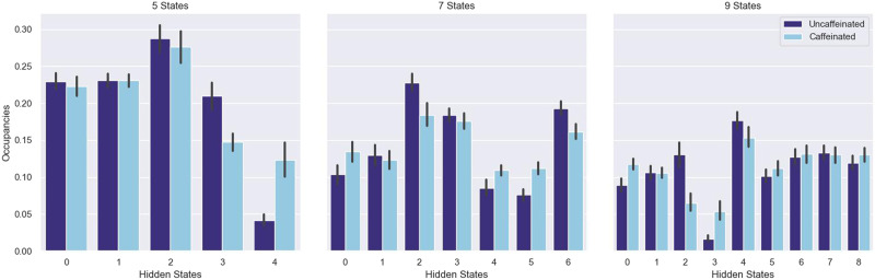
Neuroimaging researchers have made substantial progress in using brain data to predict psychological and behavioral variables, like personality, cognitive abilities, and neurological and psychiatric diagnoses. In general, however, these prediction approaches do not take account of how brain activity changes over time. In this study, we use hidden Markov models, a simple and generic model for dynamic processes, to perform brain-based prediction. We show that hidden Markov models can successfully distinguish whether a single individual had eaten and consumed caffeine before his brain scan. These models also show some promise for “fingerprinting,” or identifying individuals solely on the basis of their brain scans. This study demonstrates that hidden Markov models are a promising tool for neuroimaging-based prediction.


Conflict of interest statement

Competing Interests: The authors have declared that no competing interests exist.

Figures

<b>Figure 1.</b>
Figure 1.
The classification performance of various HMM-based approaches on the MyConnectome dataset. Results using the Yeo et al. (2011) 7-network parcellation are shown on the left; results using the 17-network parcellation are on the right. Error bars are 95% confidence intervals over 100 runs of cross-validation. Chance performance is 50%.
<b>Figure 2.</b>
Figure 2.
The classification performance of various HMM-based approaches on the 12 held-out runs from the MyConnectome dataset. Error bars are 95% confidence intervals over 100 runs of testing.
<b>Figure 3.</b>
Figure 3.
Transition matrices from ARHMMs fit to both uncaffeinated and caffeinated datasets, as well as the absolute value of their difference. Results are shown for 5-, 7-, and 9-state ARHMMs. Rows correspond to the origin states (i.e., the state from which the HMM is transitioning), and columns correspond to the destination states (i.e., the state to which the HMM is transitioning). States are zero-indexed, for example, for the 5-state model, states have been assigned numbers 0–4. Elements along the diagonal—that is, the probability of remaining in a given hidden state—have been set to 0 for visualization purposes.
<b>Figure 4.</b>
Figure 4.
The fraction of time spent in each hidden state (“state occupancy”) for the 5-, 7-, and 9-state ARHMMs. Error bars are 95% confidence intervals computed across the state occupancy times for the 33 uncaffeinated and 26 caffeinated runs.
<b>Figure 5.</b>
Figure 5.
The average network activity for each hidden state in the 5-state ARHMM. The radius of each plot represents the average z-scored activity of the network in each state across all caffeinated and uncaffeinated runs.
<b>Figure 6.</b>
Figure 6.
Classification performance on the MyConnectome dataset with high-motion volumes simply censored (“Head Motion Censored”) or with high-motion volumes censored and then interpolated over (“Head Motion Interpolated”). The left panel shows results from the ARHMM-based approach and the right from the Gaussian HMM. All models were fully trained. Chance performance is 50%.
<b>Figure 7.</b>
Figure 7.
The fingerprinting performance of various HMM-based approaches on the HCP dataset. Results using the Yeo et al. (2011) 7-network parcellation are shown on the left; results using the 17-network parcellation are on the right. Baseline fingerprinting results evaluated on the 512 ROI data (“Baseline (512-D)”) are shown in both panels as a performance ceiling, whereas the individual “Baseline” results in each panel were computed using 7- and 17-network data, respectively. To evaluate performance, models were trained using a “leave-one-run-out” regime—models were trained on three runs from an individual and tested on the fourth. Error bars are 95% confidence intervals over 100 repetitions of training and testing on randomly selected subsets of 100 subjects from the core dataset of 149 subjects. Chance performance is 1%.
<b>Figure 8.</b>
Figure 8.
The fingerprinting performance of various HMM-based approaches on the 47 individuals held out from the HCP dataset. Error bars are 95% confidence intervals over 100 runs of testing.
<b>Figure 9.</b>
Figure 9.
The fraction of misidentification errors that occurred within families, for the four model types, with two to six hidden states. Models were trained with the 7-network data. The blue series indicate the results of the same analysis performed after family membership labels were permuted, in order to create an empirical null distribution. Error bars are 95% confidence intervals over repetitions of model training/testing and, where relevant, family label permutation (5 repetitions for the unpermuted data and 20 for the permuted data).
<b>Figure 10.</b>
Figure 10.
Age distributions for both the primary and validation HCP samples.
<b>Figure 11.</b>
Figure 11.
A schematic of the HMMs used in this paper. The Markov chain at the top of the image—the hidden states and the transition matrices—is common to all models, but the emission distributions vary based on the model type. The Gaussian models have no autoregressive weights.
<b>Figure 12.</b>
Figure 12.
A schematic of the transition matrix-only training regime. Elements indicated in blue—the emission distribution and autoregressive weights—are trained on the full dataset, whereas the transition matrix, which is indicated in gold, is fine-tuned on the specific classes of training data. For the MyConnectome dataset, those classes are the caffeinated runs and the uncaffeinated runs. For the HCP dataset, each class is the set of runs from a single subject.
<b>Figure 13.</b>
Figure 13.
A schematic of the leave-one-run-out classification pipeline for MyConnectome data, in the transition matrix-only training regime.
<b>Figure 14.</b>
Figure 14.
A schematic of the classification pipeline for HCP data, in the transition matrix-only training regime.

Similar articles

References

    1. Baldassano, C., Chen, J., Zadbood, A., Pillow, J. W., Hasson, U., & Norman, K. A. (2017). Discovering event structure in continuous narrative perception and memory. Neuron, 95(3), 709–721. 10.1016/j.neuron.2017.06.041, - DOI - PMC - PubMed
    1. Bondi, E., Maggioni, E., Brambilla, P., & Delvecchio, G. (2023). A systematic review on the potential use of machine learning to classify major depressive disorder from healthy controls using resting state fMRI measures. Neuroscience & Biobehavioral Reviews, 144, 104972. 10.1016/j.neubiorev.2022.104972, - DOI - PubMed
    1. Bustamante, C., Castrillón, G., & Arias-Londoño, J. (2023). Classification of focused perturbations using time-variant functional connectivity with rs-fMRI. In Communications in computer and information science (Vol. 1746, pp. 18–30). Springer. 10.1007/978-3-031-29783-0_2 - DOI
    1. Cai, W., Warren, S. L., Duberg, K., Pennington, B., Hinshaw, S. P., & Menon, V. (2021). Latent brain state dynamics distinguish behavioral variability, impaired decision-making, and inattention. Molecular Psychiatry, 26(9), 4944–4957. 10.1038/s41380-021-01022-3, - DOI - PMC - PubMed
    1. Dadi, K., Varoquaux, G., Machlouzarides-Shalit, A., Gorgolewski, K. J., Wassermann, D., Thirion, B., & Mensch, A. (2020). Fine-grain atlases of functional modes for fMRI analysis. NeuroImage, 221, 117126. 10.1016/j.neuroimage.2020.117126, - DOI - PubMed

LinkOut - more resources