. 2024 Dec 13:15:1456443.
doi: 10.3389/fimmu.2024.1456443. eCollection 2024.

NECTIN-4-redirected T cell Antigen Coupler T cells bearing CD28 show superior antitumor responses against solid tumors

Affiliations

NECTIN-4-redirected T cell Antigen Coupler T cells bearing CD28 show superior antitumor responses against solid tumors

Cheng Wei et al. Front Immunol. .

Abstract

Introduction: T cell Antigen Coupler (TAC) T cells harness all signaling subunits of endogenous T cell receptor (TCR) to trigger T-cell activation and tumor cell lysis, with minimal release of cytokines. Some of the major obstacles to cellular immunotherapy in solid tumors include inefficient cell infiltration into tumors, lack of prolonged cellular persistence, and therapy-associated toxicity.

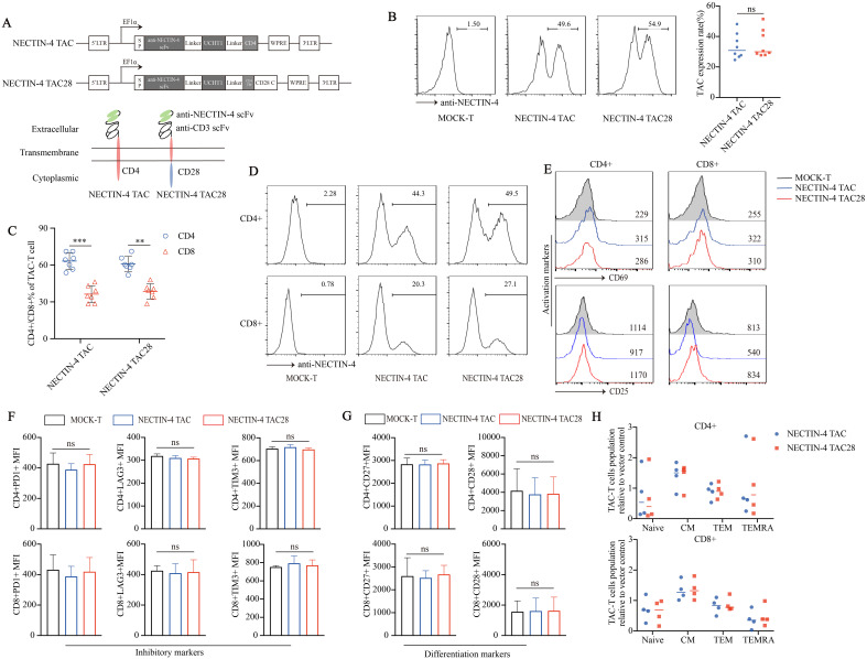
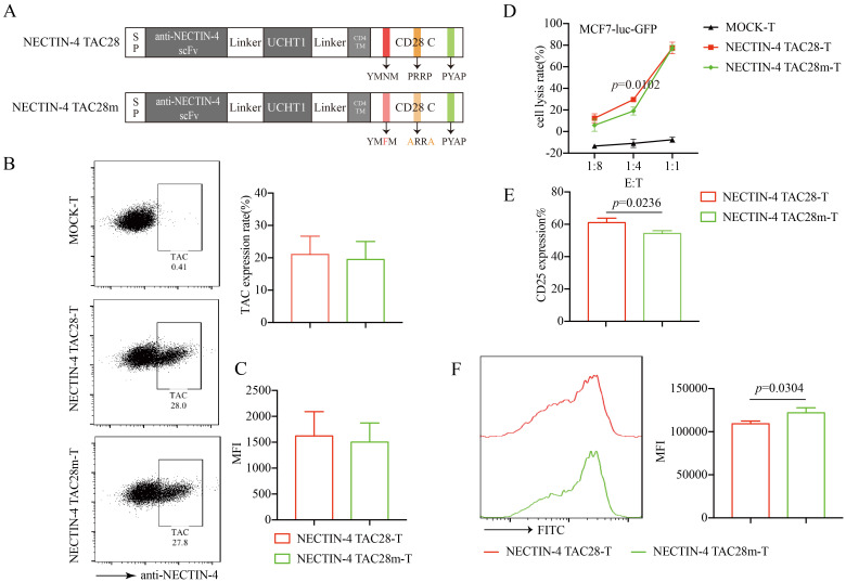
Methods: To boost the cytotoxic potential of TAC-T cells against solid tumors, we generated a novel NECTIN-4-targeted TAC-T variant, NECTIN-4 TAC28-T, which integrated the co-stimulatory CD28 cytoplasmic region, and compared the anti-tumor activities between NECTIN-4 TAC-T cells and NECTIN-4 TAC28-T cells in vitro and vivo.

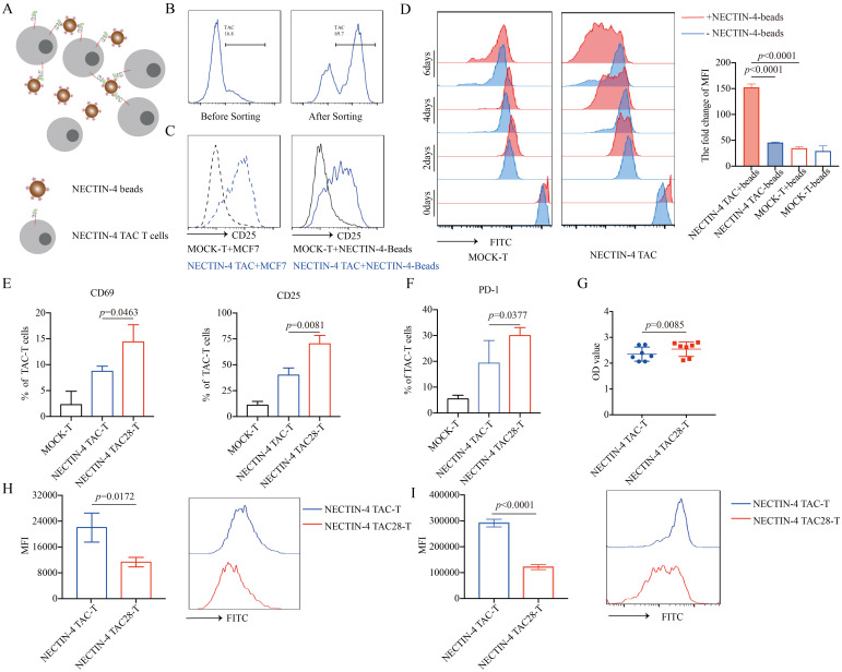
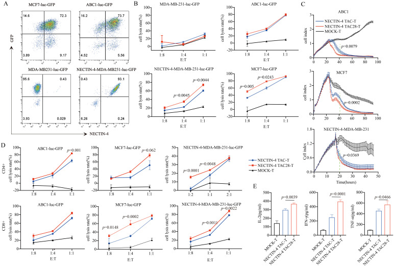
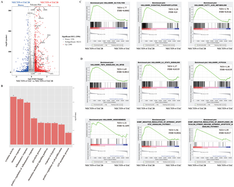
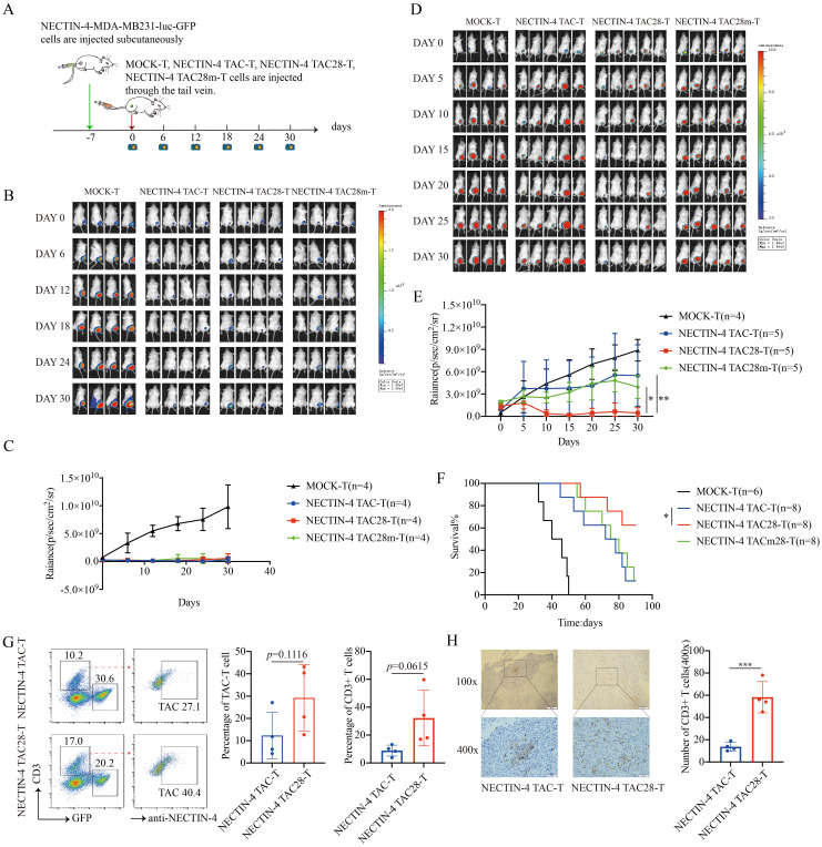
Results: We demonstrated NECTIN-4 TAC28-Tcells could be effectively activated by NECTIN-4 protein-coated magnetic beads (NECTIN-4-beads), and further revealed that the incorporated CD28 co-stimulatory domain enhanced their activation and proliferation capabilities. Notably, NECTIN-4 TAC28-T cells exhibited better anti-tumor effects both in vitro and in vivo than the original NECTIN-4 TAC-T cells.

Discussion: Our data highlighted that NECTIN-4 TAC28-T cells may represent a promising, safe and effective cell therapy for NECTIN-4-overexpressing solid tumors.

Keywords: CD28; NECTIN-4; T cell Antigen Coupler (TAC-T); adoptive cell transfer therapy; solid tumor.


Conflict of interest statement

Author JG was employed by company Zhejiang Qixin Biotech. The remaining authors declare that the research was conducted in the absence of any commercial or financial relationships that could be construed as a potential conflict of interest.

Figures

Figure 1
Figure 1
Successful generation of CD28-containing NECTIN-4 TAC28-T cells without tonic signal. (A) Schematic illustration of NECTIN-4 TAC and NECTIN-4 TAC28 constructs. (B) TAC expression in NECTIN-4 TAC-T and NECTIN-4 TAC28-T was detected by flow cytometry. (C) The ratio of CD4+ and CD8+ T cells of TAC-T cells. (D) The expression of TAC in CD4+ and CD8+ T cells. (E) CD69 and CD25 expression. (F) Expression of checkpoint receptors PD-1, LAG-3, and TIM-3. (G) CD27 and CD28 expression. (H) Memory T cells subsets of NECTIN-4 TAC-T and NECTIN-4 TAC28-T cells relative to MOCK-T cells. T cell subsets are defined as naïve (CD45RA+, CCR7+), central memory (CD45RA-, CCR7+), effector memory (CD45RA-, CCR7-), and terminal effectors (CD45RA+, CCR7-). For (E–G), the histograms represented the data from three healthy donors, and median fluorescence intensity was indicated. ns, no significant difference, **p < 0.01, ***p < 0.001, t-test. Data are presented as the mean ± SD.
Figure 2
Figure 2
NECTIN-4 TAC28-T cells showed improved activation and proliferation. (A) Schematic diagram of NECTIN-4-beads interacted with anti-NECTIN-4 TAC-T cells. (B) TAC expression after NECTIN-4-beads sorting. (C) CD25 expression of MOCK-T and NECTIN-4-redirected TAC-T cells upon stimulation with MCF-7 cells or NECTIN-4-beads. (D) MOCK-T and NECTIN-4-redirected TAC-T cells were labeled with carboxyfluorescein succinimidyl ester (CFSE), and their proliferation were assessed by flow cytometry upon stimulation with NECTIN-4-beads.The left side is the flow cytometry graph, and the right side is the ratio change of cell surface CFSE fluorescence values on day 0 and day 6. (E) CD69 and CD25 expression of MOCK-T, NECTIN-4 TAC-T and NECTIN-4 TAC28-T cells upon stimulation with NECTIN-4-beads. (F) The expression of PD-1 on TAC-T cells. (G) NECTIN-4 TAC-T and NECTIN-4 TAC28-T cells were stimulated by NECTIN-4-beads, and their proliferation were assessed through cell counting by use of CCK8 method. (H) NECTIN-4 TAC-T and NECTIN-4 TAC28-T cells were labeled with CFSE, and their proliferation were assessed by flow cytometry upon stimulation with NECTIN-4-beads. (I) NECTIN-4 TAC and NECTIN-4 TAC28 CD4+ T cells were labeled with CFSE, and their proliferation were assessed by flow cytometry upon stimulation with NECTIN-4-beads. Data came from ≥3 donors. ns, no significant difference, t-test. Data were presented as the mean ± SD.
Figure 3
Figure 3
Specific target cell lysis by NECTIN-4-redirected T cells. (A) NECTIN-4 and GFP expression in MCF7-luc-GFP, ABC1-luc-GFP, MDA-MB-231-luc-GFP and NECTIN-4-MDA-MB-231-luc-GFP cells transduced with the lentivirus encoding the Luciferase-T2A-GFP or NECTIN-4-P2A-Luciferase-T2A-GFP. (B) Cytotoxicity of NECTIN-4 TAC-T and NECTIN-4 TAC28-T cells were assessed by co-incubation with luciferase-expressing MCF7-luc-GFP, ABC1-luc-GFP, MDA-MB-231-luc-GFP and NECTIN-4-MDA-MB-231-luc-GFP cells at the indicated E/T ratio. (C) Real-time analysis (RTCA) was used to monitor the cytolysis of ABC1, MCF7 and MB231-NECTIN-4 cells by NECTIN-4 TAC-T and NECTIN-4 TAC28-T cells. Effector: target (E:T) cell ratio=1:1. (D) Cytotoxicity of NECTIN-4 TAC and NECTIN-4 TAC28 CD4+/CD8+ cells were assessed by co-incubation with luciferase-expressing NECTIN-4-MDA-MB-231-luc-GFP, MCF7-luc-GFP and ABC1-luc-GFP at the indicated E/T ratio. (E) ELISA was used to detect the secretion of IL-2, IFN-γ, and TNF-α by NECTIN-4-redirected TAC-T cells after co-culture with MCF7 cells for 24 h. Data came from ≥3 donors. ns, no significant difference, t-test. Data are presented as the mean ± SD.
Figure 4
Figure 4
The effects of incorporated CD28 cytoplasmic domain on the transcriptional profile of NECTIN-4 TAC28-T cells upon stimulation with NECTIN-4-beads. (A) Differentially expressed genes between NECTIN-4 TAC-T and NECTIN-4 TAC28-T cells. (B) Gene Ontology analysis of the different genes between NECTIN-4 TAC-T and NECTIN-4 TAC28-T cells. (C, D) Representative GSEA results from running the unfiltered NECTIN-4 TAC-T versus NECTIN-4 TAC28-T cell rank list against the MSigDB H hallmark gene sets and C5 gene ontology sets.
Figure 5
Figure 5
Effects of mutations in the incorporated CD28 cytoplasmic domain on NECTIN-4 TAC28-T function. (A) Schematic illustration of NECTIN-4 TAC28m construct design. (B) TAC expression in NECTIN-4 TAC28-T and NECTIN-4 TAC28m-T was detected by flow cytometry (C) MFI of anti-NECTIN-4 scFv on the surface of NECTIN-4-redirected TAC-T cells. (D) Cytotoxicity of NECTIN-4 TAC28-T and NECTIN-4 TAC28-T cells were assessed by co-incubation with luciferase-expressing MCF7-Luc cells at the indicated E/T ratio. (E) CD25 expression of NECTIN-4-redirected TAC-T cells was detected by flow cytometry. (F) NECTIN-4 TAC28-T and NECTIN-4 TAC28m-T cells were labeled with CFSE, and their proliferation was assessed by flow cytometry after simulation with NECTIN-4-beads. Data came from ≥3 donors. ns, no significant difference, t-test. Data are presented as the mean ± SD.
Figure 6
Figure 6
NECTIN-4 TAC28-T cells demonstrated improved efficacy over NECTIN-4 TAC-T and NECTIN-4 TAC28m-T cells in vivo. (A) Treatment scheme for NECTIN-4-MDA-MB-231-luc-GFP tumor-bearing mice. (B-E) Tumor xenografts were monitored via bioluminescence imaging. Bioluminescence images and kinetics were shown in (B, C) at E/T=3:1(3million:1million); as well as in (D, E) at E/T=1:1(1million:1million). (F) Kaplan–Meier survival curve, p=0.0471. (G) Proportion of NECTIN-4 TAC-T, NECTIN-4 TAC28-T and NECTIN-4-MDA-MB-231-luc-GFP cells in tumor lesions, detected by flow cytometry (n=4). (H) The infiltration of CD3+ T cells inside the tumor lesions of both the NECTIN-4 TAC-T cell treatment group and the NECTIN-4 TAC28-T cell treatment group was analyzed by immunohistochemistry (n=4). ns, no significant difference, *p < 0.05, **p < 0.01, ***p < 0.001, One-way ANOVA-test. Data are presented as the mean ± SD. These data are representative of three independent experiments.

Similar articles

References

    1. Rohaan, Maartje W, Wilgenhof S, Haanen JB. Adoptive cellular therapies: the current landscape. Virchows Archiv. (2019) 474:449–61. doi: 10.1007/s00428-018-2484-0 - DOI - PMC - PubMed
    1. Füchsl F, Krackhardt AM. Adoptive cellular therapy for multiple myeloma using CAR-and TCR-transgenic T cells: Response and resistance. Cells. (2022) 11.3:410. doi: 10.3390/cells11030410 - DOI - PMC - PubMed
    1. Mackensen A, Müller F, Mougiakakos D, Böltz S, Wilhelm A, Aigner M, et al. . Anti-CD19 CAR T cell therapy for refractory systemic lupus erythematosus. Nat Med. (2022) 28.10:2124–32. doi: 10.1038/s41591-022-02017-5 - DOI - PubMed
    1. Pinar Ataca A, Atilla E. Resistance against anti-CD19 and anti-BCMA CAR T cells: Recent advances and coping strategies. Trans Oncol. (2022) 22:101459. doi: 10.1016/j.tranon.2022.101459 - DOI - PMC - PubMed
    1. Sheth VS, Gauthier J. Taming the beast: CRS and ICANS after CAR T-cell therapy for ALL. Bone Marrow Transplant. (2021) 56:552–66. doi: 10.1038/s41409-020-01134-4 - DOI - PMC - PubMed

MeSH terms

LinkOut - more resources