. 2024 Dec 13:15:1488127.
doi: 10.3389/fimmu.2024.1488127. eCollection 2024.

Circulating extracellular vesicles and neutrophil extracellular traps contribute to endothelial dysfunction in preeclampsia

Affiliations

Circulating extracellular vesicles and neutrophil extracellular traps contribute to endothelial dysfunction in preeclampsia

Alex Ramos et al. Front Immunol. .

Abstract

Background: Preeclampsia (PE) is a pregnancy complication characterized by hypertension, proteinuria, endothelial dysfunction, and complement dysregulation. Placenta-derived extracellular vesicles (EVs), necessary in maternal-fetal communication, might contribute to PE pathogenesis. Moreover, neutrophil extracellular traps (NETs) play a pathogenic role in other complement-mediated pathologies, and their contribution in PE remains unexplored.

Materials and methods: EVs were isolated from PE (peEVs) and normotensive pregnant women sera. NETs were obtained incubating donor-pre-activated neutrophils with PE or control sera. Microvascular (HMEC) endothelial cells (ECs) were incubated with PE or control sera with or without (depleted sera) EVs or NETs, to assess changes in VCAM-1, ICAM-1, VE-cadherin, eNOS, VWF, ROS, and C5b-9 deposits. Results were expressed as fold increase vs. control.

Results: VWF, VCAM-1, and ROS expression was significantly higher in cells exposed to PE sera vs. control (12.3 ± 8.1, 3.6 ± 2.3, and 1.8 ± 0.2, respectively, p < 0.05), though significantly lower in cells exposed to depleted PE (dPE) sera (6.1 ± 2.7, 0.7 ± 0.6, and 1.2 ± 0.1, respectively, vs. control, p < 0.05). EC exposure to depleted control sera supplemented with peEVs (dC+peEVs) significantly increased VWF, VCAM-1, and ROS compared to non-supplemented sera (4.5 ± 0.3, 2.8 ± 2.0, and 1.4 ± 0.2, respectively, p < 0.05). ICAM-1, VE-cadherin, and C5b-9 did not differ among groups. ECs incubated with PE-NETs increased VWF and VCAM-1 and decreased VE-cadherin expression vs. control (4 ± 1.6, 5.9 ± 1.2, and 0.5 ± 0.1, respectively, p < 0.05), and notably increased C5b-9 deposit (7.5 ± 2.9, p < 0.05). ICAM-1 and ROS did not differ.

Conclusions: Both circulating EVs and NETs from PE pregnant women exhibit a deleterious effect on ECs. Whereas EVs trigger a pro-oxidant and proinflammatory state, NETs potentiate the activation of the complement system, as already described in PE.

Keywords: complement membrane attack complex; endothelium; exosome; neutrophil activation; oxidative stress; pre-eclampsia.


Conflict of interest statement

The authors declare that the research was conducted in the absence of any commercial or financial relationships that could be construed as a potential conflict of interest.

Figures
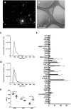
Figure 1
Figure 1
Characterization of EVs isolated from control and preeclampsia pool serums. Upper images show EV characterization by (A) nanotracking analysis with the absence of the characteristic fog pattern of contaminated samples and (B) electron microscopy (scale bars, 0.5 μm). Cryo-electron microscopy allows one to visualize the grid with an irregular distribution of hole sizes and shapes containing EVs of various sizes. (C, D) NTA particle concentration and size distribution of one control pool and three preeclampsia sera pools, respectively. (E) MFI of EV-markers CD9, CD63, and CD81 (control in black and PE in blue), obtained from MACSPlex analysis of EVs surface markers, and (F) phenotypic signature of EVs quantified by the MACSPlex Exosome Kit in conjunction with flow cytometry. Black bars correspond to PE pools and white bars correspond to control pools. *p <  0.05 and **p <  0.01 compared to the control group.
Figure 2
Figure 2
The exposure of endothelial cells to PE-EVs increased the expression of dysfunction endothelial markers. Representative microscopy image (40×) of VWF, VCAM-1, and ROS (red: VWF; ROS: green; and VCAM-1: green) on endothelial cells (4′,6-Diamidino-2-phenylindole-stained nuclei, blue) induced by exposure (48 h) to control, preeclampsia, depleted preeclampsia (without PE-EVs), and control sera pool supplemented with PE-EVs. The bar graph indicates the average fold increase of the different conditions compared to control. The vertical line indicates the standard deviation. **p < 0.01 compared to the control condition, ##p < 0.01 compared to the PE condition.
Figure 3
Figure 3
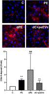
The complement system dysregulation is not mediated by EVs. Representative microscopy image (40×) of C5b-9 deposit (red) on endothelial cells (4′,6-diamidino-2-phenylindole-stained nuclei, blue) induced by exposure (4 h) to control, preeclampsia, depleted preeclampsia (without PE-EVs), and control sera pool supplemented with PE-EVs. The bar graph indicates the average fold increase of the different conditions under study compared to control. The vertical line indicates the standard deviation. **p < 0.01 compared to control, ##p < 0.01 compared to PE.
Figure 4
Figure 4
PE sera induce NET generation in neutrophils from a healthy donor. Micrographs of SYTOX green staining showed an increase in NET production by isolated donor neutrophils preactivated with TNFalpha incubated with PE sera compared to preactivated donor neutrophils incubated with sera from healthy pregnant women (micrographs taken at 40×). The bar graph indicates the result of DNA quantification (ng/mL). The vertical line depicts the standard deviation. **p < 0.01 compared to the control group.
Figure 5
Figure 5
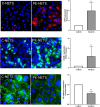
PE-NETs induce endothelial damage in the in vitro model compared to control NETs. Representative microscopy images of VWF (red, 40× micrographs), VCAM-1 (green, 40× micrographs), and VE-cadherin (100× micrographs) on endothelial cells (4′,6-diamidino-2-phenylindole-stained nuclei, blue) induced by exposure (48 h) to C-NETs and PE-NETS. The bar indicates the average fold increase compared to control. The vertical lines indicate the standard deviation. **p <  0.01 compared to the control group.
Figure 6
Figure 6
PE-NETs activate the complement system with an increase of the lytic complex C5b-9. Representative microscopy image (40×) of C5b-9 deposit stained with red fluorochrome on endothelial cells (4′,6-diamidino-2-phenylindole-stained nuclei, blue) induced by exposure (4 h) to control activated plasma (C) and this condition supplemented with PE-NETs (C+PE-NETS). The bar indicates the average fold increase compared to control. The vertical lines indicate the standard deviation. **p <  0.01 compared to the control group.

References

    1. ACOG Practice Bulletin no. 202: Gestational hypertension and preeclampsia. Obstet Gynecol. (2019) 133:1. doi: 10.1097/AOG.0000000000003018 - DOI - PubMed
    1. Goel A, Maski MR, Bajracharya S, Wenger JB, Zhang D, Salahuddin S, et al. . Epidemiology and mechanisms of DE Novo and persistent hypertension in the postpartum period. Circulation. (2015) 132:1726–33. doi: 10.1161/CIRCULATIONAHA.115.015721 - DOI - PMC - PubMed
    1. Maynard S, Epstein FH, Karumanchi SA. Preeclampsia and angiogenic imbalance. Annu Rev Med. (2008) 59:61–78. doi: 10.1146/annurev.med.59.110106.214058 - DOI - PubMed
    1. Rana S, Burke SD, Karumanchi SA. Imbalances in circulating angiogenic factors in the pathophysiology of preeclampsia and related disorders. Am J Obstet Gynecol. (2020) 226:S1019–34. doi: 10.1016/j.ajog.2020.10.022 - DOI - PMC - PubMed
    1. Zhang J, Li S, Li L, Li M, Guo Chongye and Yao J, Mi S. Exosome and exosomal microRNA: trafficking, sorting, and function. Genomics Proteomics Bioinf. (2015) 13:17–24. doi: 10.1016/j.gpb.2015.02.001 - DOI - PMC - PubMed

MeSH terms

Substances

LinkOut - more resources