

Temporal changes of spinal microglia in murine models of neuropathic pain: a scoping review
- PMID: 39735541
- PMCID: PMC11671780
- DOI: 10.3389/fimmu.2024.1460072
Temporal changes of spinal microglia in murine models of neuropathic pain: a scoping review
Abstract
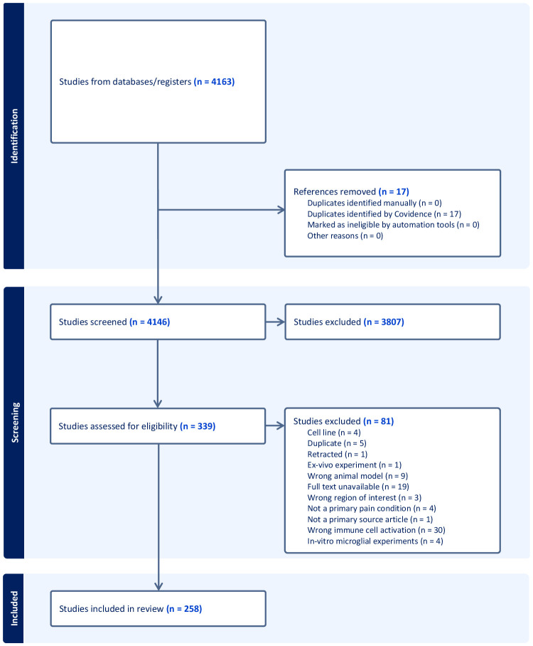
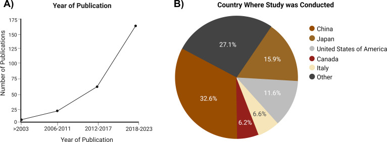
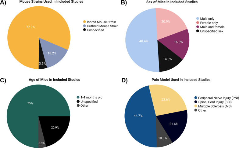
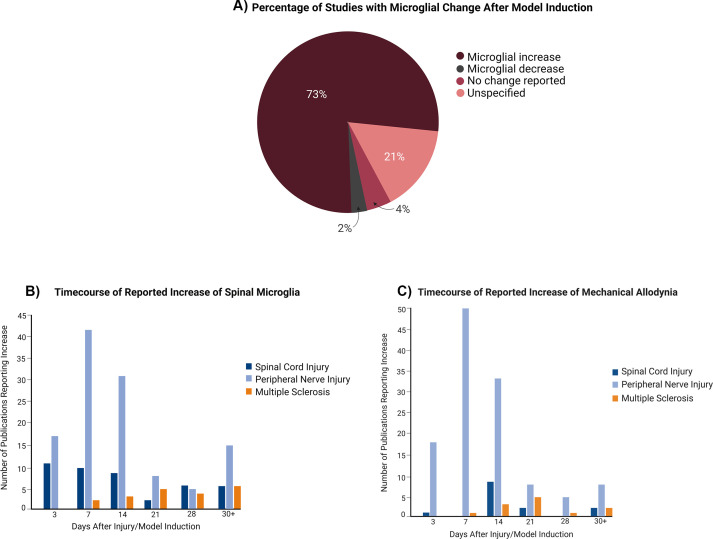
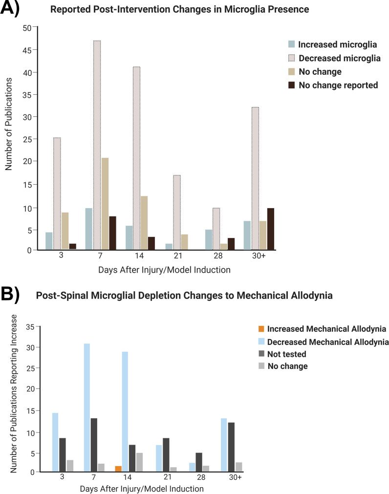
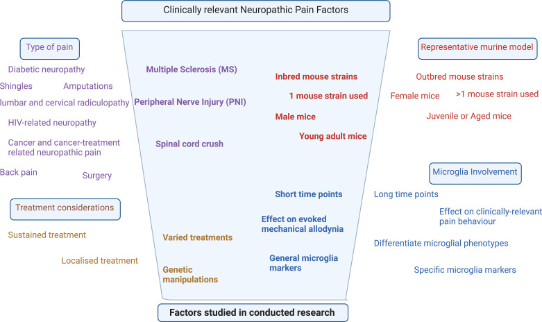
Neuropathic pain (NP) is an ineffectively treated, debilitating chronic pain disorder that is associated with maladaptive changes in the central nervous system, particularly in the spinal cord. Murine models of NP looking at the mechanisms underlying these changes suggest an important role of microglia, the resident immune cells of the central nervous system, in various stages of disease progression. However, given the number of different NP models and the resource limitations that come with tracking longitudinal changes in NP animals, many studies fail to truly recapitulate the patterns that exist between pain conditions and temporal microglial changes. This review integrates how NP studies are being carried out in murine models and how microglia changes over time can affect pain behavior in order to inform better study design and highlight knowledge gaps in the field. 258 peer-reviewed, primary source articles looking at spinal microglia in murine models of NP were selected using Covidence. Trends in the type of mice, statistical tests, pain models, interventions, microglial markers and temporal pain behavior and microglia changes were recorded and analyzed. Studies were primarily conducted in inbred, young adult, male mice having peripheral nerve injury which highlights the lack of generalizability in the data currently being collected. Changes in microglia and pain behavior, which were both increased, were tested most commonly up to 2 weeks after pain initiation despite aberrant microglia activity also being recorded at later time points in NP conditions. Studies using treatments that decrease microglia show decreased pain behavior primarily at the 1- and 2-week time point with many studies not recording pain behavior despite the involvement of spinal microglia dysfunction in their development. These results show the need for not only studying spinal microglia dynamics in a variety of NP conditions at longer time points but also for better clinically relevant study design considerations.
Keywords: microglia; mouse model; neuropathic pain (NP); spinal cord; temporal changes.
Copyright © 2024 Dhir, Derue and Ribeiro-da-Silva.
Conflict of interest statement
The authors declare that the research was conducted in the absence of any commercial or financial relationships that could be construed as a potential conflict of interest.
Figures
References
Publication types
MeSH terms
LinkOut - more resources
Full Text Sources
Miscellaneous