. 2024 Dec 20;33(1):171-184.
doi: 10.32604/or.2024.047626. eCollection 2025.

Long noncoding RNA LINC01106 promotes lung adenocarcinoma progression via upregulation of autophagy

Affiliations

Long noncoding RNA LINC01106 promotes lung adenocarcinoma progression via upregulation of autophagy

Gengyun Sun et al. Oncol Res. .

Abstract

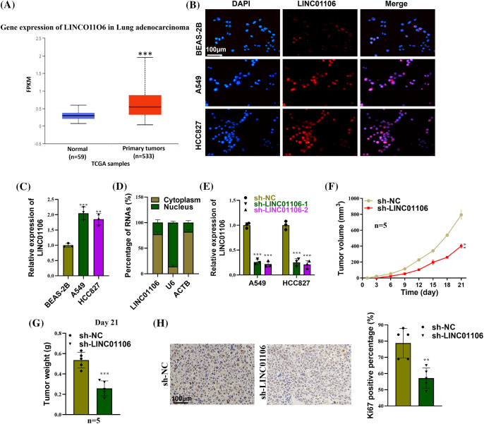
Background: Long noncoding RNA, LINC01106 exhibits high expression in lung adenocarcinoma (LUAD) tumor tissues, but its functional role and regulatory mechanism in LUAD cells remain unclear.

Methods: LINC01106 expression was analyzed in LUAD tissues and its functional impact on LUAD cells was assessed. LUAD cells were silenced with sh-LINC01106 and injected into nude mice to investigate tumor growth. The downstream transcription factors and molecular mechanism were determined using the Human transcription factor database (TFDB) database and Gene Expression Profiling Interactive Analysis (GEPIA) database. Additionally, the impact of linc01106 on autophagy was analyzed by determining the expression of autophagy-related genes (ATGs) in LUAD cells.

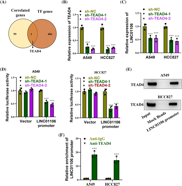
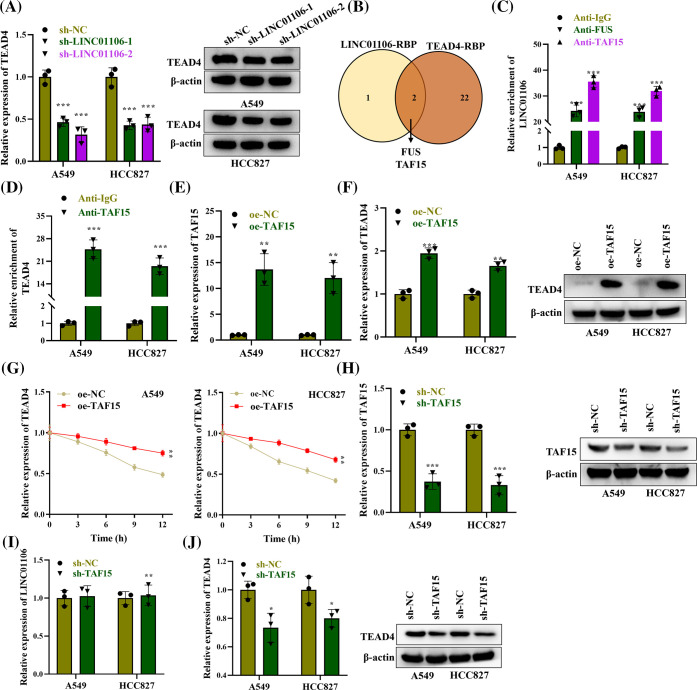
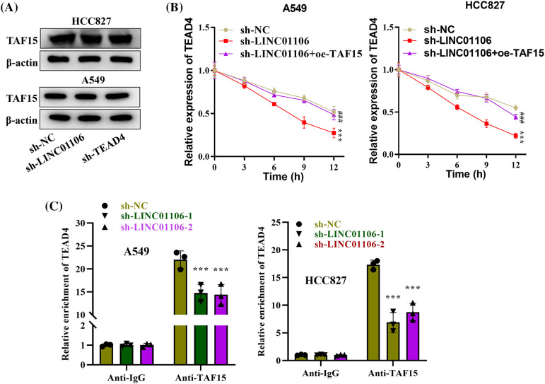
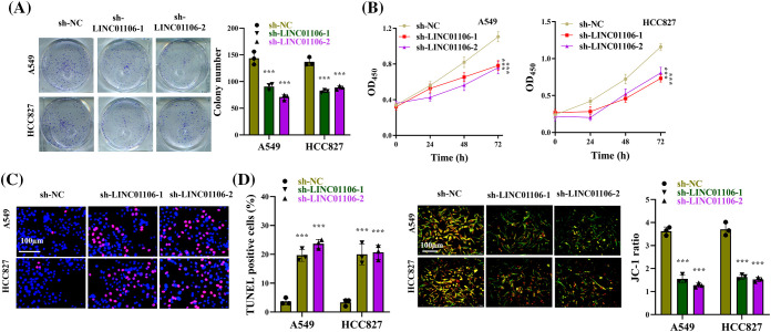
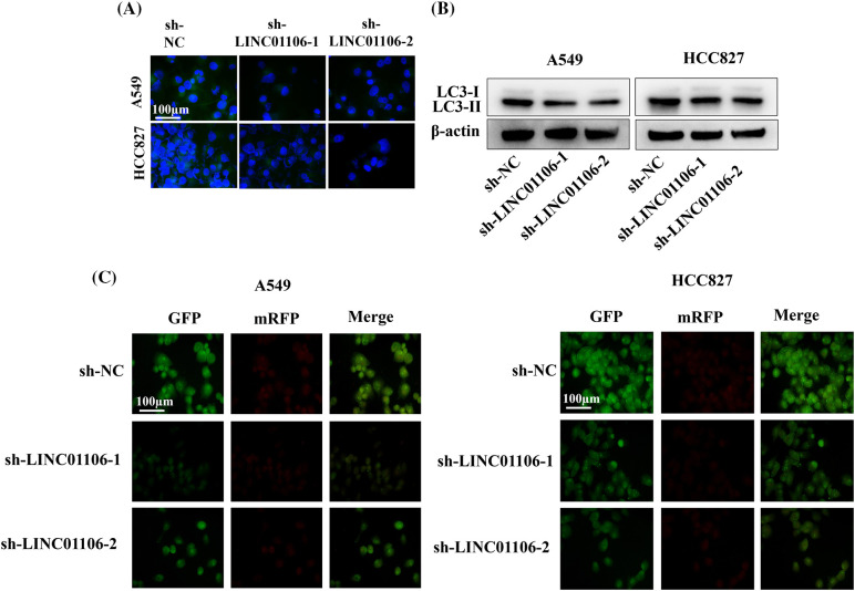
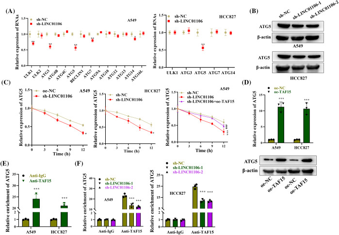
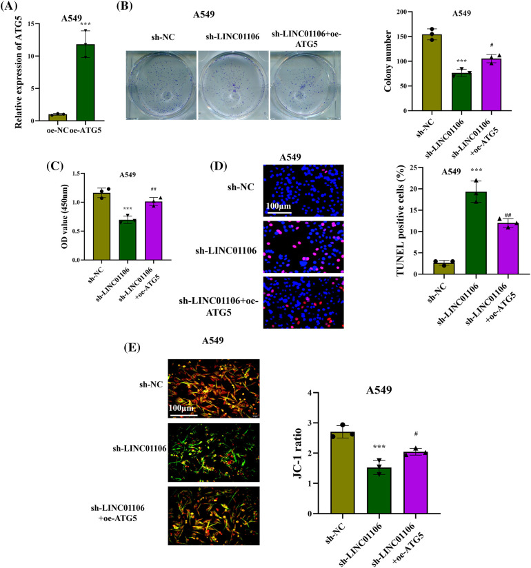
Results: Our results showed that LINC01106 exhibited upregulation in both LUAD tissues and cell lines. The silencing of LINC01106 demonstrated a suppressive effect on tumorigenesis in a xenograft mouse model of LUAD. Additionally, LINC01106 was found to recruit TATA-binding protein-associated factor 15 (TAF15), an RNA-binding protein, thereby enhancing the mRNA stability of TEA domain transcription factor 4 (TEAD4). In turn, TEAD4 served as a transcription factor that bound to the LINC01106 promoter and regulated its expression. Further assays indicated that LINC01106 promoted autophagy in LUAD cells by upregulating the expression of autophagy-related genes (ATGs). The silencing of LINC01106 in LUAD cells inhibited autophagy, and cell proliferation, and promoted apoptosis, which all were effectively reversed by ATG5 overexpression.

Conclusions: Overall, LINC01106, transcriptionally activated by TEAD4, interacts with TAF15 to promote the stability of TEAD4 and upregulates the expression of ATGs, promoting malignancy of LUAD cells.

Keywords: ATG5; LINC01106; Lung adenocarcinoma (LUAD); Non-small cell lung cancer (NSCLC); TAF15; TEAD4.


Conflict of interest statement

The authors declare no conflicts of interest to report regarding the present study.

Figures

Figure 1
Figure 1. LINC01106 facilitates lung cancer tumor growth in vivo. (A) The expression profile of LINC01106 in LUAD tissues (n = 533) and normal tissues (n = 59) was analyzed using the UALCAN database (http://ualcan.path.uab.edu/). (B and C) The expression of Linc01106in LUAD cell lines (A549, HCC827) and non-cancerous BEAS-2B cells was detected using FISH assay and RT-qPCR (n = 3). (D) Subcellular fractionation assays were conducted to determine the subcellular location of LINC01106 (n = 3). (E) The efficiency of LINC01106 knockdown in LUAD cells was assessed using RT-qPCR analysis (n = 3). (F) The measurement of mouse tumor volume at specified time points in each group (n = 5). (G) Comparison of mouse tumor weight on day 21 between the sh-NC and sh-LINC01106 groups (n = 5). (H) Immunohistochemical (IHC) assays were performed to evaluate Ki67 protein expression in mouse tumor tissues from the indicated groups (n = 3). **p < 0.01, ***p < 0.001.
Figure 2
Figure 2. TEAD4 transcriptionally activates LINC01106. (A) Venn diagram illustrating the identification of potential transcription factors positively correlated with LINC01106 expression. This was achieved by intersecting the top 100 genes most significantly correlated with LINC01106 expression from the GEPIA database and the potential transcription factors from the Human TFDB database. (B) RT-qPCR analysis confirming the efficacy of TEAD4 knockdown in LUAD cells (n = 3). (C) RT-qPCR analysis assessing the expression of LINC01106 in LUAD cells transfected with sh-TEAD4-1/-2 (n = 3). (D) Dual-luciferase reporter assays conducted to investigate the interaction between TEAD4 and the LINC01106 promoter (n = 3). (E) DNA pull-down assay and Western blot analysis performed to examine the enrichment of TEAD4 in the complex pulled down by the LINC01106 promoter (n = 3). (F) ChIP assays employed to explore the binding between the LINC01106 promoter and TEAD4 in LUAD cells (n = 3). ***p < 0.001.
Figure 3
Figure 3. TAF15 enhances the stability of TEAD4. (A) The mRNA and protein expression of TEAD4 in LUAD cells transfected with sh-LINC01106-1/-2 was examined using RT-qPCR and western blot, respectively. β-actin was used as a loading control (n = 3). (B) Venn diagram showing the shared RNA-binding proteins (RBPs) for LINC01106 and TEAD4 based on the ENCORI database, under the screening condition of ClusterNumber ≥5. (C) RNA immunoprecipitation (RIP) assays were performed to evaluate the binding between LINC01106 and FUS or TAF15 in LUAD cells (n = 3). (D) RIP assays were conducted to detect the binding relation between TEAD4 and TAF15 in LUAD cells (n = 3). (E) RT-qPCR analysis was employed to examine the efficiency of TAF15 overexpression in LUAD cells (n = 3). (F) RT-qPCR analysis and western blot were used to detect TEAD4 mRNA and protein expression in LUAD cells after TAF15 overexpression. β-actin was used as a loading control (n = 3). (G) RT-qPCR analysis assessed TEAD4 mRNA levels in LUAD cells at different time points (0, 3, 6, 9, 12 h) after ActD treatment and indicated transfection (n = 3). (H) Knockdown efficacy of sh-TAF15 was validated by western blotting (n = 3). (I) RT-qPCR was conducted to assess influence of sh-TAF15 on LINC01106 expression (n = 3). (J) Effects of sh-TAF15 on TEAD4 expression were evaluated by western blotting (n = 3). *p < 0.05, **p < 0.01, ***p < 0.001.
Figure 4
Figure 4. LINC01106 recruits TAF15 to stabilize TEAD4. (A) Protein expression of TAF15 was assessed by Western blotting in LUAD cells following LINC01106 or TEAD4 knockdown. β-actin was used as a loading control (n = 3). (B) mRNA expression of TEAD4 was measured using RT-qPCR analysis in LUAD cells after the indicated transfection (n = 3). (C) RIP assays were performed to examine the impact of LINC01106 silencing on the binding between TEAD4 and TAF15 in LUAD cells (n = 3). ***p < 0.001; ###p < 0.001.
Figure 5
Figure 5. LINC01106 promotes LUAD cell growth in vitro. (A) Clonogenic assays were performed to assess the proliferation of LUAD cells following transfection with sh-LINC01106-1/-2 (n = 3). (B) Cell viability of LUAD cells in each group was measured using CCK-8 assays. (C) TUNEL assays were conducted to evaluate the extent of apoptosis in LUAD cells after the indicated transfection (n = 3). (D) JC-1 staining was performed to quantify the proportion of apoptotic cells (green) in LUAD cells (n = 3). ***p < 0.001.
Figure 6
Figure 6. LINC01106 regulates autophagy in LUAD cells. (A) Immunofluorescence assays were performed to assess the expression of LC3 protein in LUAD cells following LINC01106 knockdown (n = 3). (B) Western blot analysis was conducted to measure the protein levels of LC3-I and LC3-II after silencing LINC01106 in LUAD cells. β-actin was used as a loading control (n = 3). (C) Autophagic flux in LUAD cells was visualized by transfecting cells with mRFP-GFP-LC3 adenovirus (n = 3).
Figure 7
Figure 7. LINC01106 interacts with TAF15 to regulate ATG5 expression. (A) RT-qPCR analysis of ATG expression in LUAD cells after LINC01106 knockdown (n = 3). (B) Western blot analysis of ATG5 protein levels in LUAD cells after indicated transfections. β-actin is used as a loading control (n = 3). (C) RT-qPCR analysis of ATG5 mRNA expression and stability in LUAD cells treated with ActD and transfected with indicated constructs (n = 3). (D) RT-qPCR and Western blot analysis of ATG5 mRNA and protein levels after TAF15 overexpression in LUAD cells (n = 3). (E) RIP assays demonstrating the interaction between ATG5 and TAF15 in LUAD cells (n = 3). (F) RIP assays investigating the impact of LINC01106 knockdown on the binding between ATG5 and TAF15 in LUAD cells (n = 3). *p < 0.05, **p < 0.01, ***p < 0.001; ###p < 0.001.
Figure 8
Figure 8. LINC01106 regulates LUAD cell growth by modulating ATG5. (A) RT-qPCR analysis confirms the overexpression efficacy of ATG5 in LUAD cells (n = 3). (B) Colony formation assays and (C) CCK-8 assays assess the proliferation of LUAD cells in each group (n = 3). (D) TUNEL assays and (E) JC-1 staining assays were performed to evaluate LUAD cell apoptosis after the indicated transfection (n = 3). Statistical significance is denoted by ***p < 0.001; #p < 0.05, ##p < 0.01.

Similar articles

Cited by

References

    1. Siegel, R. L., Miller, K. D., Wagle, N. S., Jemal, A. (2023). Cancer statistics, 2023. CA: A Cancer Journal for Clinicians , 73(1), 17–48. 10.3322/caac.21763; - DOI - PubMed
    1. Vicidomini, G. (2023). Current challenges and future advances in lung cancer: Genetics, instrumental diagnosis and treatment. Cancers , 15(14), 3710. 10.3390/cancers15143710; - DOI - PMC - PubMed
    1. Wang, W., Xia, X., Chen, K., Chen, M., Meng, Y.et al. (2021). Reduced PHLPP expression leads to EGFR-TKI resistance in lung cancer by activating PI3K-AKT and MAPK-ERK dual signaling. Frontiers in Oncology , 11, 665045. 10.3389/fonc.2021.665045; - DOI - PMC - PubMed
    1. Thai, A. A., Solomon, B. J., Sequist, L. V., Gainor, J. F., Heist, R. S. (2021). Lung cancer. Lancet , 398(10299), 535–554. 10.1016/S0140-6736(21)00312-3; - DOI - PubMed
    1. Liu, S. M., Zheng, M. M., Pan, Y., Liu, S. Y., Li, Y.et al. (2023). Emerging evidence and treatment paradigm of non-small cell lung cancer. Journal of Hematology & Oncology , 16(1), 40. 10.1186/s13045-023-01436-2; - DOI - PMC - PubMed

MeSH terms

LinkOut - more resources