. 2024 Dec 13:15:1489439.
doi: 10.3389/fphys.2024.1489439. eCollection 2024.

Nutraceuticals silybin B, resveratrol, and epigallocatechin-3 gallate-bind to cardiac muscle troponin to restore the loss of lusitropy caused by cardiomyopathy mutations in vitro, in vivo, and in silico

Affiliations

Nutraceuticals silybin B, resveratrol, and epigallocatechin-3 gallate-bind to cardiac muscle troponin to restore the loss of lusitropy caused by cardiomyopathy mutations in vitro, in vivo, and in silico

Zeyu Yang et al. Front Physiol. .

Abstract

Introduction: Adrenergic activation of protein kinase A (PKA) in cardiac muscle targets the sarcolemma, sarcoplasmic reticulum, and contractile apparatus to increase contractile force and heart rate. In the thin filaments of the contractile apparatus, cardiac troponin I (cTnI) Ser22 and Ser23 in the cardiac-specific N-terminal peptide (NcTnI: residues 1 to 32) are the targets for PKA phosphorylation. Phosphorylation causes a 2-3 fold decrease of affinity of cTn for Ca2+ associated with a higher rate of Ca2+ dissociation from cTnC leading to a faster relaxation rate of the cardiac muscle (lusitropy). Cardiomyopathy-linked mutations primarily affect Ca2+ regulation or the PKA-dependent modulatory system, such that Ca2+-sensitivity becomes independent of phosphorylation level (uncoupling) and this could be sufficient to induce cardiomyopathy. A drug that could restore the phosphorylation-dependent modulation of Ca2+-sensitivity could have potential for treatment of these pathologies. We have found that a number of small molecules, including silybin B, resveratrol and EGCG, can restore coupling in single filament assays.

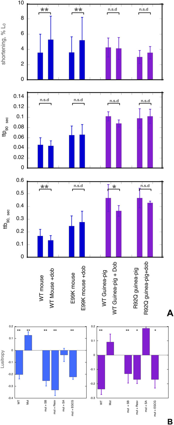
Methods: We did molecular dynamics simulations (5x1500ns for each condition) of the unphosphorylated and phosphorylated cardiac troponin core with the G159D DCM mutation in the presence of the 5 ligands and analysed the effects on several dynamic parameters. We also studied the effect of the ligands on the contractility of cardiac muscle myocytes with ACTC E99K and TNNT2 R92Q mutations in response to dobutamine.

Results: Silybin B, EGCG and resveratrol restored the phosphorylation-induced change in molecular dynamics to wild-type values, whilst silybin A, an inactive isomer of silybin B, and Epicatechin gallate, an EGCG analogue that does not recouple, did not. We analysed the atomic-level changes induced by ligand binding to explain recoupling. Mutations ACTC E99K and TNNT2 R92Q blunt the increased relaxation speed response to β1 adrenergic stimulation of cardiac myocytes and we found that resveratrol, EGCG and silybin B could restore the β1 adrenergic response, whereas silybin A did not.

Discussion: The uncoupling phenomenon caused by cardiomyopathy-related mutations and the ability of small molecules to restore coupling in vitro and lusitropy in myocytes is observed at the cellular, molecular and atomistic levels therefore, restoring lusitropy is a suitable target for treatment. Further research on compounds that restore lusitropy is thus indicated as treatments for genetic cardiomyopathies. Further molecular dynamics simulations could define the specific properties needed for recoupling and allow for the prediction and design of potential new drugs.

Keywords: EGCG; PKA phosphorylation; cardiomyopathy; lusitropy; molecular dynamics simulation; myocyte contraction; silybin; troponin (cTnI).


Conflict of interest statement

The authors declare that the research was conducted in the absence of any commercial or financial relationships that could be construed as a potential conflict of interest.

Figures

FIGURE 1
FIGURE 1
(A) Effects of dobutamine on key parameters of cardiac myocyte contractility. Data from experiments where wild-type and mutant cells were measured together. Shortening amplitude, ttp90 and ttb90 parameters are shown before and after addition of dobutamine; mean and Standard error are shown for 40–60 myocytes. Blue- WT and E99K mouse compared. Purple- WT and R92Q guinea-pig compared. The Incubation medium was Krebs-Hensleit buffer with added 1mM CaCl2, oxygenated with 95% oxygen/5% CO2. The myocytes were incubated at 37° and continuously stimulated a 1Hz. 10 s of contractility was collected and analysed for each cell. *, ** indicate significant differences due to adding dobutamine at 0.05 and 0.01 levels respectively using paired t-test. For numerical data and statistical analysis see Supplementary Material 3. (B) Lusitropy and the effect of small molecules measured in cardiomyocytes. Lusitropy is defined as the fractional change of ttb90 due to dobutamine [ 1-(ttb90+Dob/ttb90-Dob)]. Lusitropy (shorter relaxation time) corresponds to a decrease in this parameter. Blue, mouse experiments, Purple, Guinea-pig experiments. Mean, sem and significance in paired t-test is shown as for (A). Lusitropy is lost in the mutant myocytes but is restored by EGCG, Silybin B and resveratrol but not by Silybin A. Numerical data and statistical analysis are presented in Supplementary Material 4.
FIGURE 2
FIGURE 2
The distribution of a helix A/B and Interdomain angles determined by molecular dynamics simulations. (A) Left:Model defining the location of the hinge between N-terminal and C-terminal domains of troponin C. Right: Model showing the orientation of helices A and B in the N-terminus of troponin C. (B) The effects of the G159D mutation and phosphorylation on the distribution of the interdomain angle and the helix A/B angle. Unphosphorylated, uP, phosphorylated, SEP. (Yang et al., 2023). (C) The effects of phosphorylation and small molecules on the distribution of the interdomain angle (left) and the helix A/B angle (right) For G159D. Tables 2, 3 show the quantification of the distributions. Distribution plots for troponin in the presence of ECG are shown in Supplementary Material 6.
FIGURE 3
FIGURE 3
Parameterised solution configurations Silybin A (magenta) silybin B (green), EGCG (yellow) and resveratrol (blue). A and D rings of silybin A and B and A and C rings of EGCG are aligned. See Supplementary Material 5 for identification of the atoms and rings.
FIGURE 4
FIGURE 4
Ligands bound to unphosphorylated G!59D troponin in their most probable state. (A) Stick representation of the ligand interacting with TnC(light grey) and TnI(blue). TnT is light brown. The ligand is red The molecules are orientated to give the best view of the interaction. For EGCG and SilybinB parts of TnC not involved in ligation are hidden for clarity. (B) Whole troponin surface representations. TnI is blue, TnC is grey and TnT is light blue or cyan. The ligand is red. Single frames are selected from the full trajectory (3750 frames) to illustrate the most common positions of the ligands. Models are orientated to show the ligand binding optimally. Examples of complete trajectories are illustrated in Supplementary Material 8.
FIGURE 5
FIGURE 5
Comparison of the effect of phosphorylation of cardiac troponin on its biochemical, physiological and molecular dynamics parameters, its suppression by the TnC G159D DCM-related mutation and its restoration by small molecules. Parameters compared are lusitropy in intact myocytes (from Figure 1B), Fixed [Ca2+] screen for coupling in thin filaments measured by IVMA from (Sheehan et al., 2018) and Supplementary Material 1 and the effect of phosphorylation on EC50, (EC50P/EC50uP) in thin filaments measured by IVMA (from Table 1), Change on A/B angle on phosphorylation and change in hinge angle upon phosphorylation from Molecular dynamics simulations (from Figure 2; Tables 2, 3). The recouplers are highlighted in blue.

Similar articles

Cited by

References

    1. Baell J., Walters M. A. (2014). Chemistry: Chemical con artists foil drug discovery. Nature 513 (7519), 481–483. 10.1038/513481a - DOI - PubMed
    1. Baryshnikova O. K., Li M. X., Sykes B. D. (2008). Modulation of cardiac troponin C function by the cardiac-specific N-terminus of troponin I: influence of PKA phosphorylation and involvement in cardiomyopathies. J. Mol. Biol. 375 (3), 735–751. 10.1016/j.jmb.2007.10.062 - DOI - PubMed
    1. Bayly C. I., Cieplak P., Cornell W., Kollman P. A. (1993). A well-behaved electrostatic potential based method using charge restraints for deriving atomic charges: the RESP model. J. Phys. Chem. 97 (40), 10269–10280. 10.1021/j100142a004 - DOI
    1. Becke A. D. (1993). Density‐functional thermochemistry. III. The role of exact exchange. J. Chem. Phys. 98 (7), 5648–5652. 10.1063/1.464913 - DOI
    1. Biesiadecki B. J., Kobayashi T., Walker J. S., John Solaro R., de Tombe P. P. (2007). The troponin C G159D mutation blunts myofilament desensitization induced by troponin I Ser23/24 phosphorylation. Circ. Res. 100 (10), 1486–1493. 10.1161/01.RES.0000267744.92677.7f - DOI - PubMed

LinkOut - more resources