Review
. 2024 Oct 25;2(12):784-807.
doi: 10.1021/cbmi.4c00060. eCollection 2024 Dec 23.

Sequencing and Optical Genome Mapping for the Adventurous Chemist

Affiliations
Review

Sequencing and Optical Genome Mapping for the Adventurous Chemist

Elizabete Ruppeka Rupeika et al. Chem Biomed Imaging. .

Abstract

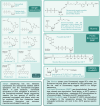
This review provides a comprehensive overview of the chemistries and workflows of the sequencing methods that have been or are currently commercially available, providing a very brief historical introduction to each method. The main optical genome mapping approaches are introduced in the same manner, although only a subset of these are or have ever been commercially available. The review comes with a deck of slides containing all of the figures for ease of access and consultation.


Conflict of interest statement

The authors declare the following competing financial interest(s): Johan Hofkens and Volker Leen are co-founders of Perseus Biomics.

Figures

Figure 1
Figure 1
DNA structure. From left: nucleobase position numbering, purines and pyrimidines, and sugar phosphate backbone and hydrogen bonds between the two antiparallel strands.
Figure 2
Figure 2
DNA elongation, triphosphate addition, release of a pyrophosphate, and formation of the phosphodiester bond of a DNA strand.
Figure 3
Figure 3
Overview of early sequencing methods: Radiolabeled Sanger sequencing (left and center) and (right) gel electrophoresis readout for (1) Maxam–Gilbert sequencing, (2) Sanger sequencing with radioactively labeled ddNTPs, and (3) Sanger sequencing with chain terminating radioactively labeled ddNTPs, and (4) Sanger sequencing with fluorescently labeled chain terminating ddNTPs.
Figure 4
Figure 4
Structures of various dNTPs and ddNTPs used for various sequencing technology generations.
Figure 5
Figure 5
Bacterial artificial chromosome generation process.
Figure 6
Figure 6
Sequencing depth and coverage explained in an illustrated example. The missing 10% may look random in this example but in reality can be represented by regions that are prone to double-stranded breaks and fragmentation, homopolymeric regions or regions of high or low GC content impacting the local accuracy of enzymes like polymerase., The missing 10% are shown as a single contiguous region for mere illustrative purposes as it is much more likely that problematic regions would be scattered across the genome. Even so, the cumulative amount of 10% in the example is very high for contemporary sequencing approaches.
Figure 7
Figure 7
Emulsion PCR workflow.
Figure 8
Figure 8
Roche 454 sequencing workflow.
Figure 9
Figure 9
An early approach to the Sequencing by Oligo Ligation Detection method.
Figure 10
Figure 10
Sequencing by Oligo Ligation Detection (SOLiD) workflow.
Figure 11
Figure 11
Solid-state (bridge) PCR workflow.
Figure 12
Figure 12
Sequencing by synthesis: Illumina workflow following the bridge PCR step.
Figure 13
Figure 13
Illumina single-dye sequencing modification workflow.
Figure 14
Figure 14
DNA nanoball (DNB) sequencing workflow: library preparation (top), base calling in DNBseq (lower left), and CoolMPS workflow (lower right).
Figure 15
Figure 15
Reversible terminators used by Helicos Biosciences.
Figure 16
Figure 16
Single-molecule real-time (SMRT) sequencing workflow.
Figure 17
Figure 17
Oxford Nanopore (ONP) sequencing workflow.
Figure 18
Figure 18
Optical genome mapping: mechanisms of action for denaturation mapping and competitive binding (CB).
Figure 19
Figure 19
Optical genome mapping: restriction mapping workflow.
Figure 20
Figure 20
Optical genome mapping: Fluorocode mechanism of action, a doubly activated synthetic analogue of methyltransferase cofactor, labeling process. Reproduced with permission from ref (181). Copyright 2024 ACS Omega.

Similar articles

References

    1. Franklin R. E.; Gosling R. G. Molecular Configuration in Sodium Thymonucleate. Nature 1953, 171, 740–741. 10.1038/171740a0. - DOI - PubMed
    1. Watson J. D.; Crick F. H. C. Molecular Structure of Nucleic Acids: A Structure for Deoxyribose Nucleic Acid. Nature 1953, 171, 737–738. 10.1038/171737a0. - DOI - PubMed
    1. Alberts B.; et al.DNA Replication Mechanisms. Molecular Biology of the Cell, 4th ed.; Garland Science, 2002.
    1. Burgers P. M. Solution to the 50-year-old Okazaki-fragment problem. Proc. Natl. Acad. Sci. U. S. A. 2019, 116, 3358–3360. 10.1073/pnas.1900372116. - DOI - PMC - PubMed
    1. Okazaki R.; Okazaki T.; Sakabe K.; Sugimoto K.; Sugino A. Mechanism of DNA chain growth. I. Possible discontinuity and unusual secondary structure of newly synthesized chains. Proc. Natl. Acad. Sci. U. S. A. 1968, 59, 598–605. 10.1073/pnas.59.2.598. - DOI - PMC - PubMed

LinkOut - more resources