. 2024 Nov 22;2(12):835-841.
doi: 10.1021/cbmi.4c00070. eCollection 2024 Dec 23.

Laser-Treated Screen-Printed Carbon Electrodes for Electrochemiluminescence imaging

Affiliations

Laser-Treated Screen-Printed Carbon Electrodes for Electrochemiluminescence imaging

Claudio Ignazio Santo et al. Chem Biomed Imaging. .

Abstract

Electrochemiluminescence (ECL) is nowadays a powerful technique widely used in biosensing and imaging, offering high sensitivity and specificity for detecting and mapping biomolecules. Screen-printed electrodes (SPEs) offer a versatile and cost-effective platform for ECL applications due to their ease of fabrication, disposability, and suitability for large-scale production. This research introduces a novel method for improving the ECL characteristics of screen-printed carbon electrodes (SPCEs) through the application of CO2 laser treatment following fabrication. Using advanced ECL microscopy, we analyze three distinct carbon paste-based electrodes and show that low-energy laser exposure (ranging from 7 to 12 mJ·cm-2) enhances the electrochemical performance of the electrodes. This enhancement results from the selective removal of surface binders and contaminants achieved by the laser treatment. We employed ECL microscopy to characterize the ECL emission using a bead-based system incorporating magnetic microbeads, like those used in commercial platforms. This approach enabled high-resolution spatial mapping of the electrode surface, offering valuable insights into its electrochemical performance. Through quantitative assessment using a photomultiplier tube (PMT), it was observed that GST electrodes could detect biomarkers with high sensitivity, achieving an approximate detection limit (LOD) of 11 antibodies per μm2. These findings emphasize the potential of laser-modified GST electrodes in enabling highly sensitive electrochemiluminescent immunoassays and various biosensing applications.


Conflict of interest statement

The authors declare no competing financial interest.

Figures
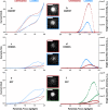
Figure 1
Figure 1
Single-bead ECL intensity collected during the cyclic voltammetry for Gwent, Henkel and GST electrodes before and after the laser treatment. In graphs a, c and e, the current trend for the three electrodes before and after laser treatment, red and blue lines respectively, are shown. Dotted lines, graphs b, d and f, represent the single-bead ECL intensity at each 200 ms. Green dotted lines correspond to the ECL intensity of the best lasered electrode. The inset images show the ECL emission of the individual beads taken at the maximum of the cyclic voltammetry ECL emission (Scale bar 5 μm). Potential scan from 0 V vs OCP to 2 V vs ref, ref (reference electrode) is Ag/AgCl (KCl sat.), the counter electrode is Pt wire, scan rate 50 mV·s–1, EM-CCD integration time 200 ms, sensitivity gain 800, gain 5, magnification 100x. Image processing involves integrating a 50 × 50 pixel square area centered on the magnetic bead across all analysis frames (one image captured every 200 ms). The background is calculated from the same area on a bead that is not functionalized with ruthenium and subtracted from the signal of the functionalized bead. Finally, each image is correlated with the applied potential at that moment to obtain a trend of the ECL intensity as a function of the scanned potential. Each measurement is an average of at least N = 5 magnetic beads.
Figure 2
Figure 2
Comparison between single-bead current a) and ECL signal b) during cyclic voltammetry of laser-treated SPEs produced with different pastes. Scan rate 50 mV·s–1, EM-CCD integration time 200 ms, sensitivity gain 800, gain 5, magnification 100×. Each measurement is an average of at least N = 5 magnetic beads.
Figure 3
Figure 3
a) Quantitative beads ECL intensity during cyclic voltammetry for different [Ru(bpy)3]2+ concentrations: 4392 (black line); 2196 (red line); 1098 (blue line); 549 (pink line); 275 (dark green line); 137 (light green line); 69 (purple line) Ru dyes·μm–2 quantified by previous ICP-MS analysis. b) Calibration curve obtained after the integration of the area below the ECL curve plotted as a function of the Ru amount for each bead. Scan rate 50 mV·s–1, PMT current amplification 000.0 nA. The equation of the linear calibration curve is Y = (1.7 × 10⁻⁴ ± 1.0 × 10⁻⁵)X + (3.4 × 10⁻² ± 2.1 × 10⁻²), R2 = 0.98, where Y is the integrated ECL signal and X is the number of Ru dye per μm2 of bead’s surface. Each datum is the mean ± SD of three independent experiments.

Similar articles

References

    1. Khanmohammadi A.; Aghaie A.; Vahedi E.; Qazvini A.; Ghanei M.; Afkhami A.; Hajian A.; Bagheri H. Electrochemical Biosensors for the Detection of Lung Cancer Biomarkers: A Review. Talanta 2020, 206, 120251.10.1016/j.talanta.2019.120251. - DOI - PubMed
    1. Miao W. Electrogenerated Chemiluminescence and Its Biorelated Applications. Chem. Rev. 2008, 108, 2506–2553. 10.1021/cr068083a. - DOI - PubMed
    1. Miao W.; Choi J. P.; Bard A. J. Electrogenerated Chemiluminescence 69: The Tris(2,2′-bipyridine)Ruthenium(II), (Ru(bpy)32+)/Tri-n-Propylamine (TPrA) System Revisited - A New Route Involving TPrA•+ Cation Radicals. J. Am. Chem. Soc. 2002, 124 (48), 14478–14485. 10.1021/ja027532v. - DOI - PubMed
    1. Zanut A.; Fiorani A.; Canola S.; Saito T.; Ziebart N.; Rapino S.; Rebeccani S.; Barbon A.; Irie T.; Josel H. P.; Negri F.; Marcaccio M.; Windfuhr M.; Imai K.; Valenti G.; Paolucci F. Insights into the Mechanism of Coreactant Electrochemiluminescence Facilitating Enhanced Bioanalytical Performance. Nat. Commun. 2020, 11 (1), 1–9. 10.1038/s41467-020-16476-2. - DOI - PMC - PubMed
    1. Zhang J.; Zhu J.; Guo F.; Jiang J.; Xie M.; Hao L.; Chao J. Reusable Electrochemiluminescence Biosensor Based on Tetrahedral DNA Signal Amplification for Ultrasensitive Detection of MicroRNAs. Chem. Comm 2023, 59 (45), 6869–6872. 10.1039/D3CC01830J. - DOI - PubMed

LinkOut - more resources