

In Depth Mapping of Mesoporous Silica Nanoparticles in Malignant Glioma Cells Using Scattering-Type Scanning Near-Field Optical Microscopy
- PMID: 39735833
- PMCID: PMC11672216
- DOI: 10.1021/cbmi.4c00053
In Depth Mapping of Mesoporous Silica Nanoparticles in Malignant Glioma Cells Using Scattering-Type Scanning Near-Field Optical Microscopy
Abstract
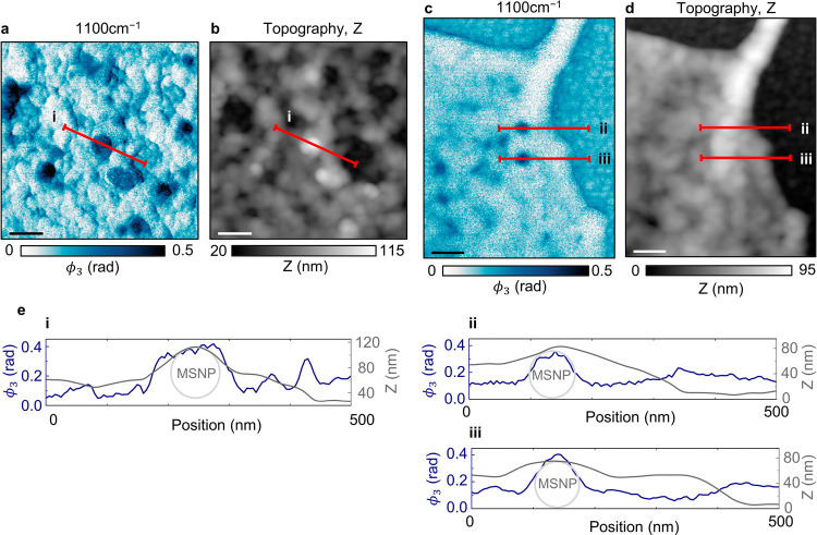
Mesoporous silica nanoparticles (MSNPs) are promising nanomedicine vehicles due to their biocompatibility and ability to carry large cargoes. It is critical in nanomedicine development to be able to map their uptake in cells, including distinguishing surface associated MSNPs from those that are embedded or internalized into cells. Conventional nanoscale imaging techniques, such as electron and fluorescence microscopies, however, generally require the use of stains and labels to image both the biological material and the nanomedicines, which can interfere with the biological processes at play. We demonstrate an alternative imaging technique for investigating the interactions between cells and nanostructures, scattering-type scanning near-field optical microscopy (s-SNOM). s-SNOM combines the chemical sensitivity of infrared spectroscopy with the nanoscale spatial resolving power of scanning probe microscopy. We use the technique to chemically map the uptake of MSNPs in whole human glioblastoma cells and show that the simultaneously acquired topographical information can provide the embedding status of the MSNPs. We focus our imaging efforts on the lamellipodia and filopodia structures at the peripheries of the cells due to their significance in cancer invasiveness.
© 2024 The Authors. Co-published by Nanjing University and American Chemical Society.
Conflict of interest statement
The authors declare no competing financial interest.
Figures


Similar articles
-
Mesoporous silica nanoparticle nanocarriers: biofunctionality and biocompatibility.Acc Chem Res. 2013 Mar 19;46(3):792-801. doi: 10.1021/ar3000986. Epub 2013 Feb 6. Acc Chem Res. 2013. PMID: 23387478 Free PMC article.
-
Nanoscale infrared absorption spectroscopy of individual nanoparticles enabled by scattering-type near-field microscopy.ACS Nano. 2011 Aug 23;5(8):6494-9. doi: 10.1021/nn2017638. Epub 2011 Jul 27. ACS Nano. 2011. PMID: 21770439
-
Nanoscale Optical Microscopy and Spectroscopy Using Near-Field Probes.Annu Rev Chem Biomol Eng. 2018 Jun 7;9:365-387. doi: 10.1146/annurev-chembioeng-060817-084150. Epub 2018 Mar 29. Annu Rev Chem Biomol Eng. 2018. PMID: 29596000 Review.
-
Tomographic and multimodal scattering-type scanning near-field optical microscopy with peak force tapping mode.Nat Commun. 2018 May 21;9(1):2005. doi: 10.1038/s41467-018-04403-5. Nat Commun. 2018. PMID: 29784951 Free PMC article.
-
Functionalized mesoporous silica nanoparticles in anticancer therapeutics.Semin Cancer Biol. 2021 Feb;69:365-375. doi: 10.1016/j.semcancer.2019.08.022. Epub 2019 Aug 20. Semin Cancer Biol. 2021. PMID: 31442571 Review.
Cited by
-
Remote Control of Gold-Iron Nanowires Using Low-Frequency 1 Hz Magneto-Mechanical Therapy and Cesium 137 0.662 MeV Radiotherapy for Treatment of Glioblastoma Multiforme.ACS Appl Mater Interfaces. 2025 Jun 11;17(23):33569-33580. doi: 10.1021/acsami.5c05004. Epub 2025 May 28. ACS Appl Mater Interfaces. 2025. PMID: 40434050 Free PMC article.
References
-
- Ocelic N.; Huber A.; Hillenbrand R. Pseudoheterodyne detection for background-free near-field spectroscopy. Appl. Phys. Lett. 2006, 89, 101124.10.1063/1.2348781. - DOI
-
- Keilmann F.; Amarie S. Mid-infrared frequency comb spanning an octave based on an Er fiber laser and difference-frequency generation. Journal of Infrared, Millimeter, and Terahertz Waves 2012, 33, 479–484. 10.1007/s10762-012-9894-x. - DOI
Grants and funding
LinkOut - more resources
Full Text Sources