Case Reports
. 2024 Dec 24:17:11577-11585.
doi: 10.2147/JIR.S489178. eCollection 2024.

Human Herpesvirus 6A Infection-Associated Acute Anterior Uveitis

Affiliations
Case Reports

Human Herpesvirus 6A Infection-Associated Acute Anterior Uveitis

Huiling Ma et al. J Inflamm Res. .

Abstract

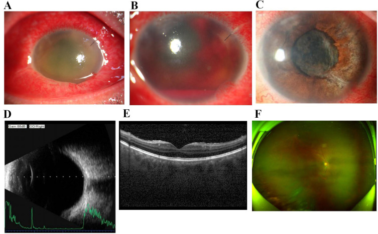
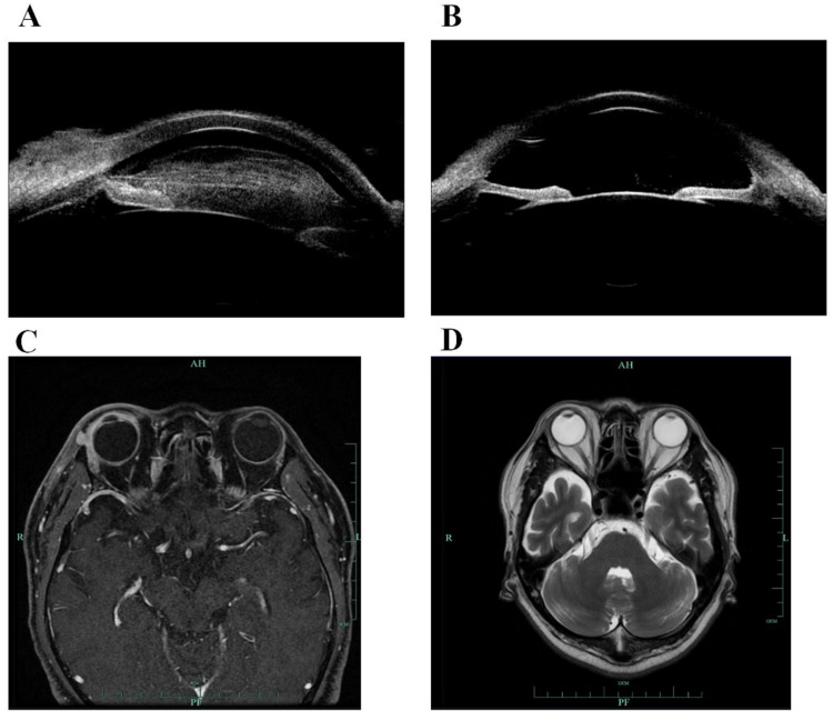
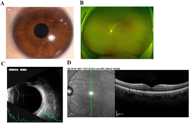
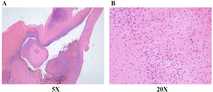
Human herpesvirus 6 (HHV-6) infection can cause ophthalmic diseases in immunocompetent patients, recipients of bone marrow transplants, and patients with acquired immunodeficiency syndrome (AIDS). This study describes the case of a healthy 37-year-old male who presented with unilateral anterior uveitis (AU), significant anterior chamber exudation, pupillary membrane closure, increased intraocular pressure, and eyelid edema. Notably, HHV-6A was the only pathogenic agent identified in the blood and aqueous humor. The patient was treated with foscarnet sodium and ganciclovir, showing effective results. Additionally, based on the literature review, the hypothesized mechanism underlying HHV-6A-associated AU was discussed. To the best of our knowledge, this is the first case report of HHV-6A involvement in ocular inflammation and may provide a theoretical basis for further investigations of occurrences of HHV-6A-associated acute AU in clinical settings.

Keywords: acute anterior uveitis; human herpesvirus 6A; secondary glaucoma.


Conflict of interest statement

The authors report no conflicts of interest in this work.

Figures

Figure 1
Figure 1
Images of the anterior segment of the right eye at initial diagnosis (A), the 1st day post-surgery (B), and 2 months post-treatment (C). The results of the B-ultrasound examination (D), optical coherence tomography (E), and fundoscopy (F) of the right eye, showing no significant abnormality.
Figure 2
Figure 2
Ultrasound Biomicroscope images showing the change of the anterior chamber inflammation before (A) and after (B) antiviral treatment. The ocular magnetic resonance imaging showing the changes in the eyelids and lacrimal glands before (C) and after (D) antiviral treatment.
Figure 3
Figure 3
Images of the anterior segment (A), fundoscopy (B), B-ultrasound examination (C), and optical coherence tomography (D) of the left eye, showing no significant abnormality.
Figure 4
Figure 4
Representative 5x (A) and 20x (B) images of the removed exudate stained with hematoxylin and eosin.

Similar articles

References

    1. Wakefield D, Dunlop I, McCluskey PJ, Penny R. Uveitis: aetiology and disease associations in an Australian population. Aust NZ J Ophthalmol. 1986;14(3):181–187. doi:10.1111/j.1442-9071.1986.tb00034.x - DOI - PubMed
    1. Chan NS, Chee SP. Demystifying viral anterior uveitis: a review. Clin Exp Ophthalmol. 2019;47(3):320–333. doi:10.1111/ceo.13417 - DOI - PubMed
    1. Sugita S, Shimizu N, Watanabe K, et al. Virological analysis in patients with human herpes virus 6-associated ocular inflammatory disorders. Invest Ophthalmol Vis Sci. 2012;53(8):4692–4698. doi:10.1167/iovs.12-10095 - DOI - PubMed
    1. Okuno T, Hooper LC, Ursea R, et al. Role of human herpes virus 6 in corneal inflammation alone or with human herpesviruses. Cornea. 2011;30(2):204–207. doi:10.1097/ICO.0b013e3181e2e9be - DOI - PubMed
    1. de Groot-Mijnes JD, de Visser L, Zuurveen S, et al. Identification of new pathogens in the intraocular fluid of patients with uveitis. Am J Ophthalmol. 2010;150(5):628–636. doi:10.1016/j.ajo.2010.05.015 - DOI - PMC - PubMed

Publication types

LinkOut - more resources