. 2024 Dec 24:17:11469-11483.
doi: 10.2147/JIR.S489087. eCollection 2024.

Macrophage Infiltration Correlated with IFI16, EGR1 and MX1 Expression in Renal Tubular Epithelial Cells Within Lupus Nephritis-Associated Tubulointerstitial Injury via Bioinformatics Analysis

Affiliations

Macrophage Infiltration Correlated with IFI16, EGR1 and MX1 Expression in Renal Tubular Epithelial Cells Within Lupus Nephritis-Associated Tubulointerstitial Injury via Bioinformatics Analysis

Ming Tian et al. J Inflamm Res. .

Abstract

Objective: A comprehensive bioinformatics analysis was conducted to investigate potential new diagnostic biomarkers and immune infiltration characteristics associated with tubulointerstitial injury in lupus nephritis (LN), and to examine possible correlations between key genes and infiltrating immune cells.

Methods: The GSE32591, GSE113342, and GSE200306 datasets were downloaded from the Gene Expression Omnibus database and differentially expressed genes (DEGs) were identified in the pooled dataset. Support vector machine-recursive feature elimination analysis and the least absolute shrinkage and selection operator regression model were used to screen for possible markers, and the compositional patterns of the 22 types of immune cell fractions in LN were determined using CIBERSORT. Finally, Western blotting, quantitative real-time polymerase chain reaction, and multiple immunofluorescence methods were used to confirm the significance of these feature genes in MRL/lpr mice and patients with LN.

Results: Seventeen DEGs were identified, of which 11 were considerably upregulated and six were markedly downregulated. Kyoto Encyclopedia of Genes and Genomes pathway analysis revealed significant enrichment in pertussis, complement and coagulation cascades, systemic lupus erythematosus, and other pathways. Based on the machine learning results, we identified IFI16, EGR1 and MX1 were key diagnostic genes for tubulointerstitial injury associated with LN. Immune cell infiltration analysis revealed that IFI16, EGR1 and MX1 were associated with M1 macrophages. Finally, the association between IFI16, EGR1, MX1 and macrophages in MRL/lpr mice and patients with LN were verified.

Conclusion: This study suggests that IFI16, EGR1 and MX1 which are highly expressed in renal tubular epithelial cells in LN and are associated with macrophage infiltration, may be a novel diagnostic and therapeutic target.

Keywords: EGR1; IFI16; MX1; immune infiltration; lupus nephritis; tubulointerstitial injury.


Conflict of interest statement

The authors declared that they have no conflicts of interest to disclose.

Figures

Figure 1
Figure 1
Heatmap displaying the expressions of the DEGs between normal and LN specimens. Blue corresponds to lower gene expression and red to higher gene expression.
Figure 2
Figure 2
GO and KEGG enrichment analysis of DEGs related to LN. (A) Bubble plot of enriched GO terms showing DEGs; (B) Bubble plot of enriched KEGG pathways showing DEGs. A redder color and a larger bubble denote a more significant difference.
Figure 3
Figure 3
Selection of diagnosis marker candidates for LN. (A) Tuning feature screening in the LASSO model. The selection process of the optimum value of the parameter λ in the Lasso regression model by cross-validation method, The solid vertical lines represent the partial likelihood deviance SE, and the number of genes (n = 7) corresponding to the lowest point of the curve is the most suitable for LASSO. (B) A plot of biological marker screening via the SVM-RFE arithmetic. The ordinate represents the error rate of the curve change after ten cross-validation, and the number of genes (n = 8) indicates the lowest error rate which is the most suitable for SVM-RFE.
Figure 4
Figure 4
The expression and diagnosis significance of IFI16, EGR1 and MX1 in LN. (A) IFI16 expression was distinctly upregulated in LN samples; (C) EGR1 expression was distinctly downregulated in LN samples; (E) MX1 expression was distinctly upregulated in LN samples. (B/D/F) Receiver operating characteristic (ROC) curves for IFI16, EGR1 and MX1 in LN. Normal samples n=35, LN samples n=91.
Figure 5
Figure 5
Distribution of 22 kinds of immune cells and heatmap of correlations between 22 immune cell subtypes. (A) Bar charts of 22 immune cell proportions in normal and LN tubulointerstitium tissues; (B) Heatmap of correlations between 22 immune cell subtypes. Both horizontal and vertical axes show immune cell subtypes, and the values inside represent the correlation coefficients of immune cells. Red represents positive correlation and blue represents negative correlation.
Figure 6
Figure 6
Immune cell infiltration analysis of lupus nephrities. (A) Violin diagram of 22 types of immune cells between normal and LN specimens. Correlation of core genes IFI16 (B), EGR1 (C), MX1 (D) with infiltrating immune cells; the vertical ordinate represents the name of the immune cell and the abscissa represents the correlation coefficient; the circle size represents the absolute value size of the correlation coefficient.
Figure 7
Figure 7
Renal pathology and expression of IFI204, EGR1 and MX1 in tubular epithelial cells in LN. (A) HE staining of control mice kidney tissue sections. (B) HE staining of MRL/lpr mice kidney tissue sections. (C) The protein of IFI204, EGR1 and MX1 levels were examined in protein extracts from tubular epithelial cells of MRL/lpr or C57BL/6 mice, and (D-F) RT-qPCR for the levels of mRNA levels of EGR1, MX1 and IFI204 of tubular epithelial cells isolated from MRL/lpr or C57BL/6 mice. All data were shown as mean ± SD.
Figure 8
Figure 8
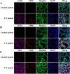
Expression of target protein in renal tubular with LN patients. Co-localisation of target proteins (pink) (A) EGR1, (B) IFI16 and (C) MX1, F4/80 (red) (marker of macrophage), and Megalin (green) (marker of the renal tubules). DAPI was used for nuclear staining. LN, lupus nephritis; DAPI, 4′,6-diamidino-2-phenylindole.
Figure 9
Figure 9
Expression of target protein in renal tubular within MRL/lpr. Co-localisation of target proteins (pink) (A) EGR1, (B): IFI204 and (C): MX1, F4/80 (red) (marker of macrophage), and Megalin (green) (marker of the renal tubules). DAPI was used for nuclear staining. LN, lupus nephritis; DAPI, 4′,6-diamidino-2-phenylindole.

Similar articles

Cited by

References

    1. Kolios AGA, Tsokos GC. Skin-kidney crosstalk in SLE. Nat Rev Rheumatol. 2021;17(5):253–254. doi:10.1038/s41584-021-00588-0 - DOI - PubMed
    1. Davidson A. What is damaging the kidney in lupus nephritis? Nat Rev Rheumatol. 2016;12(3):143–153. doi:10.1038/nrrheum.2015.159 - DOI - PMC - PubMed
    1. Anders HJ, Saxena R, Zhao MH, et al. Lupus nephritis. Nat Rev Dis Primers. 2020;6(1):7. doi:10.1038/s41572-019-0141-9 - DOI - PubMed
    1. Bajema IM, Wilhelmus S, Alpers CE, et al. Revision of the international society of nephrology/renal pathology society classification for lupus nephritis: clarification of definitions, and modified national institutes of health activity and chronicity indices. Kidney Int. 2018;93(4):789–796. doi:10.1016/j.kint.2017.11.023 - DOI - PubMed
    1. Yu F, Haas M, Glassock R, et al. Redefining lupus nephritis: clinical implications of pathophysiologic subtypes. Nat Rev Nephrol. 2017;13(8):483–495. doi:10.1038/nrneph.2017.85 - DOI - PubMed

LinkOut - more resources