Lighting Up and Identifying Metal-Binding Proteins in Cells
- PMID: 39735929
- PMCID: PMC11672145
- DOI: 10.1021/jacsau.4c00879
Lighting Up and Identifying Metal-Binding Proteins in Cells
Abstract
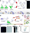
Metal ions, either essential or therapeutic, play critical roles in life processes or in the treatment of diseases. Proteins and enzymes are involved in metal homeostasis and the action of metallodrugs. Imaging and identifying these metal-binding proteins will facilitate the elucidation of metal-mediated life processes. The emerging research field of metallomics and metalloproteomics has significantly advanced our understanding of metal homeostasis and the roles that metals play in biology and medicine. Fluorescence-based metalloproteomics offers the possibility of not only visualization but also identification of metal-binding proteins in living cells and tissues. Herein, we summarize different strategies of labeling and tracking of metal-binding proteins with the aid of fluorescent probes. We highlight several examples as showcases of how this fluorescence-based metalloproteomics approach could be utilized in metallobiology and chemical biology. In conclusion, we also discuss the advantages and limitations of fluorescence-based metalloproteomics approaches and point out future directions of metalloproteomics including development of more sensitive and selective fluorescence probes, integration with other omics approaches, as well as application of emerging advanced super-resolution imaging techniques that utilize fluorescent molecules or proteins. We aim to attract more scientists to engage in this exciting field.
© 2024 The Authors. Published by American Chemical Society.
Conflict of interest statement
The authors declare no competing financial interest.
Figures


Similar articles
-
Metalloproteomics for Biomedical Research: Methodology and Applications.Annu Rev Biochem. 2022 Jun 21;91:449-473. doi: 10.1146/annurev-biochem-040320-104628. Epub 2022 Mar 18. Annu Rev Biochem. 2022. PMID: 35303792 Review.
-
Emerging Strategies in Metalloproteomics.In: Nriagu JO, Skaar EP, editors. Trace Metals and Infectious Diseases [Internet]. Cambridge (MA): MIT Press; 2015. Chapter 18. In: Nriagu JO, Skaar EP, editors. Trace Metals and Infectious Diseases [Internet]. Cambridge (MA): MIT Press; 2015. Chapter 18. PMID: 33886190 Free Books & Documents. Review.
-
Systems Approaches for Unveiling the Mechanism of Action of Bismuth Drugs: New Medicinal Applications beyond Helicobacter Pylori Infection.Acc Chem Res. 2019 Jan 15;52(1):216-227. doi: 10.1021/acs.accounts.8b00439. Epub 2018 Dec 31. Acc Chem Res. 2019. PMID: 30596427
-
Recognition of Proteins by Metal Chelation-Based Fluorescent Probes in Cells.Front Chem. 2019 Aug 9;7:560. doi: 10.3389/fchem.2019.00560. eCollection 2019. Front Chem. 2019. PMID: 31448265 Free PMC article. Review.
-
Metalloproteomics in conjunction with other omics for uncovering the mechanism of action of metallodrugs: Mechanism-driven new therapy development.Curr Opin Chem Biol. 2020 Apr;55:171-179. doi: 10.1016/j.cbpa.2020.02.006. Epub 2020 Mar 19. Curr Opin Chem Biol. 2020. PMID: 32200302 Review.
References
-
- Zhou Y.; Yuan S.; Xiao F.; Li H.; Ye Z.; Cheng T.; Luo C.; Tang K.; Cai J.; Situ J.; Sridhar S.; Chu W.-M.; Tam A. R.; Chu H.; Che C.-M.; Jin L.; Hung I. F.-N.; Lu L.; Chan J. F.-W.; Sun H. Metal-coding assisted serological multi-omics profiling deciphers the role of selenium in COVID-19 immunity. Chem. Sci. 2023, 14 (38), 10570–10579. 10.1039/D3SC03345G. - DOI - PMC - PubMed
-
- Zhou Y.; Cheng T.; Tang K.; Li H.; Luo C.; Yu F.; Xiao F.; Jin L.; Hung I. F.-N.; Lu L.; Yuen K.-Y.; Chan J. F.-W.; Yuan S.; Sun H. Integration of metalloproteome and immunoproteome reveals a tight link of iron-related proteins with COVID-19 pathogenesis and immunity. Clin. Immunol. 2024, 263, 110205.10.1016/j.clim.2024.110205. - DOI - PubMed
Publication types
LinkOut - more resources
Full Text Sources
