Case Reports
. 2024 Dec 30:25:e945804.
doi: 10.12659/AJCR.945804.

B-Acute Lymphoblastic Leukemia/Lymphoblastic Lymphoma Mimicking Fibrosing Mediastinitis: A Case Report and Diagnostic Insight

Affiliations
Case Reports

B-Acute Lymphoblastic Leukemia/Lymphoblastic Lymphoma Mimicking Fibrosing Mediastinitis: A Case Report and Diagnostic Insight

Aya Kitamura et al. Am J Case Rep. .

Abstract

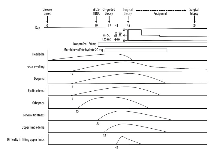
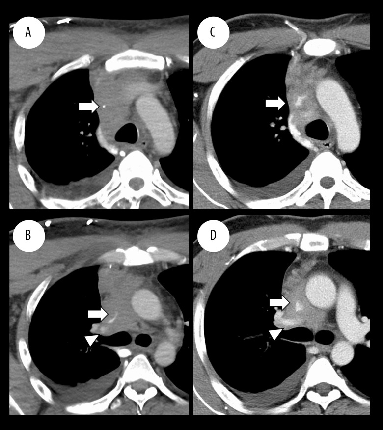
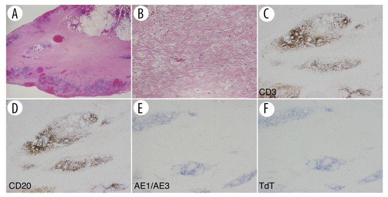
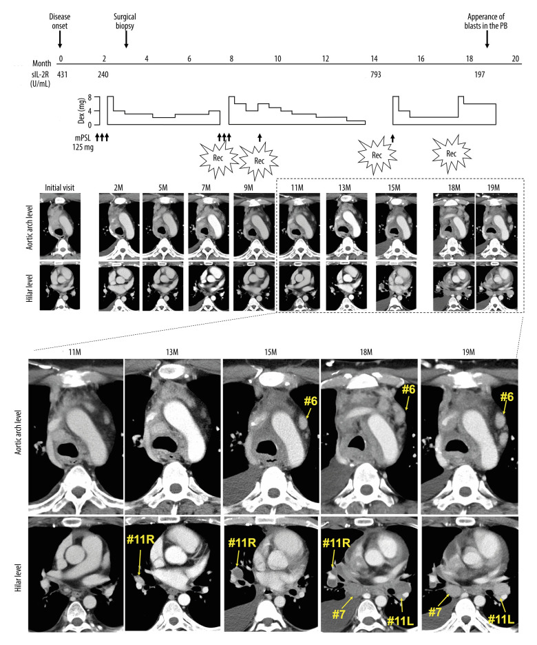
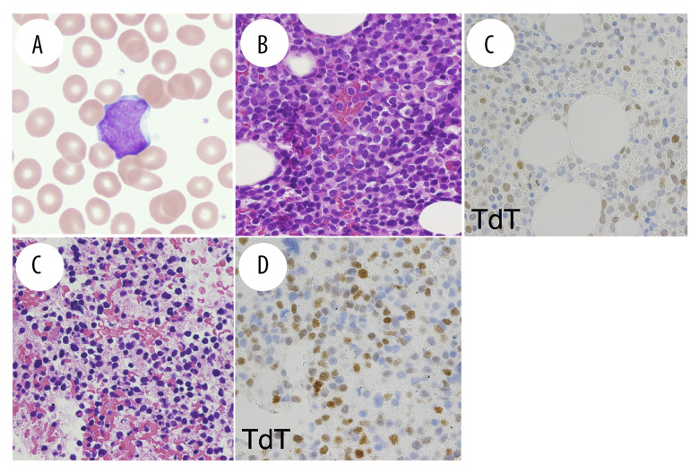
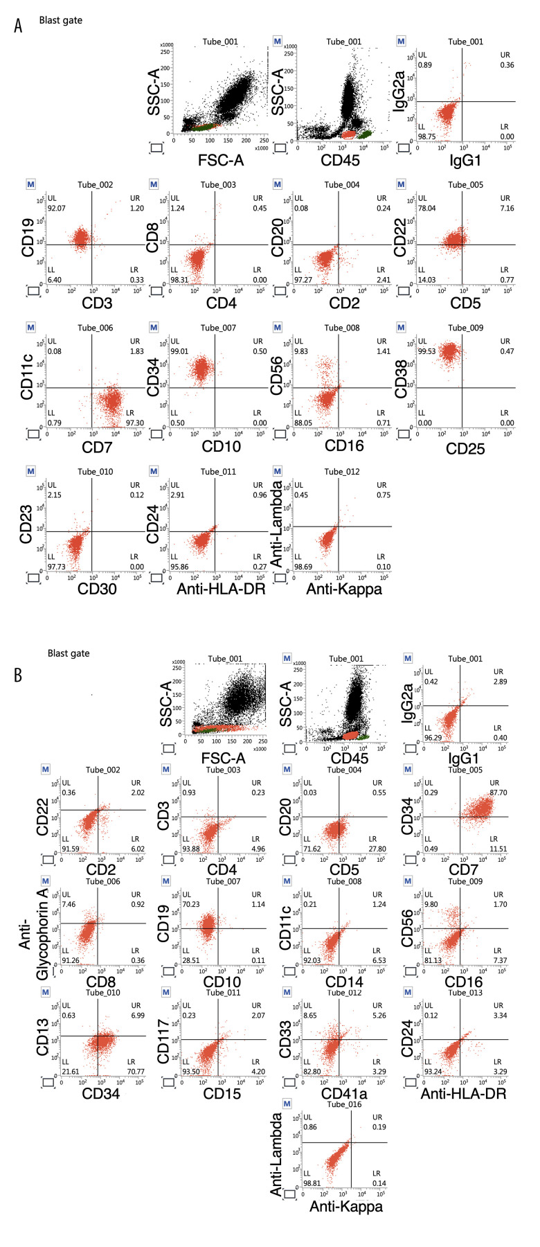
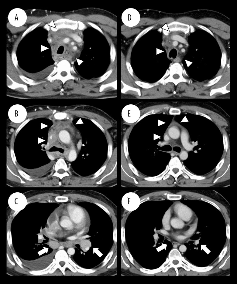
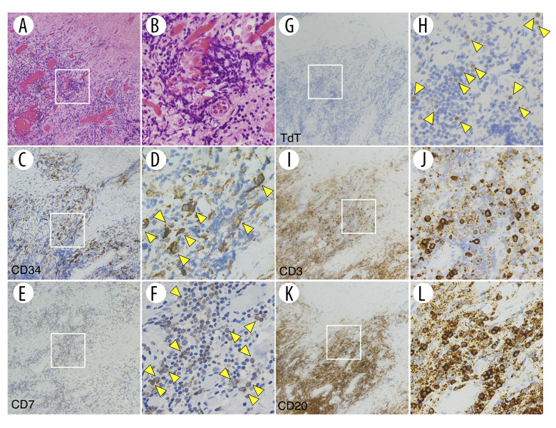
BACKGROUND Fibrosing mediastinitis (FM) is a rare, fibroproliferative disorder within the mediastinum. It is extremely rare for hematologic malignancies to develop as FM. CASE REPORT A 32-year-old Japanese man with a 1-month history of headache and 2-week history of facial swelling underwent chest computed tomography (CT); a diffuse mass-like lesion was revealed in the anterior mediastinum with severe stenosis of vital mediastinal organs. After a surgical biopsy, an initial diagnosis of idiopathic FM was made. The FM lesions responded mildly to corticosteroids but recurred repeatedly. Sixteen months after the treatment initiation, blasts appeared in the peripheral blood (PB), and the patient was diagnosed with B-acute lymphoblastic leukemia/lymphoblastic lymphoma (B-ALL/LBL). Chemotherapy led to complete remission of the B-ALL/LBL and almost complete disappearance of FM-like lesions. Immunohistochemistry of the mediastinal biopsy specimen taken before the blasts' appearance in PB demonstrated a CD34/CD7/terminal deoxynucleotidyl transferase-positive population, an identical pattern of expression common to the blasts in the patient's PB and bone marrow. CONCLUSIONS This is the first case report of B-ALL/LBL presenting as FM. This case underscores the importance of considering the possibility of latent hematologic malignancy even in the absence of new symptoms other than those caused by FM lesions for a long period of time. This is the first demonstration that leukemia cells may be present in the FM lesions from the initial stage of disease onset. Even if a diagnosis of idiopathic FM is confirmed, continued suspicion of the presence of hematologic malignancy is vital for improving patient outcomes.


Conflict of interest statement

Conflict of interest: None declared

Figures

Figure 1.
Figure 1.
The clinical course of the early stage of the patient’s disease onset. The course of each symptom is shown in the lower column. Loxoprofen and morphine sulfate hydrate were administered to relieve cervical tightness and dyspnea, respectively. EBUS-TBNA on day 29 of onset and CT-guided biopsy on day 37 failed to reach a pathological diagnosis. A surgical biopsy scheduled on day 45 was postponed and was performed on day 84 when symptoms were relieved by steroid administration. CT – computed tomography; EBUS-TBNA – endobronchial ultrasound-guided transbronchial needle aspiration; Dex – dexamethasone; mPSL – methylprednisolone.
Figure 2.
Figure 2.
Chest computed tomography (CT) at the patient’s first visit (A, B) showed a diffuse soft tissue mass-like lesion of the anterior mediastinum with severe stenosis of the superior vena cava (arrows) and right pulmonary artery (arrowhead). (C, D) CT at 1 week after the corticosteroid treatment. The size of the mediastinal mass-like lesion was mildly reduced, and the severe stenosis of the superior vena cava (allows) and right pulmonary artery (arrowhead) was slightly mitigated.
Figure 3.
Figure 3.
Thoracic biopsy findings of the mediastinal mass-like lesion. The specimen comprised an undemarcated dense fibrotic lesion (A, B). There was mild infiltration of small- to medium-sized lymphocytes. (C–F) Immunohistochemical findings. The infiltrating lymphocytes were composed of CD3-positive T lymphocytes and CD20-positive B lymphocytes (C, D). No apparent neoplastic proliferations of cytokeratin AE1/AE3-positive cells or terminal deoxynucleotidyl transferase (TdT)-positive cells was detected (E, F). A: 12.5×, B: 200×, C–F: 40× magnification. A, B: Hematoxylin and eosin (H&E) stain.
Figure 4.
Figure 4.
Clinical course. Steroid administration resulted in mild reduction of mediastinal mass-like lesion. However, as the steroid dose was tapered off, there were 4 relapses with re-enlargement of the mediastinal mass-like lesion. At 13 months after the disease onset, the hilar and mediastinal lymph nodes gradually became enlarged. Eighteen months after the disease onset, a routine blood test detected abnormal cells incidentally on a hemogram. Dex – dexamethasone; M – month; mPSL – methylprednisolone; PB – peripheral blood; Rec – recurrence.
Figure 5.
Figure 5.
The onset of B-acute lymphoblastic leukemia/lymphoblastic lymphoma (B-ALL/LBL). (A) Blast in the peripheral blood. (B, C) Bone marrow (BM) biopsy. BM hypercellularity with increased lymphocyte-like blasts which exhibited immunoreactivity for terminal deoxynucleotidyl transferase (TdT) was observed. (D, E) Left hilar lymph node (#11L) biopsy. Infiltration of TdT-positive blasts was observed. A: 1,000×, B–E: 400× magnification. A: May-Giemsa stain, B, D: Hematoxylin and eosin (H&E) stain.
Figure 6.
Figure 6.
Flow cytometry plots of the patient’s peripheral blood (PB) (A) and bone marrow (BM) (B). Blasts in the PB were positive for CD7, CD19, CD22, CD34, and CD38 and negative for CD2, CD3, CD4, CD5, CD8, CD10, CD11c, CD16, CD20, CD23, CD24, CD25, CD30, CD56, IgG1, IgG2a, HLA-DR, Kappa, and Lambda. Blasts in the BM were positive for CD7, CD19, and CD34 and negative for CD2, CD3, CD4, CD5, CD8, CD10, CD11c, CD13, CD14, CD15, CD16, CD20, CD22, CD24, CD33, CD41a, CD56, CD117, IgG1, IgG2a, Glycophorin A, HLA-DR, Kappa, and Lambda.
Figure 7.
Figure 7.
Chest computed tomography (CT) at the onset of B-acute lymphoblastic leukemia/lymphoblastic lymphoma (B-ALL/LBL) (A–C) and at 6 months after the end of the induction (D–F). The mediastinal mass-like lesion (arrowheads) as well as mediastinal and hilar lymph nodes (arrows) showed significant shrinkage after the B-ALL/LBL treatment. The right pleural effusion also disappeared.
Figure 8.
Figure 8.
Thoracic biopsy findings of the mediastinal mass-like lesion. Relatively large cell clusters were present nearby the fibrotic lesion (A, B). These cells were positive for CD34 (C, D), CD7 (E, F), and terminal deoxynucleotidyl transferase (TdT) (G, H, arrowheads). CD3 (I, J) and CD20 (K, L) were positive in small-sized lymphocytes with no atypia. The images in (B, D, F, H, J, L) are ×4 magnifications of the indicated regions in (A, C, E, G, I, K). A, C, E, G, I, K: 100×, B, D, F, H, J, L: 400× magnification. A, B: Hematoxylin and eosin (H&E) stain.

Similar articles

References

    1. Goodwin RA, Nickell JA, Des Prez RM. Mediastinal fibrosis complicating healed primary histoplasmosis and tuberculosis. Medicine. 1972;51:227–46. - PubMed
    1. Peikert T, Colby TV, Midthun DE, et al. Fibrosing mediastinitis: Clinical presentation, therapeutic outcomes, and adaptive immune response. Medicine. 2011;90:412–23. - PubMed
    1. Rossi SE, McAdams HP, Rosado-de-Christenson ML, et al. Fibrosing mediastinitis. Radiographics. 2001;21:737–57. - PubMed
    1. Rossi GM, Emmi G, Corradi D, et al. Idiopathic mediastinal fibrosis: A systemic immune-mediated disorder. A case series and a review of the literature. Clinic Rev Allerg Immunol. 2017;52:446–59. - PubMed
    1. Lindholm KE, de Groot P, Moran CA. Fibrosing/sclerosing lesions of the mediastinum: A review. Adv Anat Pathol. 2019;26:235–40. - PubMed

Publication types

Supplementary concepts

LinkOut - more resources