. 2024 Dec 31;19(1):100.
doi: 10.1186/s13024-024-00786-w.

Distinct regulation of Tau Monomer and aggregate uptake and intracellular accumulation in human neurons

Affiliations

Distinct regulation of Tau Monomer and aggregate uptake and intracellular accumulation in human neurons

Amir T Marvian et al. Mol Neurodegener. .

Abstract

Background: The prion-like spreading of Tau pathology is the leading cause of disease progression in various tauopathies. A critical step in propagating pathologic Tau in the brain is the transport from the extracellular environment and accumulation inside naïve neurons. Current research indicates that human neurons internalize both the physiological extracellular Tau (eTau) monomers and the pathological eTau aggregates. However, similarities or differences in neuronal transport mechanisms between Tau species remain elusive.

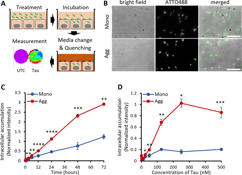
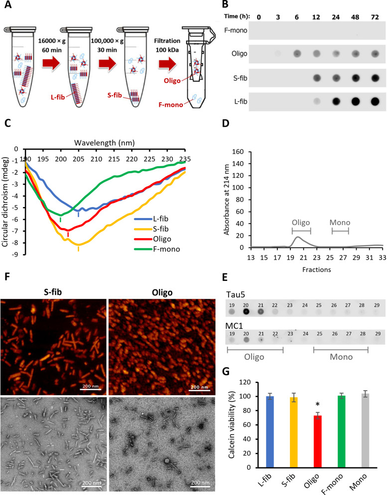
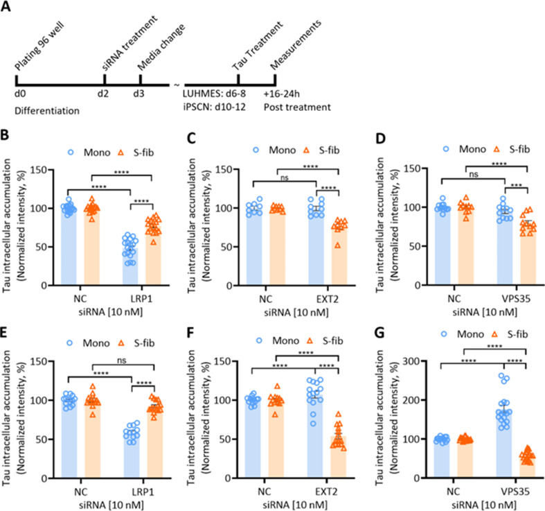
Method: Monomers, oligomers, and fibrils of recombinant 2N4R Tau were produced and characterized by biochemical and biophysical methods. A neuronal eTau uptake and accumulation assay was developed for human induced pluripotent stem cell-derived neurons (iPSCNs) and Lund human mesencephalic cells (LUHMES)-derived neurons. Mechanisms of uptake and cellular accumulation of eTau species were studied by using small molecule inhibitors of endocytic mechanisms and siRNAs targeting Tau uptake mediators.

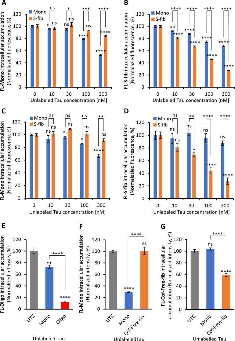
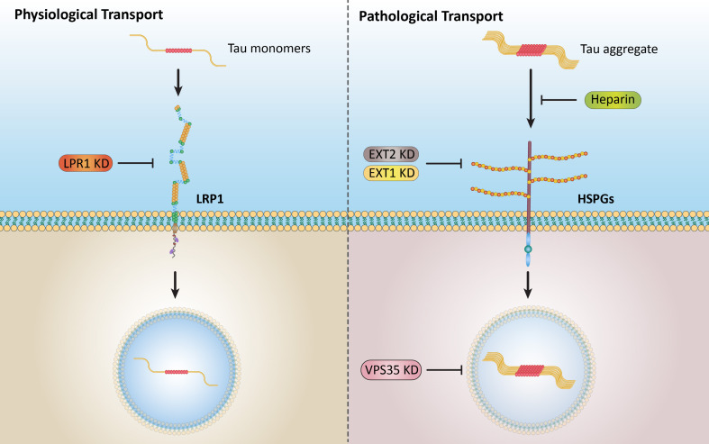
Results: Extracellular Tau aggregates accumulated more than monomers in human neurons, mainly due to the higher efficiency of small fibrillar and soluble oligomeric aggregates in intraneuronal accumulation. A competition assay revealed a distinction in the neuronal accumulation between physiological eTau Monomers and pathology-relevant aggregates, suggesting differential transport mechanisms. Blocking heparan sulfate proteoglycans (HSPGs) with heparin only inhibited the accumulation of eTau aggregates, whereas monomers' uptake remained unaltered. At the molecular level, the downregulation of genes involved in HSPG synthesis exclusively blocked neuronal accumulation of eTau aggregates but not monomers, suggesting its role in the transport of pathologic Tau. Moreover, the knockdown of LRP1, as a receptor of Tau, mainly reduced the accumulation of monomeric form, confirming its involvement in Tau's physiological transport.

Conclusion: These data propose that despite the similarity in the cellular mechanism, the uptake and accumulation of eTau Monomers and aggregates in human neurons are regulated by different molecular mediators. Thus, they address the possibility of targeting the pathological spreading of Tau aggregates without disturbing the probable physiological or non-pathogenic transport of Tau Monomers.

Keywords: Cell-to-cell spreading; Extracellular Tau; HSPGs; LRP1; Neurodegeneration; Uptake; VPS35.


Conflict of interest statement

Declarations. Ethics approval: The experiments were approved by the Ethics Committee of the Technical University of Munich (21/19 S-AS). Consent for publication: N/A. Competing interests: The authors declare that they have no competing interests to disclose.

Figures

Fig. 1
Fig. 1
Characterization of recombinant 2N4R Tau aggregates. A Fibril formation kinetics of recombinant human 2N4R Tau by Thioflavin T (ThT) fluorescence, fitted with the Finke-Watzky model of two-step nucleation-autocatalysis. B Dot-blot analysis of Tau aggregates at different time points during the fibrillization process in A by three different conformation-sensitive Tau antibodies (MC1, TNT-1 and TOMA). C Atomic-force microscopic image of a mixture of aggregates after 72 h of fibrillization, including large fibrils (filled arrowheads), small fibrils (open arrowheads), and oligomers (arrows). Scale bar: 200 nm. D Size-exclusion chromatography (SEC) analysis of the soluble fraction of aggregates after removing insoluble fibrils by ultracentrifugation, showing the absorbance at 214 nm in the eluting fractions, including Tau oligomers (Oligo) and monomers (Mono). E Dot-blot analysis of SEC fractions in D using the antibodies Tau5 (total Tau) and MC1 (conformationally altered Tau). F Dynamic light scattering measurements of Mono and Oligo Tau showing the hydrodynamic size distribution of soluble Tau species obtained from SEC (d.nm: diameter in nanometers)
Fig. 2
Fig. 2
Extracellular Tau aggregates accumulate more than monomers in human iPSC-derived neurons. A Schematic representation of the uptake and accumulation assay. First, cells were treated with fluorescently ATTO488-labeled Tau. After a defined incubation time, the culture medium was changed to a quenching medium to eliminate the extracellular but not the intracellular fluorescence. Finally, a fluorescence plate reader quantified the well surface fluorescence in a 96-well–plate with a transparent bottom. UTC: untreated control. B Live images of cells (DIV 15) treated with 100 nM ATTO488-labeled Tau Monomers (Mono) or aggregates (Agg) for 24 h in the presence of the quencher. Scale bar: 100 μm. C Time-dependent uptake of 100 nM ATTO488-labeled Tau Monomers and aggregates, quantified on a fluorescence plate-reader in the presence of the quencher. D Concentration-dependent uptake of ATTO488-labeled Tau Monomers and aggregates (monomer equivalent) after 24 h on a fluorescence plate-reader in the presence of the quencher. Fluorescence values are normalized by dividing by the background. Error bars represent SD; n = 3 per experimental condition. One-way ANOVA followed by post-hoc test; ns: not significant, *p < 0.05, **p < 0.01, ***p < 0.001, ****p < 0.0001 vs. Mono
Fig. 3
Fig. 3
Characterization of different biochemical fractions of recombinant 2N4R Tau aggregates. A Schematic representation of the fractionation procedure including two sequential centrifugations of low (16,000 ×g) and high (100,000 ×g) gravitational force to sediment large insoluble fibrils (L-fib) and small fibrils (S-fib), respectively, followed by a 100 kDa filtration step separating the soluble oligomers (Oligo) that are retained on the filter from fibrillization-derived monomers (F-mono) that pass through the filter. B Dot-blot analysis of the content of fibrillization mixture and fractions during the fibrillization process using MC1 conformational antibody. C Circular dichroism spectrum showing the conformational status of Tau in each fraction (minimum of peaks showed with short lines). mdeg: millidegrees. D Size-exclusion chromatography (SEC) spectrum of Oligo fraction showing the absorbance at 214 nm of the eluting fractions with lines indicating the expected fractions for oligomers (Oligo) and monomers (Mono). E Dot-blot analysis of SEC fractions in D using the antibodies Tau5 (total Tau) and MC1 (conformationally altered Tau). F Atomic force microscopy (upper row) and transmission electron microscopy (lower row) images of small fibrils and soluble aggregates or oligomers. G Viability of iPSC-derived neurons treated for 24 h with 250 nM of either the Tau fractions mentioned above or recombinant Tau Monomers (Mono). Error bars represent SEM; n = 3 per experimental condition. One-way ANOVA followed by post-hoc test; *p < 0.05 vs. Mono
Fig. 4
Fig. 4
Intracellular accumulation, escape to the cytosol and endogenous aggregation of various recombinant 2N4R Tau species. A The kinetics of intracellular accumulation of different Tau species, including large fibrils (L-fib), small fibrils (S-fib), oligomers (Oligo), fibrillization-derived monomers (F-mono), and recombinant monomers (Mono) within 48 h with 100 nM labeled Tau species. B Titration curve for intracellular accumulation of different Tau species after 20 h of incubation. Error bars represent SD; n = 3 per experimental condition. One-way ANOVA followed by a post-hoc test; ns: not significant, **p < 0.01, ***p < 0.001, ****p < 0.0001. C Schematic of the Tau entry assay. 0N4R P301S-Tau-HiBiT assemblies were added to cells expressing cytosolic LgBiT. Uptake of Tau assemblies may lead to cytosolic entry, resulting in Tau-HiBiT-mediated Nluc reconstitution by LgBiT binding. The addition of cell-permeable substrate results in the Nluc-mediated production of photons, which are readily quantifiable. Cytosolic entry is, therefore, proportional to the detected luminescent signal. D Percent of Tau-HiBiT that enters the cytosol of GPLN neurons following exposure to 2 ug/ml Tau-HiBiT monomers or Tau aggregated species (L-fib, S-fib, Oligo, Mono). Error bars denote SD. n = 3 per experimental condition. **p < 0.01; ***p < 0.001 by two-way ANOVA with Dunnett’s multiple comparisons. E Fluorescence microscopic images of HEK293-biosensor cells expressing P301S Tau-venus, either left untreated or treated with 200 nM unlabeled Tau small fibrils (S-fib). Arrows showing the inclusions of endogenous P301S Tau-venus. Scale bar: 61.7 nm. F Fluorescence analysis of cells treated with 200 nM Tau fractions and monomer using a fluorescence plate reader. Error bars represent SEM; n = 3 per experimental condition. One-way ANOVA followed by a post-hoc test; ns: not significant, **p < 0.01, ***p < 0.001 vs. Mono
Fig. 5
Fig. 5
The competition between labeled and unlabeled extracellular Tau Monomers and fibrils for intraneuronal accumulation. The uptake and accumulation of 50 nM fluorescently labeled monomers (FL-Mono, A) and small fibrils (FL-S-fib, B) in the presence of increasing concentrations of unlabeled Tau Monomers (Mono, blue bars) and small fibrils (S-fib, orange bars) after 20 h of incubation in iPSCNs (A, B), and in LUHMES neurons (C, D). Significance was calculated by comparing Mono and S-fib at each concentration versus “0” and versus each other. E The neuronal accumulation of fluorescently labeled oligomers (50 nM) in iPSCNs neurons after 20 h of incubation in the presence of a 5-fold higher concentration of unlabeled monomers and oligomers (250 nM). F The uptake and accumulation of 25 nM FL-Mono in the presence of a 4-fold higher concentration of unlabeled Tau Mono and cofactor-free fibrils (Cof-free-fib) after 20 h of incubation. G The uptake and accumulation of 25 nM fluorescently labeled Cof-free-fib (FL-Cof-free-fib) in the presence of a 4-fold higher concentration of unlabeled Tau Mono and Cof-free-fib after 20 h of incubation. Significance compared to the untreated control (UTC). Error bars represent SEM; n ≥ 3 independent experiments per experimental condition. Two-way ANOVA followed by Tukey post-hoc test; ns: not significant, *p < 0.05, **p < 0.01, ***p < 0.001, ****p < 0.0001
Fig. 6
Fig. 6
The impact of small molecule inhibitors on the intracellular accumulation of Tau. The intracellular level of Tau in cells left untreated as control (UTC) or treated with 50 µM Chlorpromazine (CPZ), 20 µM Cytochalasin D (CD), 30 µM 5-N-ethyl-N-isopropyl amiloride (EIPA), 75 µM Dyngo-4a (DYNGO), 200 µM Genistein (GEN), or 10 µM Nystatin (NYST) for 30 min before incubation with A fluorescently labeled monomers (FL-Mono), and B fluorescently labeled small fibrils (FL-S-fib), both at 250nM concentration for 3 h (exceptionally, EIPA were present during the incubation with Tau). Error bars represent SEM; n = 9–14. One-way ANOVA ****p < 0.0001. Fluorescence measurement of cells treated with 25 nM fluorescently labeled Tau C FL-Mono, and D FL-S-fib in the presence of 100 nM bafilomycin A1 (Baf), 30 µM chloroquine (CQ), 100 nM MG132, 200 µM Atropine (Atr) or 20 µM Pirenzepine (Pirz) for 20 h. Error bars represent SEM; n = 9–14 independent experiments per experimental condition. One-way ANOVA ****p < 0.0001, *p < 0.05 vs. UTC. E Fluorescence analysis of iPSCNs treated with 25 nM labeled Tau Monomers and small fibrils for 20 h in the presence of 2 µM Heparin. Error bars represent SEM; n = 3. One-way ANOVA followed by post-hoc test; ****p < 0.0001 vs. UTC. ns: not significant. Kinetics of intracellular Tau accumulation in LUHMES neurons pre-treated with 100 µM Heparin for 2 h before exposure to F 250 nM fluorescently labeled Mono (FL-Mono) and G 150 nM fluorescently labeled-small fibrils (FL-S-fib). The significance was calculated between “No pretreat” and “Pretreat” at each time point (Only significant points were shown). Error bars represent SD. n = 3 per experimental condition. One-way ANOVA followed by posthoc test; *p < 0.05, **p < 0.01, ***p < 0.001 vs. “No pretreatment”. H Intracellular accumulation in LUHMES neurons pretreated with 100 µM Heparin for 2 h before 9 h treatment with 100 nM fluorescently labeled Tau Monomers (Mono), 50 nM Oligomers (Oligo), or 50 nM mixture of aggregates including large fibrils, small fibrils, and oligomers. Error bars represent SEM. n = 3 per experimental condition. One-way ANOVA followed by posthoc test; ***p < 0.001, ****p < 0.0001 vs. UTC. ns: not significant. I Representative images of H. Scale bar: 25 nm
Fig. 7
Fig. 7
siRNA-mediated downregulation of some molecular mediators differentially impacts the intracellular accumulation of Tau Monomers and small fibrils. A Timeline of the experimental scheme. iPSC-derived neuronal progenitor cells or LUHMES cells were seeded in 96 well-plates in the differentiation medium. Cells were treated with 10nM siRNA at day 2 of differentiation. iPSCNSs and LUHMES neurons were treated with fluorescently labeled Tau at days 10 to 12 and 6 to 8, respectively. Fluorescence measurements were implemented after 16 to 24 h of treatment. Intracellular accumulation of labeled Tau Monomers and small fibrils were shown in B, C,D iPSCNs, and E, F,G LUHMES neurons that were treated with siRNAs of LRP1, EXT2, and VPS35. Error bars represent SEM; n = 9–18. One-way ANOVA ***p < 0.001, ****p < 0.0001. Error bars represent SEM. NC: negative control siRNA
Fig. 8
Fig. 8
Model of differential uptake and accumulation of Tau Monomers and aggregates in human neurons. Under physiological conditions, Tau Monomers exist in the extracellular environment and internalize neurons via LRP1-mediated endocytosis, which can be inhibited by LRP1 knockdown. Under pathological conditions, Tau aggregates in the extracellular environment internalize neurons mainly via HSPGs mediated endocytosis, which can be blocked by heparin or knockdown of HSPGs synthetizing enzymes such as EXT1 and EXT2. LRP1 may be partially involved in aggregate uptake in some types of neurons, which needs further investigation. The downregulation of VPS35, as a critical component of the retromer complex, reduced the accumulation of aggregates in both models of human neurons in this study. The endocytic vesicles inside the cells are depicted with faded colors since the internalization of Tau might be via endocytic vesicles and/or direct cytosol entry, which was not investigated in this study

References

    1. Rosler TW, Tayaranian Marvian A, Brendel M, Nykanen NP, Hollerhage M, Schwarz SC, et al. Four-repeat tauopathies. Prog Neurobiol. 2019;180:101644. - PubMed
    1. Grundke-Iqbal I, Iqbal K, Quinlan M, Tung YC, Zaidi MS, Wisniewski HM. Microtubule-associated protein tau. A component of Alzheimer paired helical filaments. J Biol Chem. 1986;261(13):6084–9. - PubMed
    1. Kosik KS, Joachim CL, Selkoe DJ. Microtubule-associated protein tau (tau) is a major antigenic component of paired helical filaments in Alzheimer disease. Proc Natl Acad Sci U S A. 1986;83(11):4044–8. - PMC - PubMed
    1. Goedert M, Falcon B, Clavaguera F, Tolnay M. Prion-like mechanisms in the pathogenesis of tauopathies and synucleinopathies. Curr Neurol Neurosci Rep. 2014;14(11):495. - PubMed
    1. Braak H, Alafuzoff I, Arzberger T, Kretzschmar H, Del Tredici K. Staging of Alzheimer disease-associated neurofibrillary pathology using paraffin sections and immunocytochemistry. Acta Neuropathol. 2006;112(4):389–404. - PMC - PubMed

LinkOut - more resources