

The effect of Miya on skeletal muscle changes by regulating gut microbiota in rats with osteoarthritis through AMPK pathway
- PMID: 39736635
- PMCID: PMC11684039
- DOI: 10.1186/s12891-024-08203-5
The effect of Miya on skeletal muscle changes by regulating gut microbiota in rats with osteoarthritis through AMPK pathway
Abstract
Background: The study aimed to explore whether Miya (MY), a kind of Clostridium butyricum, regulated osteoarthritis (OA) progression through adenosine 5'-monophosphate-activated protein kinase (AMPK) pathway.
Methods: The OA rats were orally given MY daily for 4 weeks and were intramuscularly injected with AMPK inhibitor once a week for 4 weeks. Hematoxylin eosin (HE) staining was used to observe the histological morphology of the knee joint. The levels of succinate dehydrogenase (SDH) and muscle glycogen (MG) in the tibia muscle of rats were detected by the corresponding kits, as well as the expression of related genes/proteins were assessed by real-time quantitative PCR (RT-qPCR) and western blot.
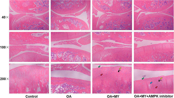
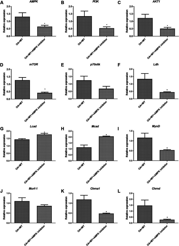
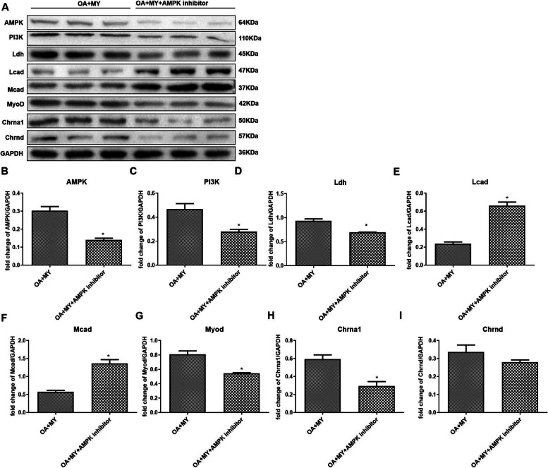
Results: HE staining suggested that MY suppressed the symptoms of OA, which was abolished by AMPK inhibitor. Furthermore, the SDH and MG contents in the OA + MY + AMPK inhibitor group were lower than in the OA + MY group. At last, the levels of AMPK, PI3K, AKT1, Ldh, Myod, Chrna1, and Chrnd were notably decreased after AMPK inhibitor treatment, while the levels of Lcad and Mcad were up-regulated by AMPK inhibitor. Furthermore, their protein expression levels detected by western blot were consistent with those from RT-qPCR.
Conclusion: MY may partially regulate skeletal muscle changes and prevente OA development through the AMPK pathway.
Keywords: AMPK; Gut microbiota; Miya; Osteoarthritis.
© 2024. The Author(s).
Conflict of interest statement
Declarations. Ethics approval and consent to participate: All animal studies were conducted in compliance with guidelines and regulations. All the animal experiments were approved by the Ethics Committee of Shanghai Tenth People's Hospital, Tongji University School of Medicine. And the study is reported in accordance with the relevant ARRIVE guidelines. Consent for publication: Not applicable. Competing interests: The authors declare no competing interests.
Figures

References
-
- Glyn-Jones S, Palmer AJ, Agricola R, Price AJ, Vincent TL, Weinans H, Carr AJ. Osteoarthritis. Lancet (London, England). 2015;386(9991):376–87. - PubMed
-
- Abramoff B, Caldera FE. Osteoarthritis: pathology, diagnosis, and treatment options. Med Clin North Am. 2020;104(2):293–311. - PubMed
-
- Martel-Pelletier J, Barr AJ, Cicuttini FM, Conaghan PG, Cooper C, Goldring MB, Goldring SR, Jones G, Teichtahl AJ, Pelletier JP. Osteoarthritis. Nat Rev Dis Primers. 2016;2:16072. - PubMed
-
- Jiang Y. Osteoarthritis year in review 2021: biology. Osteoarthritis Cartilage. 2022;30(2):207–15. - PubMed
-
- Zhao X, Shah D, Gandhi K, Wei W, Dwibedi N, Webster L, Sambamoorthi U. Clinical, humanistic, and economic burden of osteoarthritis among noninstitutionalized adults in the United States. Osteoarthritis Cartilage. 2019;27(11):1618–26. - PubMed
MeSH terms
Substances
Grants and funding
LinkOut - more resources
Full Text Sources
Miscellaneous
