

The utility of customised tissue probability maps and templates for patients with idiopathic normal pressure hydrocephalus: a computational anatomy toolbox (CAT12) study
- PMID: 39736638
- PMCID: PMC11687168
- DOI: 10.1186/s12987-024-00611-y
The utility of customised tissue probability maps and templates for patients with idiopathic normal pressure hydrocephalus: a computational anatomy toolbox (CAT12) study
Erratum in
-
Correction: The utility of customised tissue probability maps and templates for patients with idiopathic normal pressure hydrocephalus: a computational anatomy toolbox (CAT12) study.Fluids Barriers CNS. 2025 Apr 22;22(1):40. doi: 10.1186/s12987-025-00655-8. Fluids Barriers CNS. 2025. PMID: 40264152 Free PMC article. No abstract available.
Abstract
Background: Disproportionately enlarged subarachnoid space hydrocephalus (DESH) is one of the neuroradiological characteristics of idiopathic normal pressure hydrocephalus (iNPH), which makes statistical analyses of brain images difficult. This study aimed to develop and validate methods of accurate brain segmentation and spatial normalisation in patients with DESH by using the Computational Anatomy Toolbox (CAT12).
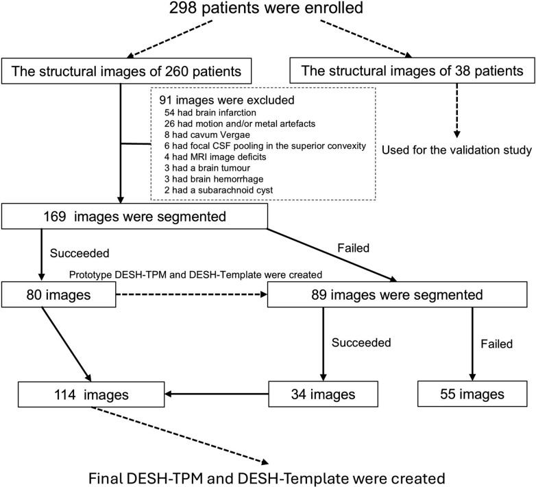
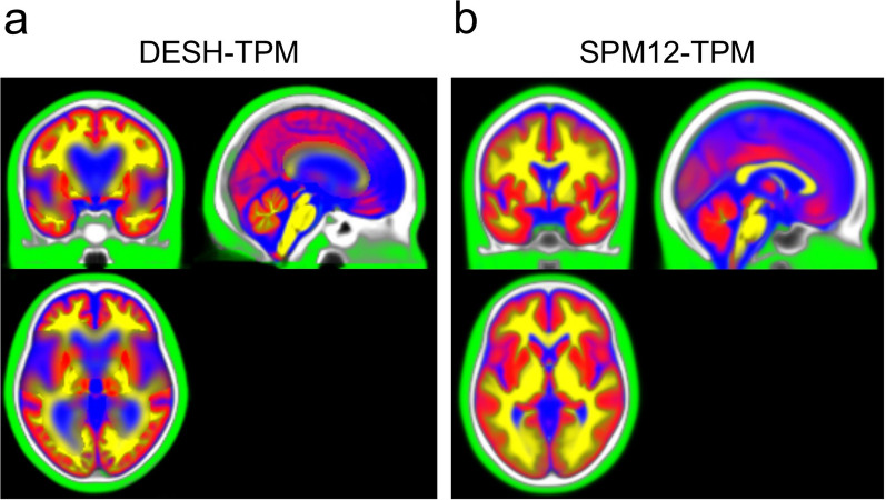
Methods: Two hundred ninety-eight iNPH patients with DESH and 25 healthy controls (HCs) who underwent cranial MRI were enrolled in this study. We selected the structural images of 169 patients to create customised tissue probability maps and diffeomorphic anatomical registration through exponentiated Lie algebra (DARTEL) templates for patients with DESH (DESH-TPM and DESH-Template). The structural images of 38 other patients were used to evaluate the validity of the DESH-TPM and DESH-Template. DESH-TPM and DESH-Template were created using the 114 well-segmented images after the segmentation processing of CAT12. In the validation study, we compared the accuracy of brain segmentation and spatial normalisation among three conditions: customised condition, applying DESH-TPM and DESH-Template to CAT12 and patient images; standard condition, applying the default setting of CAT12 to patient images; and reference condition, applying the default setting of CAT12 to HC images.
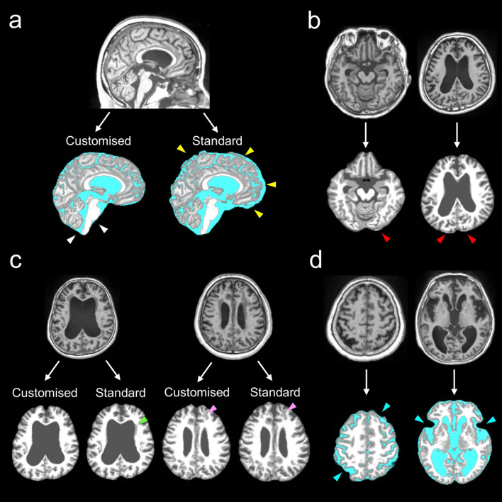
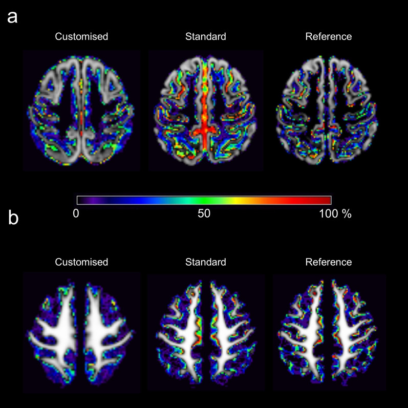
Results: In the validation study, we identified three error types during segmentation. (1) The proportions of misidentifying the dura and/or extradural structures as brain structures in the customised, standard, and reference conditions were 10.5%, 44.7%, and 13.6%, respectively; (2) the failure rates of white matter hypointensity (WMH) cancellation in the customised, standard, and reference conditions were 18.4%, 44.7%, and 0%, respectively; and (3) the proportions of cerebrospinal fluid (CSF)-image deficits in the customised, standard, and reference conditions were 97.4%, 84.2%, and 28%, respectively. The spatial normalisation accuracy of grey and white matter images in the customised condition was the highest among the three conditions, especially in terms of superior convexity.
Conclusions: Applying the combination of the DESH-TPM and DESH-Template to CAT12 could improve the accuracy of grey and white matter segmentation and spatial normalisation in patients with DESH. However, this combination could not improve the CSF segmentation accuracy. Another approach is needed to overcome this challenge.
Keywords: Brain tissue segmentation; CAT12 = computational anatomy toolbox; DESH = disproportionately enlarged subarachnoid space hydrocephalus; INPH = idiopathic normal pressure hydrocephalus; Spatial normalisation; Template.
© 2024. The Author(s).
Conflict of interest statement
Declarations. Ethics approval and consent to participate: The protocol of this study was approved by the ethics committees of Tohoku University (approval numbers: 2006-195, 2010-505, and 2020-1-285) and South Miyagi Medical Centre (approval numbers: 28-7 and 29-1) and was therefore performed in accordance with the ethical standards expressed in the 1964 Declaration of Helsinki and its later amendments. Written informed consent was obtained from patients and their families on the first admission. This study was registered at the Japan Registry of Clinical Trials as jRCT1021230047 ( https://jrct.niph.go.jp/latest-detail/jRCT1021230047 ). Consent for publication: Not applicable. Competing interests: The authors declare no competing interests.
Figures
References
-
- Kanno S, Abe N, Saito M, Takagi M, Nishio Y, Hayashi A, et al. White matter involvement in idiopathic normal pressure hydrocephalus: a voxel-based diffusion tensor imaging study. J Neurol. 2011;258:1949–57. - PubMed
Publication types
MeSH terms
Grants and funding
LinkOut - more resources
Full Text Sources
Medical
