Review
. 2024 Dec 31;22(1):617.
doi: 10.1186/s12964-024-01980-5.

The therapeutic potential of RNA m(6)A in lung cancer

Affiliations
Review

The therapeutic potential of RNA m(6)A in lung cancer

Jingran Yu et al. Cell Commun Signal. .

Abstract

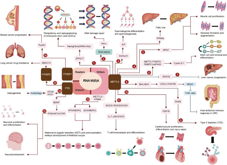
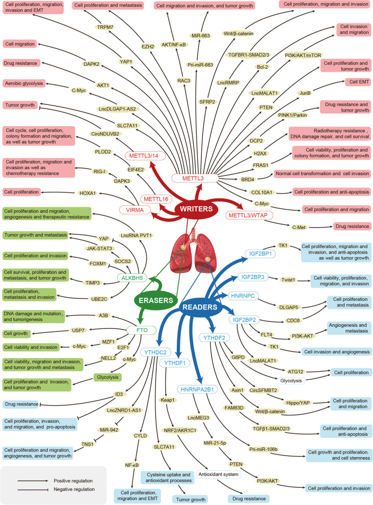
Lung cancer (LC) is a highly malignant and metastatic form of cancer. The global incidence of and mortality from LC is steadily increasing; the mean 5-year overall survival (OS) rate for LC is less than 20%. This frustrating situation may be attributed to the fact that the pathogenesis of LC remains poorly understood and there is still no cure for mid to advanced LC. Methylation at the N6-position of adenosine (N6mA) of RNA (m(6)A) is widely present in human tissues and organs, and has been found to be necessary for cell development and maintenance of homeostasis. However, numerous basic and clinical studies have demonstrated that RNA m(6)A is deregulated in many human malignancies including LC. This can drive LC malignant characteristics such as proliferation, stemness, invasion, epithelial-mesenchymal transition (EMT), metastasis, and therapeutic resistance. Intriguingly, an increasing number of studies have also shown that eliminating RNA m(6)A dysfunction can exert significant anti-cancer effects on LC such as suppression of cell proliferation and viability, induction of cell death, and reversal of treatment insensitivity. The current review comprehensively discusses the therapeutic potential of RNA m(6)A and its underlying molecular mechanisms in LC, providing useful information for the development of novel LC treatment strategies.

Keywords: Epigenetics; Lung cancer; M(6)A; Target therapy; Tumorigenesis.


Conflict of interest statement

Declarations. Ethical approval and consent to participate: Not applicable. Consent for publication: All authors consent to publication. Competing interests: The authors declare no competing interests.

Figures

Fig. 1
Fig. 1
Schematic diagram of m(6)A biological functions. As the most common RNA modification, m(6)A is involved in a variety of normal physiological and disease pathological processes: ① Transcriptor zinc finger protein 217 (ZFP217) can reduce the binding of METTL3 to other RNAs by interacting with METTL3, thereby reducing m(6)A modification of the mRNA of key stem cell regulatory factors including Nanog, Sox2, Klf4, and c-Myc. This promotes their translation and increases protein levels, ultimately promoting and maintaining embryonic stem cell (ESC) pluripotency and somatic cell reprogramming. ② METTL3-mediated m(6)A modification of intracellular transcripts can recruit Pol κ to DNA damage sites caused by UV, thereby accelerating DNA damage repair to promote cell survival. ③ METTL3-mediated m(6)A modification can regulate the selective splicing of spermatogenesis-related transcription factor SOHLH1 mRNA, which increases its protein expression and up-regulates Kit protein expression, ultimately activating spermatogonia differentiation and spermatogenesis-related signal transduction. ④ The increase in intracellular ROS mediated by loss of expression of the circadian clock determinant Bmal1 can increase PPAR-α mRNA m(6)A through METTL3-YTHDF2, promoting its degradation and reducing protein expression, ultimately damaging liver lipid metabolism and leading to lipid accumulation. ⑤ METTL3 can promote the translation and stability of nephronectin (NPNT) mRNA by increasing NPNT mRNA m(6)A, thereby up-regulating NPNT protein levels. Elevated NPNT can further increase the number of muscle cells, expand the diameter of myotubes, and delay skeletal muscle atrophy and myotube aging, thereby maintaining the stability of muscle tissue and conferring skeletal muscle resistance to degeneration caused by aging. ⑥ High expression of METTL3 promotes m(6)A modification of Cyclin D1 (CCND1) mRNA. YTHDF3 recognizes and binds to the m(6)A modification site of CCND1 mRNA, recruiting cytoplasmic 1 like protein (PABPC1L) and eIF4G2 to promote its translation. This increases its protein level, ultimately promoting hematopoietic stem cell remodeling, differentiation, and self-renewal. ⑦ METTL3 overexpression can increase suppressor of cytokine signaling 2(SOCS2) mRNA m(6)A, induce its degradation, and inhibit protein expression in a YTHDF2-dependent manner. This promotes proliferation, migration, and colony formation in liver cancer cells, thereby accelerating the progression of liver cancer. ⑧ METTL3 overexpression can promote BHLHE41 mRNA m(6)A and translation in colorectal cancer (CRC) cells, thereby up-regulating expression of the basic helix-loop-helix family member e41 (Bhlhe41) and inducing CXCL1 transcription, expression, and secretion. Increased CXCL1 then recruits MDSC and inhibits anti-tumor immune responses of CD8( +) T cells and NK cells. ⑨ The increased expression of METTL3 in hepatocytes can promote the stability of fatty acid synthase (Fasn) mRNA m(6)A and up-regulate its protein level. This enhances fatty acid metabolism and reduces insulin sensitivity, ultimately contributing to obesity and type 2 diabetes (T2D). ⑩ Increased expression of METTL3 in cardiomyocytes can promote m(6)A- and YTHDF2-dependent degradation of FGF16 mRNA, down-regulate its protein expression, and ultimately inhibit cardiomyocyte proliferation and differentiation, which is detrimental to myocardial injury repair and heart regeneration. ⑪ Insufficient expression of METTL 3 in naive T cells can reduce the levels of m(6)A in mRNAs including SOCS1, SOCS3, and CISH. It also delays their decay, increases their protein levels, which inhibits IL-7-mediated JAK-STAT5 activation. This ultimately impairs T cell proliferation and differentiation and disrupts T cell homeostasis. ⑫ During embryonic development, METTL3-mediated zygotic genome activation (ZGA) and m(6)A enrichment of murine ERV-like (MERVL, 2-cell stage transposon), VIRMA-mediated maternal transposon (MTA), and MERVL m(6)A are increased, promoting the maternal-to-zygotic transition (MZT) process. As a result, the oocyte completes cell fate transition and ultimately maintains normal development of the preimplantation embryo. ⑬ After the fetus is born, the high expression of FTO in its brain tissue can maintain the m(6)A modification of brain-derived neurotrophic factor (BDNF) mRNA and up-regulate its protein expression. This activates the PI3K/AKT/mTOR signaling pathway, ultimately promoting the proliferation of neural stem cells, neuronal differentiation, and maintenance of normal brain development and intellectual development. ⑭ FTO can induce m(6)A in the mRNA of key autophagy regulators such as ATG5 and ATG7, increasing their mRNA stability and protein expression and ultimately promoting autophagy and adipogenesis. ⑮ Overexpression of FTO in lung cancer tissue can enhance the removal of m(6)A from ABCC10 mRNA, thereby promoting its stability and protein expression. This ultimately accelerates cell growth and enhances gefitinib resistance. ⑯ Overexpressed YTHDF1 in breast cancer tissues recognizes and binds to m(6)A-labeled FoxM1 mRNA and promotes its translation. Elevated FoxM1 then promotes cell proliferation, EMT, metastasis, and invasion, ultimately leading to cancer progression
Fig. 2
Fig. 2
Deregulated RNA m(6)A signaling pathway network in lung cancer

Similar articles

Cited by

References

    1. Sung H, Ferlay J, Siegel RL, Laversanne M, Soerjomataram I, Jemal A, Bray F. Global Cancer Statistics 2020: GLOBOCAN Estimates of Incidence and Mortality Worldwide for 36 Cancers in 185 Countries. CA Cancer J Clin. 2021;71(3):209–49. - PubMed
    1. Herbst RS, Morgensztern D, Boshoff C. The biology and management of non-small cell lung cancer. Nature. 2018;553(7689):446–54. - PubMed
    1. Siegel RL, Miller KD, Fuchs HE, Jemal A. Cancer Statistics, 2021. CA-Cancer J Clin. 2021;71(1):7–33. - PubMed
    1. Bailey-Wilson JE, Amos CI, Pinney SM, Petersen GM, de Andrade M, Wiest JS, Fain P, Schwartz AG, You M, Franklin W, Klein C, Gazdar A, Rothschild H, Mandal D, Coons T, Slusser J, Lee J, Gaba C, Kupert E, Perez A, Zhou X, Zeng D, Liu Q, Zhang Q, Seminara D, Minna J, Anderson MW. A major lung cancer susceptibility locus maps to chromosome 6q23-25. Am J Hum Genet. 2004;75(3):460–74. - PMC - PubMed
    1. Amos CI, Wu X, Broderick P, Gorlov IP, Gu J, Eisen T, Dong Q, Zhang Q, Gu X, Vijayakrishnan J, Sullivan K, Matakidou A, Wang Y, Mills G, Doheny K, Tsai YY, Chen WV, Shete S, Spitz MR, Houlston RS. Genome-wide association scan of tag SNPs identifies a susceptibility locus for lung cancer at 15q25.1. Nat Genet. 2008;40(5):616–22. - PMC - PubMed

LinkOut - more resources