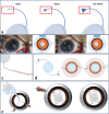
Recent advances and current challenges in suture and sutureless scleral fixation techniques for intraocular lens: a comprehensive review
- PMID: 39736769
- PMCID: PMC11684149
- DOI: 10.1186/s40662-024-00414-0
Recent advances and current challenges in suture and sutureless scleral fixation techniques for intraocular lens: a comprehensive review
Erratum in
-
Correction: Recent advances and current challenges in suture and sutureless scleral fixation techniques for intraocular lens: a comprehensive review.Eye Vis (Lond). 2025 Dec 26;12(1):51. doi: 10.1186/s40662-025-00468-8. Eye Vis (Lond). 2025. PMID: 41449468 Free PMC article. No abstract available.
Abstract
Over the past two decades, both suture and sutureless techniques for scleral fixation of intraocular lenses have seen significant advancement, driven by improvements in methodologies and instrumentation. Despite numerous reports demonstrating the effectiveness, safety, and superiority of these techniques, each approach carries with it its own drawbacks, including an elevated risk of certain postoperative complications. This article delves into various surgical techniques for scleral fixation of posterior chamber intraocular lenses, discussing their procedural nuances, benefits, drawbacks, postoperative complications, and outcomes. Furthermore, a comparative analysis between suture and sutureless fixation methods is presented, elucidating their respective limitations and associated factors. It is hoped that this comprehensive review will offer clinicians guidance on how to individualize procedural selection and mitigate surgical risks, and thus achieve optimal visual outcomes. This review will also endeavor to provide guidance for future advancements in intraocular lens fixation techniques.
Keywords: Aphakia; Intraocular lens implantation; Intrascleral fixation; Modified Yamane technique; Sutureless fixation; Transscleral suture fixation.
© 2024. The Author(s).
Conflict of interest statement
Declarations. Ethics approval and consent to participate: Not applicable. Consent for publication: Not applicable. Competing interests: The authors declare that they have no competing interests.
Figures


References
-
- Por YM, Lavin MJ. Techniques of intraocular lens suspension in the absence of capsular/zonular support. Surv Ophthalmol. 2005;50(5):429–62. - PubMed
-
- Sekundo W, Bertelmann T, Schulze S. Retropupillary iris claw intraocular lens implantation technique for aphakia. Ophthalmologe. 2014;111(4):315–9. - PubMed
Publication types
LinkOut - more resources
Full Text Sources
Miscellaneous

