. 2024 Oct 9:24:100163.
doi: 10.1016/j.mbplus.2024.100163. eCollection 2024 Dec.

Bone quality relies on hyaluronan synthesis - Insights from mice with complete knockout of hyaluronan synthase expression

Affiliations

Bone quality relies on hyaluronan synthesis - Insights from mice with complete knockout of hyaluronan synthase expression

A Saalbach et al. Matrix Biol Plus. .

Abstract

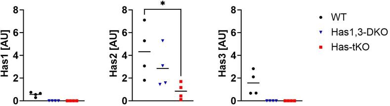
Bone consists of a complex mineralised matrix that is maintained by a controlled equilibrium of synthesis and resorption by different cell types. Hyaluronan (HA) is an important glycosaminoglycan in many tissues including bone. Previously, the importance of HA synthesis for bone development during embryogenesis has been shown. We therefore investigated whether HA synthesis is involved in adult bone turnover and whether abrogation of HA synthesis in adult mice would alter bone quality. To achieve complete abrogation of HA synthesis in adult mice, we generated a novel Has-total knockout (Has-tKO) mouse model in which a constitutive knockout of Has1 and Has3 was combined with an inducible, Ubc-Cre-driven Has2 knockout. By comparing bone tissue from wild-type, Has1,3 double knockout and Has-tKO mice, we demonstrate that Has2-derived HA mainly contributes to the HA content in bone. Furthermore, Has-tKO mice show a significant decrease of bone integrity in trabecular and cortical bone, as shown by µ-CT analysis. These effects are detectable as early as five weeks after induced Has2 deletion, irrespective of sex and progress with age. Mesenchymal stem cells (MSC) during osteogenic differentiation in vitro showed that Has2 expression is increased while Has3 expression is decreased during differentiation. Furthermore, the complete abrogation of HA synthesis results in significantly reduced osteogenic differentiation as indicated by reduced marker gene expression (Runx-2, Tnalp, Osterix) as well as alizarin red staining. RNAseq analysis revealed that MSC from Has-tKO are characterised by decreased expression of genes annotated for bone and organ development, whereas expression of genes associated with chemokine related interactions and cytokine signalling is increased. Taken together, we present a novel mouse model with complete deletion of HA synthases in adult mice which has the potential to study HA function in different organs and during age-related HA reduction. With respect to bone, HA synthesis is important for maintaining bone integrity, presumably based on the strong effect of HA on osteogenic differentiation.

Keywords: Bone; Hyaluronan synthase; Osteogenic differentiation.


Conflict of interest statement

The authors declare that they have no known competing financial interests or personal relationships that could have appeared to influence the work reported in this paper.

Figures

Fig. 1
Fig. 1
Organ distribution of HA and impact of Has knockout. (A) HA content varies in different organs of adult mice. HA was measured by ELISA after enzymatic tissue dissociation and calculated per mg of tissue weight. (B) Knockout of HA synthases reduces HA concentration in different organs. HA content was detected in the indicated organs of wild-type (WT) mice, mice with a constitutive double KO of Has1 and Has3 (Has1,3-DKO) and mice with an induced complete KO of all Has (Has-tKO). Mice of both sexes were treated with tamoxifen at 8–10 weeks and used at 15–25 weeks of age. Each point represents one mouse. ANOVA with Tukey’s multiple comparison test *p < 0.05, **p < 0.005, ***p < 0.001, ****p < 0.0001.
Fig. 2
Fig. 2
Knockout of Has-enzymes is reflected by strongly reduced Has mRNA expression in bones of adult mice. RT-qPCR from bone tissue from adult female mice after tamoxifen treatment. Has1 mRNA was not detectable in bone tissue from Has1,3-DKO and Has-tKO mice. One-way ANOVA, * p < 0.05.
Fig. 3
Fig. 3
Knockout of HA synthases leads to impaired bone quality as detected by µCT analysis of adult mouse femurs. Images of bone material loss in (A) trabecular and (B) cortical bone of femurs in male Has1,3-DKO and Has-tKO mice. (C) Quantitation of bone volume/total volume (BV/TV), bone surface/bone volume (BS/BV), trabecular thickness (Tb.Th.), separation (Tb.Sep.) and number (Tb.N) in trabecular bones. (D) Quantitation of bone surface/bone volume, cortical cross-sectional thickness (Cs.Th.), closed porosity (Po(cl)) and cortical bone density. Femurs were obtained for µCT analysis from a cohort of adult male mice treated with tamoxifen at 8–10 weeks of age. C57Bl6 mice were treated in the same way and used as wild-type controls. Ordinary one-way ANOVA, * p < 0.05, *** p < 0.0005, **** p < 0.0001.
Fig. 4
Fig. 4
Loss of HA synthesis leads to reduced bone quality and progresses with age. Two cohorts of age-matched female mice (A-25 weeks and B-61 weeks) treated with tamoxifen between 8 and 10 weeks of age, were used to obtain femurs. C57Bl6 mice were treated equally as wild-type controls. µCT scans were analysed for bone volume/total volume (BV/TV), bone surface/bone volume (BS/BV), trabecular thickness (Tb.Th.), separation (Tb.sep.) and number (Tb.N) in femoral trabecular bone. Bone surface/bone volume (BS/BV), cortial cross-sectional thickness (Cs.Th.) and closed porosity (Po(cl)) were determined in femoral cortical bone. Unpaired t-test with Welch’s correction, * p < 0.05, ** p < 0.01, *** p < 0.0005.
Fig. 5
Fig. 5
Abrogation of HA synthesis impairs osteogenic MSC differentiation in vitro. (A) Regulation of HA synthesis in bone marrow-derived MSC during osteogenic differentiation. Amount of HA released by MSC into the culture supernatant during 48 h before addition of differentiation medium. *p < 0.05 Mann-Whitney test. (B,C) Gene expression of MSC at different time points with control medium or osteogenic differentiation medium. One-way ANOVA *,# p < 0.05; ***,### p < 0.001; ****,#### p < 0.0001. (D) Gene expression levels of Runx2, Tnalp1, Osterix and Osteocalcin (Ocn) were quantified by RT-qPCR at day 7 and day 12 of differentiation. (E) Alizarin red staining of MSC at day 7 and day 12 of differentiation. (F) XTT assay at day 12 of differentiation. 2way ANOVA for gene expression analysis and unpaired Student’s T-test for alizarin red OD. **p < 0.01; ***p < 0.001; ****p < 0.0001, (G) Reduced phosphorylation of Smad1/5 in Has-tKO-derived MSC upon addition of osteogenic medium. Densitometry and representative blot. n = 6, One-way ANOVA, **p < 0.01. (For interpretation of the references to colour in this figure legend, the reader is referred to the web version of this article.)
Fig. 6
Fig. 6
Has knockout in MSC results in modified transcriptomes during osteogenic differentiation. Different gene expression patterns in wild-type and Has-tKO derived MSC during osteogenic differentiation (A). MSC from wild-type mice are characterised by increased expression of genes related to bone morphogenesis (B), when Has-tKO-derived MSC show increased expression of inflammation-related genes, especially chemokines (C). Differential expression of Oncostatin m, Cxcl3 and Lif was detected in vivo in tibiae of WT and Has-tKO mice. *p < 0.05, Student’s T-test with Welch’s correction (D).
Supplementary Fig. 1
Expression of hyaluronidases Hyal1, Hyal2 and Cemip in tibiae from different mouse strains. RT-qPCR data from tibiae of a cohort of adult male mice treated with tamoxifen at 8-10 weeks of age. C57Bl6 mice were treated in the same way and used as wild-type controls. n=2-4. Rplp0 was used as reference gene.
Supplementary Fig. 2
Has-knockout does not affect voluntary activity, heat production and respiratory exchange rate (RER) in adult mice. (A) Analysis of voluntary activity, heat production and respiratory exchange rate (RER) in adult mice (25weeks of age) in a PhenoMaster® metabolic screening platform for 3 nights and 3 days. Data from dark phase (nights) 2, 3 and 4 (dark) and light phase 1, 2 and 3 (light) are combined. (B) Analysis of voluntary activity, heat production and respiratory exchange rate (RER) in adult mice 61 weeks of age in a PhenoMaster® metabolic screening platform for 2 nights and 2 days. Data from dark phase 1 and 2 (dark) and light phase 2 and 3 (light) are combined. Mice were treated with tamoxifen at age of 8-10 weeks. Two way ANOVA, not significant.
Supplementary Fig. 3
Reduced Bending stiffness of femurs of adult Has tKO. A cohort of adult male mice of matched age that received tamoxifen treatment 5 weeks before was used to obtain femora biomechanical analysis. C57Bl6 mice were treated equally as wildtype controls. Student’s T-test with Welch’s correction, ** p<0.01. n=4-7 per group.
Supplementary Fig. 4
The total knockout of Has does not impair osteoclast activity. (A) Exemplary TRAP staining (purple) of femurs from wild-type and Has-tKO mice. Bar= 20 µm. (B) Bone histomorphometry of TRAP stained trabecular bone from wild-type and Has-tKO mice with number of osteoclasts per bone perimeter (N.Oc/B.Pm) and Osteoclast surface per Bone surface (Oc.S/BS). p>0.05 Mann-Whitney Test. (C) Expression of bone resorption associated genes (Nfat-c1, Oscar, RankL, Trap) in tibiae from different mouse strains. Has2 expression is shown for validation of the induced knockout in these mice. RT-qPCR data from tibiae of adult female mice (25 weeks). Mice were treated with tamoxifen at age of 8-10 weeks. n=4-7. Rplp0 was used as reference gene.* p<0.01, Mann-Whitney test. Others were not significant.

Similar articles

Cited by

References

    1. R. Florencio-Silva G.R. da S. Sasso, E. Sasso-Cerri, M.J. Simões, P.S. Cerri, Biology of Bone Tissue: Structure, Function, and Factors That Influence Bone Cells., Biomed Res Int 2015 2015 10.1155/2015/421746 421746. - PMC - PubMed
    1. Noonan K.J., Stevens J.W., Tammi R., Tammi M., Hernandez J.A., Midura R.J. Spatial distribution of CD44 and hyaluronan in the proximal tibia of the growing rat. J. Orthop. Res. 1996;14:573–581. doi: 10.1002/jor.1100140411. - DOI - PubMed
    1. Camenisch T.D., Spicer A.P., Brehm-Gibson T., Biesterfeldt J., Augustine M.L., Calabro A., Kubalak S., Klewer S.E., McDonald J.A. Disruption of hyaluronan synthase-2 abrogates normal cardiac morphogenesis and hyaluronan-mediated transformation of epithelium to mesenchyme. J Clin Invest. 2000;106:349–360. doi: 10.1172/JCI10272. - DOI - PMC - PubMed
    1. Spicer A.P., Mcdonald J.A. Characterization and Molecular Evolution of a Vertebrate Hyaluronan. Synthase Gene Family*. 1998 doi: 10.1074/jbc.273.4.1923. - DOI - PubMed
    1. Bastow E.R., Byers S., Golub S.B., Clarkin C.E., Pitsillides A.A., Fosang A.J. Hyaluronan synthesis and degradation in cartilage and bone. Cell. Mol. Life Sci. 2008;65:395–413. doi: 10.1007/s00018-007-7360-z. - DOI - PMC - PubMed

LinkOut - more resources