. 2024 Dec 11:15:1421131.
doi: 10.3389/fphar.2024.1421131. eCollection 2024.

Broccoli (Brassica oleracea var. italica) leaves exhibit significant antidiabetic potential in alloxan-induced diabetic rats: the putative role of ABC vacuolar transporter for accumulation of Quercetin and Kaempferol

Affiliations

Broccoli (Brassica oleracea var. italica) leaves exhibit significant antidiabetic potential in alloxan-induced diabetic rats: the putative role of ABC vacuolar transporter for accumulation of Quercetin and Kaempferol

Sara Latif et al. Front Pharmacol. .

Abstract

Background: The global prevalence of diabetes among adults over 18 years of age is expected to increase from 10.5% to 12.2% (between 2021 and 2045). Plants can be a cost-effective source of flavonoids like quercetin and kaempferol with anti-diabetic properties.

Methodology: We aimed to assess the antidiabetic potential of leaves of Brassica oleracea cvs. Green Sprout and Marathon. Further, flavonoid contents were measured in broccoli leaves grown under light and dark conditions. The methanolic extracts of Green Sprout (GSL-M) and Marathon (ML-M) were first evaluated in vitro for their α-amylase and α-glucosidase inhibitory potential and then for antidiabetic activity in vivo in alloxan-induced diabetic rat models.

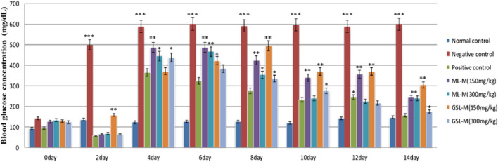
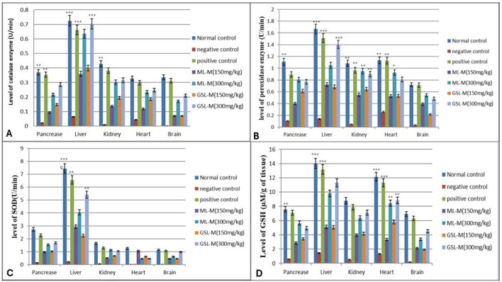
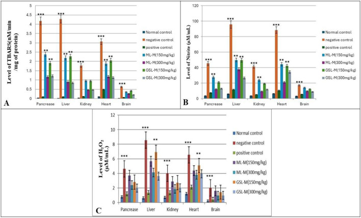
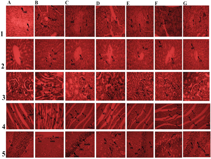
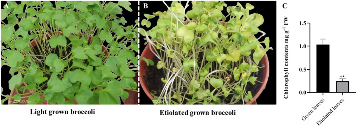
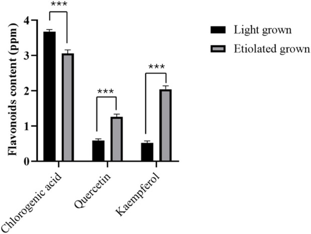
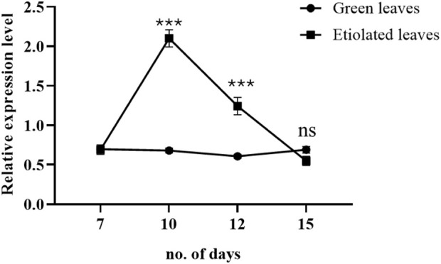
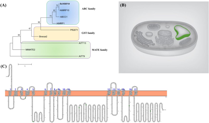
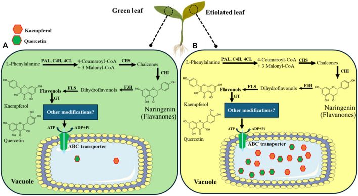
Results: Treatment with plant extracts promoted the reduced glutathione (GSH) content and CAT, POD, and SOD activities in the pancreas, liver, kidney, heart, and brain of diabetic rats, whereas lowered lipid peroxidation, H2O2, and nitrite concentrations. The histopathological studies revealed the protective effect of plant extracts at high dose (300 mg/kg), which could be due to broccoli's rich content of chlorogenic acid, quercetin, and kaempferol. Strikingly, etiolated leaves of broccoli manifested higher levels of quercetin and kaempferol than green ones. The putative role of an ABC transporter in the accumulation of quercetin and kaempferol in etiolated leaves was observed as evaluated by qRT-PCR and in silico analyses.

Conclusion: In conclusion, the present study shows a strong link between the antidiabetic potential of broccoli due to the presence of chlorogenic acid, quercetin, and kaempferol and the role of an ABC transporter in their accumulation within the vacuole.

Keywords: ABC transporter; Brassica oleracea; alloxan; antidiabetic; antioxidant enzymes; flavonoids; histopathology; lipid peroxidation.


Conflict of interest statement

The authors declare that the research was conducted in the absence of any commercial or financial relationships that could be construed as a potential conflict of interest.

Figures

FIGURE 1
FIGURE 1
Blood glucose concentration (mg/dL) at different intervals in experimental groups. Normal control: Non-diabetic healthy animals. Negative control: Untreated diabetic animals. Positive control: Glibenclamide-treated diabetic animals. ML-M: Marathon leaves methanol extract treated diabetic animals. GSL-M: Green sprout leaves methanol extract treated diabetic animals. All groups were compared to the normal control group using two-way ANOVA followed by Bonferroni’s test. *P ≤ 0.05, **P ≤ 0.01, ***P ≤ 0.001.
FIGURE 2
FIGURE 2
Effect of 150 mg/kg and 300 mg/kg doses of ML-M and GSL-M methanolic extracts on the status of antioxidants in the pancreas, liver, kidney, heart, and brain tissues of diabetic animals. (A) Catalase (CAT) activity (U/min). (B) Peroxidase (POD) activity (U/min). (C) Superoxide dismutase (SOD) activity (U/min). (D) Reduced glutathione (GSH) level (µM/g of tissue). Normal control: Non-diabetic healthy animals. Negative control: Untreated diabetic animals. Positive control: Glibenclamide-treated diabetic animals. ML-M: Marathon leaves methanol extract treated diabetic animals. GSL-M: Green sprout leaves methanol extract treated diabetic animals. All groups were compared to negative control groups using one-way ANOVA followed by Tukey’s comparison. *P ≤ 0.05, **P ≤ 0.01, ***P ≤ 0.001.
FIGURE 3
FIGURE 3
Effect of 150 mg/kg and 300 mg/kg doses of ML-M and GSL-M methanolic extracts on the status of oxidative stress markers in the pancreas, liver, kidney, heart, and brain tissues of diabetic animals. (A) Level of TBARS (nM/min/mg of protein). (B) Level of H2O2 (µM/mL). (C) Level of nitrite (µM/mL). Normal control: Non-diabetic, healthy animals. Negative control: Untreated, diabetic animals. Positive control: Glibenclamide-treated diabetic animals. ML-M: Marathon leaves methanol extract treated diabetic animals. GSL-M: Green sprout leaves methanol extract treated diabetic animals. All groups were compared to normal non-diabetic groups using one-way ANOVA followed by Tukey’s comparison. *P ≤ 0.05, **P ≤ 0.01, ***P ≤ 0.001.
FIGURE 4
FIGURE 4
Effect of 150 mg/kg and 300 mg/kg doses of ML-M and GSL-M methanolic extracts on histo-architecture of vital organs. Column A: Normal control. Column B: Negative control. Column C: Positive control. Column; D ML-M 150 mg/kg. Column E; ML-M 300 mg/kg. Column F; GSL-M 150 mg/kg. Column G; GSL-M 300 mg/kg. Row (1) Pancreas; IL: islets of Langerhans; AC: Acinar cells; MLD: mild Langerhans disruption; ALD: acute Langerhans disruption; LD: Langerhans disruption; AD: acinar disintegration; ACS: acinar cell steatosis; IC: inflammatory cells; PD: pancreatic duct. Row (2) Liver; CV: central vein; HPC: hepatocytes; DCV: damaged central vein; CT: cellular infiltration; CHT: cellular hypertrophy; N: necrosis; S: sinusoids. Row (3) Kidney; G: glomerulus; BC: Bowman’s capsule; BS: Bowman’s space; ML: mild lobulation; CI: cellular infiltration; CD: capsule distortion; Abs: alterations in Bowman’s space; TD: tubule dilation; PT: proximal convoluted tubule; DCT: dilated convoluted tubule; RBC: regenerating Bowman’s capsule; LBB: loss of brush border; DCG: degenerative changes in glomerulus; RT: renal tubule; BB: brush border. Row (4) Heart; M: myocytes; DS: damaged striations; N: nucleus; E: edema; MCN: muscle cell nucleus; BC: blood capillary; CD: capillary damage; S: striation. Row (5) Brain; PM: Pia Matter; ML: Molecular Layer; GL: Granular Layer; WM: White Matter; PL: Purkinge’s cell Layer; NWM: Narrowed White Matter; GL: Degenerated Granular Layer; DML: Distorted Molecular Layer; WMN: White matter narrowed; MDPL: Mild Distorted Purkinge’s cell Layer; MDGL: Mild Distorted Granular Layer.
FIGURE 5
FIGURE 5
Phenotype and chlorophyll contents of light and dark (etiolated) grown seedlings. The broccoli seedlings were grown under light (A) and dark (B) for 2 weeks. The chlorophyll contents were measured after 2 weeks (C). Asterisks show a significance level of P < 0.01. Data shows an average of three repeats; error bars represent ± SE.
FIGURE 6
FIGURE 6
Flavonoids level in broccoli seedlings. Chlorogenic acid, quercetin, and kaempferol were measured from 15-day-old seedlings grown in light and dark conditions for 7 days. Data represented the mean ± SE of three replications. Asterisks show a significance level of P < 0.001.
FIGURE 7
FIGURE 7
Effects of dark and light on the expression level of broccoli ABC transporter (BolC8t51326H). The dark condition was imposed on the day 7th of the broccoli seedlings. The expression was determined during days 7, 10, 12, and 15 of growing broccoli seedlings in light or dark. Data represent the mean ± SE of three independent biological repeats. Asterisks show a significance level of P < 0.001.
FIGURE 8
FIGURE 8
Phylogenetic tree construction (A), predicted subcellular localization (B), and membrane topology (C) of BoMRP10 (BolC8t51326H). The protein accessions used were BoMRP10 (BolC8t51326H), Bronze2 (X81971.1; Marrs et al., 1995), PfGST1 (AB362191; Yazaki et al., 2008), ABCC1 (JX245004; Francisco et al., 2013), AtMRP1 (AF008124; Lu et al., 1998), AtTT9 (At3g28430; Ichino et al., 2014), AtMRP10 (At3G62700; Sugiyama et al., 2006), AtTT13 (At1g17260; Appelhagen et al., 2015), MtMATE (HM856605.1; Zhao et al., 2011). Prediction shows tonoplast localization of the transporter and 17 membrane helices across the membrane.
FIGURE 9
FIGURE 9
Pathway for kaempferol and quercetin synthesis, transport, and accumulation into vacuole via vacuolar ABC transporter in broccoli. The pathway was modified and adapted from Zhao et al. (2011). The dark condition was imposed on the 7th day of the broccoli seedlings, and green (A) and etiolated (B) leaves were sampled on the 15th day. The 7-day continuous dark condition triggered a higher accumulation of flavonoids such as kaempferol and quercetin in etiolated leaves than in light-grown seedlings.

Similar articles

References

    1. Agati G., Azzarello E., Pollastri S., Tattini M. (2012). Flavonoids as antioxidants in plants: location and functional significance. Plant Sci. 196, 67–76. 10.1016/j.plantsci.2012.07.014 - DOI - PubMed
    1. Agbon A. N., Ingbian S. D., Dahiru A. U. (2014). Preliminary histological and histochemical studies on the neuroprotective effect of aqueous fruit extract of Phoenix dactylifera L. (Date Palm) on atesunate - induced cerebellar damage in wistar rats. Sub-Saharan Afr. J. Med 1, 204. 10.4103/2384-5147.144744 - DOI
    1. Alkhalidy H., Wang Y., Liu D. (2018). Dietary flavonoids in the prevention of T2D: an overview. Nutrients 10, 438. 10.3390/nu10040438 - DOI - PMC - PubMed
    1. Appelhagen I., Nordholt N., Seidel T., Spelt K., Koes R., Quattrochio F., et al. (2015). TRANSPARENT TESTA 13 is a tonoplast P3A-ATPase required for vacuolar deposition of proanthocyanidins in Arabidopsis thaliana seeds. Plant J. 82, 840–849. 10.1111/tpj.12854 - DOI - PubMed
    1. Aslam N., Sameeullah M., Yildirim M., Baloglu M. C., Yucesan B., Lössl A. G., et al. (2022). Isolation of the 3β-HSD promoter from Digitalis ferruginea subsp. ferruginea and its functional characterization in Arabidopsis thaliana . Mol. Biol. Rep. 49, 7173–7183. 10.1007/s11033-022-07634-4 - DOI - PubMed

LinkOut - more resources