. 2024 Dec 16:15:1475051.
doi: 10.3389/fimmu.2024.1475051. eCollection 2024.

CRP deposition in human abdominal aortic aneurysm is associated with transcriptome alterations toward aneurysmal pathogenesis: insights from in situ spatial whole transcriptomic analysis

Affiliations

CRP deposition in human abdominal aortic aneurysm is associated with transcriptome alterations toward aneurysmal pathogenesis: insights from in situ spatial whole transcriptomic analysis

Eun Na Kim et al. Front Immunol. .

Abstract

Background: We investigated the effects of C-reactive protein (CRP) deposition on the vessel walls in abdominal aortic aneurysm (AAA) by analyzing spatially resolved changes in gene expression. Our aim was to elucidate the pathways that contribute to disease progression.

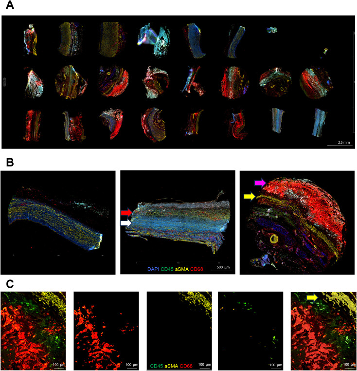
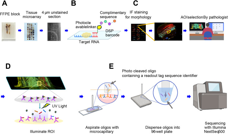
Methods: AAA specimens from surgically resected formalin-fixed paraffin-embedded tissues were categorized into the AAA-high CRP [serum CRP ≥ 0.1 mg/dL, diffuse and strong immunohistochemistry (IHC); n = 7 (12 cores)] and AAA-low-CRP [serum CRP < 0.1 mg/dL, weak IHC; n = 3 (5 cores)] groups. Normal aorta specimens obtained during heart transplantation were used as the control group [n = 3 (6 cores)]. Spatially resolved whole transcriptomic analysis was performed, focusing on CD68-positive macrophages, CD45-positive lymphocytes, and αSMA-positive vascular smooth muscle cells.

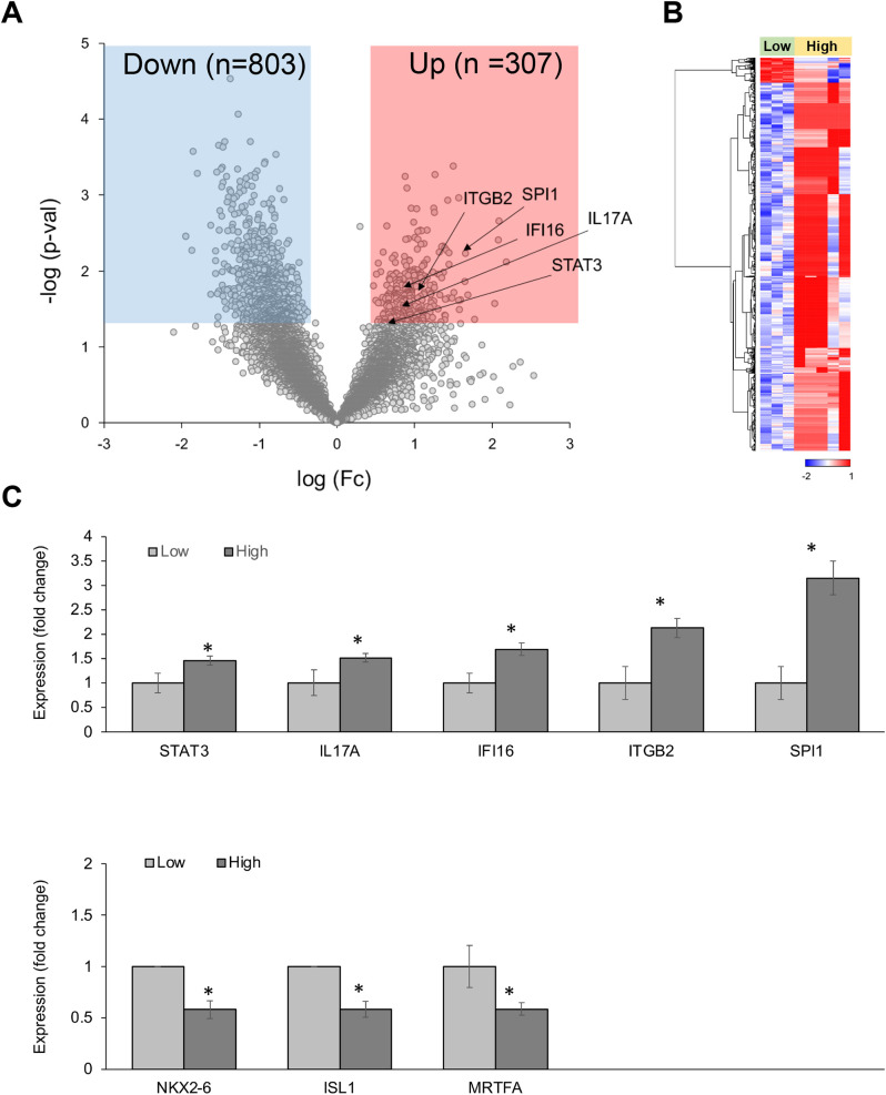
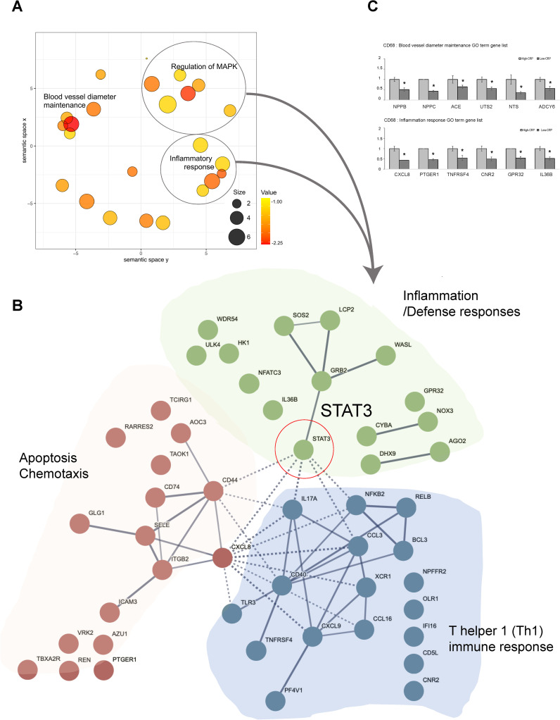
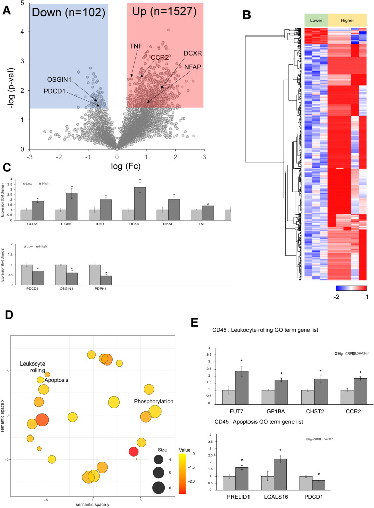
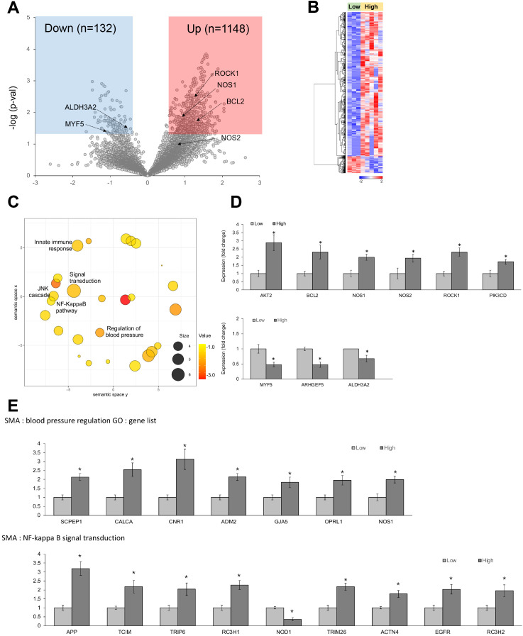
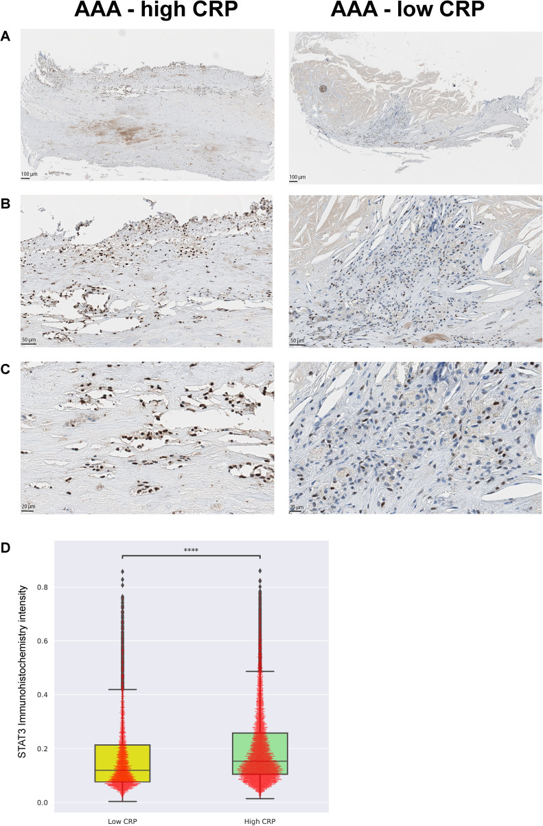
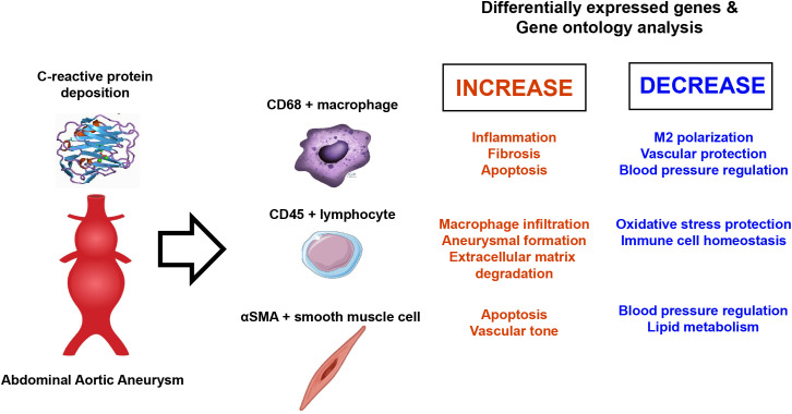
Results: Spatial whole transcriptomic analysis revealed significant differential expression of 1,086, 1,629, and 1,281 genes between high-CRP and low-CRP groups within CD68-, CD45-, and αSMA-positive cells, respectively. Gene ontology (GO) analysis of CD68-positive macrophages identified clusters related to inflammation, apoptosis, and immune response, with signal transducer and activator of transcription 3 implicated across three processes. Notably, genes involved in blood vessel diameter maintenance were significantly downregulated in the high-CRP group. GO analysis of lymphocytes showed upregulation of leukocyte rolling and the apoptosis pathway, whereas, in smooth muscle cells, genes associated with Nuclear factor kappa B (NF-κB) signaling and c-Jun N-terminal Kinase (JNK) pathway were upregulated, and those related to blood pressure regulation were downregulated in the high-CRP group.

Discussion: CRP deposition was associated with significant transcriptomic changes in macrophages, lymphocytes, and vascular smooth muscle cells in AAA, suggesting its potential role in promoting pro-inflammatory and apoptotic processes, as well as contributing to the degradation of vascular structure and elasticity.

Keywords: C-reactive protein; STAT3; abdominal aortic aneurysm; apoptosis; inflammation; spatial transcriptomics.


Conflict of interest statement

The authors declare that the research was conducted in the absence of any commercial or financial relationships that could be construed as a potential conflict of interest.

Figures

Figure 1
Figure 1
Morphologic marker staining and area of illumination selection in aortic tissue analysis. (A) Whole TMA slide scan view images for a normal aorta, AAA with low CRP, and AAA with high CRP. Each TMA section was prepared by creating 3-mm-diameter cores from the aortic wall, focusing on areas in contact with atheroma if present [AAA–low-CRP (n = 3), AAA–high-CRP (n = 7), and control group (n = 3)]. (B) Representative sections of a normal aorta, AAA–low CRP, and AAA–high CRP. The normal aorta primarily consists of αSMA-positive vascular smooth muscle cells with minimal inflammatory cell presence. In AAA–low CRP, the αSMA-positive vascular wall is thinner with fibrotic changes (white arrow), and CD68-positive macrophage infiltration is observed between fibrotic regions (red arrow). In AAA–high CRP, an extensively stained CD68-positive atherosclerotic plaque (magenta arrow) accumulates on a significantly thinned αSMA-positive vascular smooth muscle cell layer (yellow arrow). (C) Representative image of AOI segmentation using IF morphologic markers. The atheromatous plaque and vascular smooth muscle interface were stained simultaneously with CD45, αSMA, and CD68 markers. Vascular smooth muscle segmentation is highlighted (yellow arrow).
Figure 2
Figure 2
Schematic overview of the GeoMx platform for spatially resolved whole RNA transcriptome analysis. (A) Following the review of hematoxylin and eosin–stained slides, a pathologist selects regions of interest (ROIs) from the FFPE block of an aortic specimen. Tissue microarrays (TMAs) are created by punching 3-mm cores and sectioning them into micrometer-thin unstained sections. (B) A high-plex mixture of photocleavable oligo-linked probes and morphology-specific immunofluorescence (IF) antibodies is applied. (C) The pathologist uses IF morphologic markers to re-identify ROIs and defines the areas of illumination (AOIs). (D) The GeoMx instrument illuminates the AOIs with UV light, causing the photo-released oligos to detach and be deposited into a microtiter plate. (E) For NGS readout, the photo-cleaved oligos contain sequence identifiers that serve as readout tags. Each AOI is uniquely indexed during library preparation, pooled, and sequenced using the Illumina NextSeq 500 platform.
Figure 3
Figure 3
Differentially expressed genes (DEGs) in CD68-positive macrophages from spatial whole transcriptomic analysis [AAA–high CRP (n = 7) versus AAA–low CRP (n = 3)]. (A) Volcano plot displaying highly expressed DEGs in CD68-positive macrophages based on the degree of CRP deposition in AAA, using log10 fold changes. (B) Clustered heatmap of the selected ROIs showing whole transcriptomic expression fold changes (average gene expression, z-score) in relation to CRP levels in AAA. Approximately 1,100 genes were differentially expressed according to the CRP deposition level. (C) Key DEGs between the high-CRP and low-CRP groups from (A). The asterisk symbol (*) indicates statistical significance at p < 0.05.
Figure 4
Figure 4
Functional enrichment and protein–protein interaction analysis of GO terms related to CRP deposition in AAA tissues: in CD68-positive macrophages [AAA–high CRP (n = 7) versus AAA–low CRP (n = 3)]. (A) Scatter plot from REVIGO illustrating cluster representatives after redundancy reduction in a two-dimensional space. Multidimensional scaling was applied to a matrix of the 34 GO terms’ semantic similarities. Notable GO terms identified include inflammatory response and blood vessel diameter maintenance. The bubble size indicates the frequency of the GO terms in the underlying GOA database, with color shading representing user-supplied p-values (legend in the lower right corner). (B) Potential enriched GO pathways in CD68+/CRP (high/low) regions. A total of 34 statistically significant biological process GO terms were observed. Further analysis highlighted representative GO terms such as inflammatory responses and MAPK cascade. Protein–protein interaction analysis using the STRING program with high confidence revealed that “STAT3” connects two other clusters associated with apoptosis and immune responses within the inflammation cluster. (C) GO analysis results of genes associated with “blood vessel diameter maintenance,” and “inflammation response.” DEG, differentially expressed gene; Down, downregulated; Up, upregulated; AAA–high CRP, abdominal aortic aneurysm with high CRP; AAA–low CRP, abdominal aortic aneurysm with low CRP. The asterisk symbol (*) indicates statistical significance at p < 0.05.
Figure 5
Figure 5
Differentially expressed genes (DEGs) in CD45-positive lymphocytes from spatial whole transcriptomic analysis [AAA–high CRP (n = 7) versus AAA–low CRP (n = 3)]. (A) Volcano plot displaying highly expressed DEGs in CD45-positive lymphocytes based on CRP deposition in AAA, using log10 fold changes. (B) Clustered heatmap of the selected ROIs showing whole transcriptomic expression fold changes (average gene expression, z-score) in relation to CRP levels in AAA. A total of 1,602 genes were differentially expressed according to the CRP deposition level. (C) Key DEGs between AAA–high-CRP and AAA–low-CRP groups from (A). (D) Scatter plot from REVIGO illustrating cluster representatives after redundancy reduction in a two-dimensional space. Notable GO terms identified include apoptosis and leukocyte rolling. The bubble size indicates the frequency of the GO terms in the underlying GOA database, with color shading representing user-supplied p-values (legend in the lower right corner). (E) GO analysis results of genes associated with “leukocyte rolling,” and “apoptosis.”. DEG, differentially expressed gene; Down, downregulated; Up, upregulated; AAA–high CRP, abdominal aortic aneurysm with high CRP; AAA–low CRP, abdominal aortic aneurysm with low CRP. The asterisk symbol (*) indicates statistical significance at p < 0.05.
Figure 6
Figure 6
Differentially expressed genes (DEGs) in αSMA-positive vascular smooth muscle cells from spatial whole transcriptomic analysis [AAA–high CRP (n = 7) versus AAA–low CRP (n = 3)]. (A) Volcano plot displaying highly expressed DEGs in αSMA-positive vascular smooth muscle cells based on CRP deposition in AAA, using log10 fold changes. (B) Clustered heatmap of the selected ROIs showing whole transcriptomic expression fold changes (average gene expression, z-score) in relation to CRP levels in AAA. A total of 1,280 genes were differentially expressed according to the CRP deposition level. (C) Scatter plot from REVIGO illustrating cluster representatives after redundancy reduction in a two-dimensional space. Notable GO terms identified include NF-κB signal transduction, NJK cascade, and regulation of blood pressure. The bubble size indicates the frequency of the GO terms in the underlying GOA database, with color shading representing user-supplied p-values (legend in the lower left corner). (D) Key DEGs between the AAA–high-CRP and AAA–low-CRP groups from (A). (E) GO analysis results of genes associated with “blood pressure regulation” and “NF-κB signal transduction.” DEG, differentially expressed gene; Down, downregulated; Up, upregulated; AAA–high CRP, abdominal aortic aneurysm with high CRP; AAA–low CRP, abdominal aortic aneurysm with low CRP. The asterisk symbol (*) indicates statistical significance at p < 0.05.
Figure 7
Figure 7
Validation with phospho-STAT3 immunohistochemistry [AAA-high CRP (n = 7) versus AAA-low CRP (n = 3)]. To validate the transcriptome enrichment of STAT3 in AAA–high CRP compared to that in AAA–low CRP, immunohistochemical staining for phospho-STAT3 was performed. (A) Low magnification, (B) medium magnification, and (C) high magnification. (D) Intensity of nuclear pSTAT3-positive cells in the vascular tissue of the AAA-high-CRP group compared to that of the AAA–low-CRP group (Mann–Whitney–Wilcoxon test two-sided with Bonferroni correction, p < 0.0001). The symbols used to indicate levels of statistical significance are as follows: *p < 0.05, **p < 0.01, ***p < 0.001, and ****p < 0.0001.
Figure 8
Figure 8
Gene expression changes in abdominal aortic aneurysm.

Similar articles

References

    1. Aggarwal S, Qamar A, Sharma V, Sharma A. Abdominal aortic aneurysm: A comprehensive review. Exp Clin Cardiol. (2010) 16:11–5. - PMC - PubMed
    1. Golledge J. Abdominal aortic aneurysm: update on pathogenesis and medical treatments. Nat Rev Cardiol. (2019) 16:225–42. doi: 10.1038/s41569-018-0114-9 - DOI - PubMed
    1. Sampson UKA, Norman PE, Fowkes FGR, Aboyans V, Song Y, Harrell FE, et al. . Global and regional burden of aortic dissection and aneurysms mortality trends in 21 world regions, 1990 to 2010. Global Hear. (2014) 9:171–180.e10. doi: 10.1016/j.gheart.2013.12.010 - DOI - PubMed
    1. Golledge J, Moxon JV, Singh TP, Bown MJ, Mani K, Wanhainen A. Lack of an effective drug therapy for abdominal aortic aneurysm. J Intern Med. (2020) 288:6–22. doi: 10.1111/joim.12958 - DOI - PubMed
    1. Márquez-Sánchez AC, Koltsova EK. Immune and inflammatory mechanisms of abdominal aortic aneurysm. Front Immunol. (2022) 13:989933. doi: 10.3389/fimmu.2022.989933 - DOI - PMC - PubMed

MeSH terms

Substances

LinkOut - more resources