. 2024 Dec 16:15:1463224.
doi: 10.3389/fimmu.2024.1463224. eCollection 2024.

Metformin improves Mycobacterium avium infection by strengthening macrophage antimicrobial functions

Affiliations

Metformin improves Mycobacterium avium infection by strengthening macrophage antimicrobial functions

Sindre Dahl Mediaas et al. Front Immunol. .

Abstract

Introduction: The incidence and prevalence of infections with non-tuberculous mycobacteria such as Mycobacterium avium (Mav) are increasing. Prolonged drug regimens, inherent antibiotic resistance, and low cure rates underscore the need for improved treatment, which may be achieved by combining standard chemotherapy with drugs targeting the host immune system. Here, we examined if the diabetes type 2 drug metformin could improve Mav-infection.

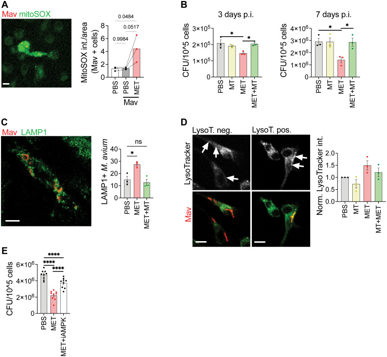
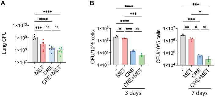
Methods: Metformin was administered to C57BL/6 mice infected intranasally with Mav and C57BL/6 mice were infected intranasally with Mav and treated with metformin over 3 weeks. Organ bacterial loads and lung pathology, inflammatory cytokines and immune cell profiles were assessed. For mechanistic insight, macrophages infected with Mav were treated with metformin alone or in combination with inhibitors for mitochondrial ROS or AMPK and assessed for bacterial burden and phagosome maturation.

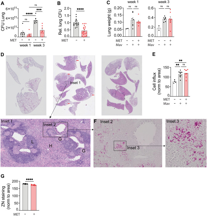
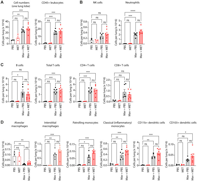
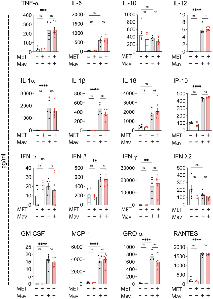
Results and discussion: Three weeks of metformin treatment significantly reduced the lung mycobacterial burden in mice infected with Mav without major changes in the overall lung pathology or immune cell composition. Metformin treatment had no significant impact on tissue inflammation except for a tendency of increased lung IFNγ and infiltration of Mav-specific IFNγ-secreting T cells. Metformin did, however, boost the antimicrobial capacity of infected macrophages directly by modulating metabolism/activating AMPK, increasing mitochondrial ROS and phagosome maturation, and indirectly by bolstering type I immunity. Taken together, our data show that metformin improved the control of Mav-infection in mice, mainly by strengthening antimicrobial defenses in macrophages, and suggest that metformin has potential as an adjunct treatment of Mav infections.

Keywords: Metformin; Mycobacterium avium; host-directed therapy; macrophage; mouse; non-tuberculous.


Conflict of interest statement

The authors declare that the research was conducted in the absence of any commercial or financial relationships that could be construed as a potential conflict of interest. The author(s) declared that they were an editorial board member of Frontiers, at the time of submission. This had no impact on the peer review process and the final decision.

Figures

Figure 1
Figure 1
Metformin reduces the lung organ bacterial load of mice infected with Mycobacterium avium. C57Bl/6 mice were infected intranasally with 5x10⁷ Mav TMC724 or mock-infected (PBS) and treated 5 times a week with 200 mg/kg metformin (MET) or PBS intraperitoneally over 1-3 weeks. n=7 mice per group in three individual experiments. (A) Lung organ bacterial load (CFU) 1 and 3 weeks post infection from one representative experiment. (B) Lung CFUs 3 weeks post infection from MET-treated mice normalized to CFUs in mock-treated mice (PBS). Data are combined from three independent experiments. (C) Lung weight from the same experiment as in (A). (D-G) Organ histology 3 weeks post infection from the same experiment as in (A). (D) Hematoxylin and eosin staining of lung tissue sections from uninfected (left) or Mav-infected and mock-treated (middle) or MET-treated (right) mice. Scalebar is xx um. Insets are marked to zoom in on lung structures and bacteria. Inset 1 shows areas of lymphocyte infiltration (L), histiocytes (H) and granulomas (G). Scalebar is xx um. (E) Morphometric analysis of lung sections shown in (D) quantified as cell infiltration relative to the lung area. (F) Inset 2 as marked in Inset 1 (D): Ziehl-Neelsen staining of acid-fast bacteria (Mav) in lung granuloma (scalebar is xx um), with magnified Inset 3 (scalebar is xx um). (G) Quantification of lung organ bacterial load from Ziehl-Neelsen staining intensity per area in Mav-infected and MET- or mock-treated mice. Significance testing was done using 1-way ANOVA with Tukey’s multiple comparison post-test (A, C, E) or students t-test (B, G). **p < 0,01, ***p < 0,005, ****p < 0,001​, ns, not significant. CFU, colony forming units; PBS, phosphate buffered saline; MET, metformin.
Figure 2
Figure 2
Metformin treatment does not significantly change cell composition in Mav-infected lungs. C57Bl/6 mice were infected with 5x10⁷ Mav TMC724 or mock-infected (PBS) and treated 5 times a week with 200 mg/kg metformin (MET) or PBS (infected: n=7, mock: n=3). Lung cell quantification and phenotyping of lung immune cells were performed 3 weeks post infection. Flow cytometry staining panels are listed in Table 1 (Methods); gating strategies are shown in Supplementary Figure S3 . (A) Total cell number and total leukocyte numbers per lung. (B) NK-cells and neutrophils (staining panel A). (C) Adaptive immune cell subsets (staining panel B). (D) Monocyte, macrophage, and myeloid dendritic cell subsets (staining panel B). Significance testing was done using 1-way ANOVA with Tukey’s multiple comparison post-test. *p < 0,05, **p < 0,01, ***p < 0,005, ****p < 0,001, ns, not significant.
Figure 3
Figure 3
Metformin treatment does not alter cytokine responses in Mav-infected lungs. C57Bl/6 mice were infected with 5x10⁷ Mav TMC724 or mock-infected (PBS) and treated 5 times a week with 200 mg/kg metformin (MET) or PBS over 3 weeks (infected: n=7, mock: n=6 (PBS) or n=3 (MET)). Multiplex cytokine analysis of lung homogenates. Some datapoints are missing due to low bead-counts. Datapoints lower than the detection limit are set to 0. Significance testing was done using 1-way ANOVA with Tukey’s multiple comparison post-test. ** p < 0,01, *** p < 0,005, **** p < 0,001, ns, not significant.
Figure 4
Figure 4
Metformin reduces Mav load in macrophages by increasing mitochondrial ROS, phagosome maturation, and activation of AMPK. (A) Mouse BMDMs were infected with Mav104-CFP at MOI 10 for 10 minutes, washed, and treated with metformin (MET, 2 mM) or PBS for 3 days before cells were stained with MitoSOX prior to confocal imaging (n = 3). (B) Mouse BMDMs were infected with MavTMC724 at MOI 10 for 10 min and treated with MET (2 mM) and/or MitoTEMPO (10 µM), for 3 (left) or 7 (right) days before cells were lysed and bacterial loads quantified from CFUs (n = 3). (C, D) Mouse BMDMs were infected with Mav104-CFP at MOI 10 and treated with MET (2 mM) and/or MitoTEMPO (10 µM) for 3 days before cells were stained with anti-LAMP1 (C) or LysoTracker (D) prior to confocal imaging (n = 3). (E) Mouse BMDMs infected with MavTMC724 at MOI 10 and treated with MET (2 mM) +/- an AMPK inhibitor, Compound C (100 nM) before cells were lysed and bacterial loads quantified from CFUs (n = 9). Significance was tested using 1-way ANOVA with Tukey’s (A, B, E), Dunnett’s (C), or Holm-Sidak’s (D) multiple comparison post-test. *p < 0,05, ****p < 0,001, ns, not significant. BMDM, bone-marrow derived macrophage; CFU, colony forming units; PBS, phosphate buffered saline; MET, metformin.
Figure 5
Figure 5
Antibiotic co-treatment with metformin. (A) Lung organ bacterial load. C57Bl/6 mice were infected intranasally with 5x10⁷ Mav TMC724 and treated 5 times a week with PBS, 200 mg/kg MET alone or in combination with 200 mg/kg Clarithromycin, 10 mg/kg Rifampicin and 50 mg/kg Ethambutol (CRE) over 3 weeks before organ bacterial loads were assessed from lung homogenate CFUs. n=7 mice per group. (B) Mouse BMDMs were infected with MavTMC724 MOI 10 for 10 minutes, washed, and treated for 3 or 7 days with 2 mM MET, 20 µg/ml Clarithromycin, 10 µg/ml Rifampicin and 20 µg/ml Ethambutol (CRE) or CRE+MET prior to cell lysis and CFU plating. Significance was tested using 1-way ANOVA with Tukey’s multiple comparison post-test. * p < 0,05, ** p < 0,01, *** p < 0,005, **** p < 0,001, ns, not significant. BMDM, bone-marrow derived macrophage; CFU, colony forming units; PBS, phosphate buffered saline; MET, metformin; CRE, Clarithromycin, Rifampicin, Ethambutol.

Similar articles

Cited by

References

    1. Adjemian J, Daniel-Wayman S, Ricotta E, Prevots DR. Epidemiology of nontuberculous mycobacteriosis. Semin Respir Crit Care Med. (2018) . 39:325–35. doi: 10.1055/s-0038-1651491 - DOI - PMC - PubMed
    1. Dahl VN, Mølhave M, Fløe A, van Ingen J, Schön T, Lillebaek T, et al. . Global trends of pulmonary infections with nontuberculous mycobacteria: a systematic review. Int J Infect Dis. (2022) . 125:120–31. doi: 10.1016/j.ijid.2022.10.013 - DOI - PubMed
    1. van Ingen J, Obradovic M, Hassan M, Lesher B, Hart E, Chatterjee A, et al. . Nontuberculous mycobacterial lung disease caused by Mycobacterium avium complex - disease burden, unmet needs, and advances in treatment developments. Expert Rev Respir Med. (2021) . 15:1387–401. doi: 10.1080/17476348.2021.1987891 - DOI - PubMed
    1. Kumar K, Loebinger MR. Nontuberculous mycobacterial pulmonary disease: clinical epidemiologic features, risk factors, and diagnosis: the nontuberculous mycobacterial series. Chest. (2022) . 161:637–46. doi: 10.1016/j.chest.2021.10.003 - DOI - PubMed
    1. Prevots DR, Marshall JE, Wagner D, Morimoto K. Global epidemiology of nontuberculous mycobacterial pulmonary disease: A review. Clin Chest Med. (2023) . 44:675–721. doi: 10.1016/j.ccm.2023.08.012 - DOI - PMC - PubMed

LinkOut - more resources