

Giant periscapular lipoma unmasked by post-bariatric surgery weight loss: a case report
- PMID: 39737213
- PMCID: PMC11683727
- DOI: 10.1093/jscr/rjae817
Giant periscapular lipoma unmasked by post-bariatric surgery weight loss: a case report
Abstract
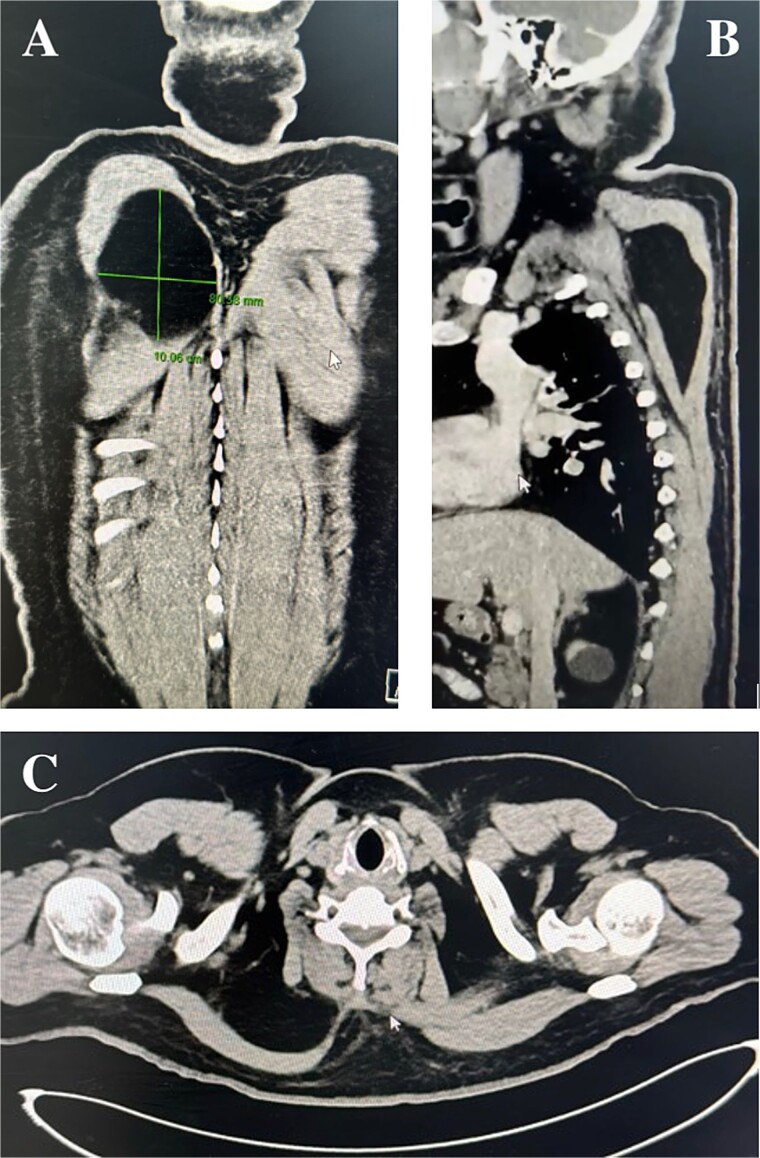
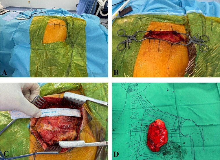
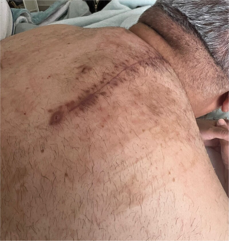
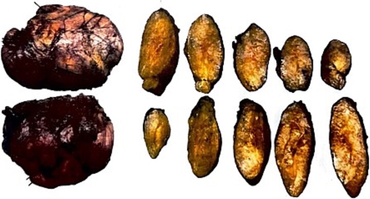
Giant lipomas, rare benign tumours composed of mature adipose tissue, represent only 1% of all lipomas, typically exceeding 10 cm in diameter or weighing over 1000 g. These tumours can cause nerve compression, discomfort, or functional impairment, necessitating surgical excision. We report a 52-year-old male with a giant intramuscular lipoma in the periscapular region, initially identified following significant weight loss after bariatric surgery. Clinical evaluation revealed a 15 × 20 cm mass, confirmed via computed tomography (CT) scan. Surgical excision was performed, followed by histopathological analysis, which confirmed a benign lipoma. Postoperative management was complicated by seroma formation, requiring drainage. This case underscores the importance of early diagnosis, imaging, and appropriate surgical management for large lipomas to prevent complications and recurrence while ensuring optimal cosmetic outcomes.
Keywords: case report; giant lipoma; intramuscular tumour; obesity; surgical excision; weight loss.
Published by Oxford University Press and JSCR Publishing Ltd. © The Author(s) 2024.
Conflict of interest statement
The authors have no financial, commercial or personal competing interests to declare.
Figures
Similar articles
-
A 5 kg giant breast lipoma in a 40-year-old woman: a case report.Ann Med Surg (Lond). 2024 Jan 4;86(2):1229-1233. doi: 10.1097/MS9.0000000000001683. eCollection 2024 Feb. Ann Med Surg (Lond). 2024. PMID: 38333323 Free PMC article.
-
Giant Lipoma in the Trapezius Muscle: A Rare Case Report.Aesthet Surg J Open Forum. 2024 Sep 26;6:ojae081. doi: 10.1093/asjof/ojae081. eCollection 2024. Aesthet Surg J Open Forum. 2024. PMID: 39430212 Free PMC article.
-
A giant multi-compartment lipoma of the hand causing median nerve compression: A case report and review of literature.Int J Surg Case Rep. 2024 Apr;117:109527. doi: 10.1016/j.ijscr.2024.109527. Epub 2024 Mar 15. Int J Surg Case Rep. 2024. PMID: 38503162 Free PMC article.
-
Axillary giant lipoma: a report of two cases and published work review.J Dermatol. 2014 Sep;41(9):841-4. doi: 10.1111/1346-8138.12598. Epub 2014 Aug 25. J Dermatol. 2014. PMID: 25156953 Review.
-
What should be the treatment modality in giant cutaneous lipomas? Review of the literature and report of 4 cases.Br J Plast Surg. 2005 Apr;58(3):394-8. doi: 10.1016/j.bjps.2004.09.005. Br J Plast Surg. 2005. PMID: 15780237 Review.
References
-
- Charifa A, Azmat CE, Badri T.. Lipoma Pathology. 2022 Dec 5. In: StatPearls [Internet]. Treasure Island (FL): StatPearls Publishing, 2024. - PubMed
Publication types
LinkOut - more resources
Full Text Sources
