

Radiological Outcomes According to the Matta Score After the Surgical Fixation of Acetabular Fractures
- PMID: 39737305
- PMCID: PMC11683425
- DOI: 10.7759/cureus.74803
Radiological Outcomes According to the Matta Score After the Surgical Fixation of Acetabular Fractures
Abstract
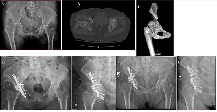
Background Acetabular fractures, a rising concern in developing countries, pose a significant challenge due to their complexity and association with post-operative complications. Often caused by high-energy mechanisms like falls and motor vehicle accidents, these fractures require accurate reduction to prevent long-term issues and the potential need for hip replacement. This study investigates the radiological outcomes of acetabular fracture surgery at six months, focusing on the effectiveness of achieving anatomical reduction using the Matta criteria in a low-and middle-income country (LMIC) setting. Methods and materials This prospective study was conducted at a tertiary care center in Pakistan from May 2023 to December 2023, with ethical approval. Patients with isolated acetabular fractures were recruited. Preoperative X-rays and CT scans classified fractures using the Judet and Letournel Classification. Six-month postoperative X-rays were assessed using Matta radiographic criteria. Appropriate statistical analysis was deployed with a significance level at p < 0.05. Results A total of 33 cases met the study criteria, and the mean average age of patients was 44.18 ±17.2 years. Males constituted 87.9% of the cases. Longer hospital stays were associated with poorer outcomes (p < 0.001). Fracture patterns were significant predictors of outcomes (p < 0.001). Six months post-surgery, 45.5% of patients had excellent results, 24.2% had good results, and 15.2% each had fair and poor results according to the Matta radiographic criteria. Avascular necrosis (AVN) developed in 9.1% of patients. Of the 10 patients with femoral head dislocation, only one developed AVN Conclusion This LMIC-based study investigated factors affecting outcomes in patients with acetabular fractures treated using Open Reduction and Internal Fixation (ORIF). We found a relatively younger patient population, and injury patterns suggested a link to the local environment (e.g., traffic accidents). Optimizing hospital stay and timely surgery improved radiological outcomes as assessed by Matta criteria. While limitations exist, the study supports using Matta criteria in LMICs. Additionally, the use of plain radiographs, rather than CT scans, offers a cost-effective and radiation-reducing alternative for post-operative evaluation in resource-constrained settings.
Keywords: acetabular fractures; demographics; developing countries; fracture patterns; hospital stay; lmic-specific classification; matta criteria; orif; radiological outcomes.
Copyright © 2024, Durrani et al.
Conflict of interest statement
Human subjects: Consent for treatment and open access publication was obtained or waived by all participants in this study. Institutional Review Board of the Agha Khan University Hospital issued approval 2022-0525-22307. Animal subjects: All authors have confirmed that this study did not involve animal subjects or tissue. Conflicts of interest: In compliance with the ICMJE uniform disclosure form, all authors declare the following: Payment/services info: All authors have declared that no financial support was received from any organization for the submitted work. Financial relationships: All authors have declared that they have no financial relationships at present or within the previous three years with any organizations that might have an interest in the submitted work. Other relationships: All authors have declared that there are no other relationships or activities that could appear to have influenced the submitted work.
Figures


References
-
- Ilioinguinal versus modified Stoppa approach for open reduction and internal fixation of displaced acetabular fractures: a systematic review and meta-analysis of 717 patients across ten studies. Srivastava A, Rajnish RK, Kumar P, Haq RU, Dhammi IK. https://link.springer.com/article/10.1007/s00402-022-04369-6. Arch Orthop Trauma Surg. 2023;143:895–907. - PubMed
-
- The value of digital 3D models in evaluating surgical outcomes using the uninjured contralateral acetabulum after acetabular fracture repair. Nijsink H, Arts E, Verhamme L, Biert J, Bemelman M, Brouwers L, van Wageningen B. Injury. 2023;54:1169–1175. - PubMed
-
- The incidence and trauma mechanisms of acetabular fractures: A nationwide study in Finland between 1997 and 2014. Rinne PP, Laitinen MK, Huttunen T, Kannus P, Mattila VM. Injury. 2017;48:2157–2161. - PubMed
LinkOut - more resources
Full Text Sources
Miscellaneous