

The Impact of Focused Hip Ultrasound Training on Imaging Quality in Infants With Hip Dysplasia
- PMID: 39737307
- PMCID: PMC11684545
- DOI: 10.7759/cureus.74787
The Impact of Focused Hip Ultrasound Training on Imaging Quality in Infants With Hip Dysplasia
Abstract
Background: The orthopedic department at Al Jalila Children's Specialty Hospital (AJCH) was opened in April 2018. A focused hip ultrasound training course was conducted in April 2019 to improve hip ultrasound imaging quality.
Objectives: This study aims to evaluate the impact of focused training courses on predefined image quality criteria of infant hip ultrasound in the context of developmental hip dysplasia. It also seeks to measure the inter- and intra-rater agreement among various disciplines.
Methods: A retrospective review of 120 hip ultrasound images (60 infants) was performed between April 2018 and April 2020. Based on internationally agreed criteria, 60 hip images obtained before the course were compared to another 60 hip images obtained after the course. Inter-rater and intra-rater agreements were also evaluated using intraclass correlation (ICC).
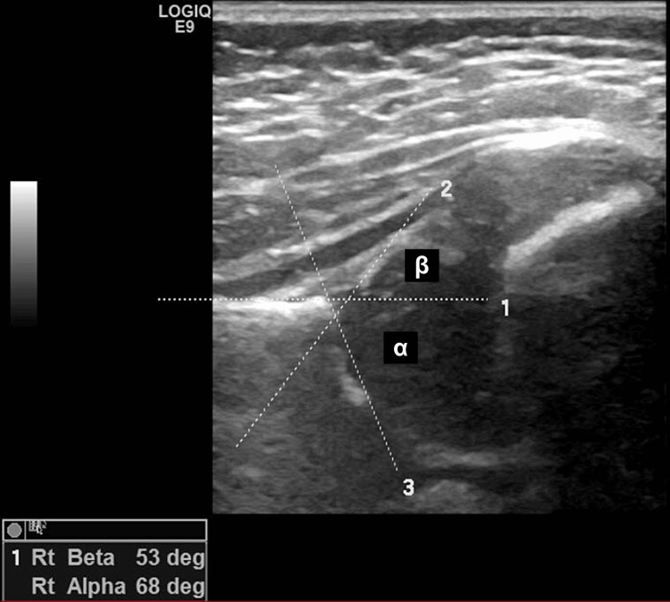
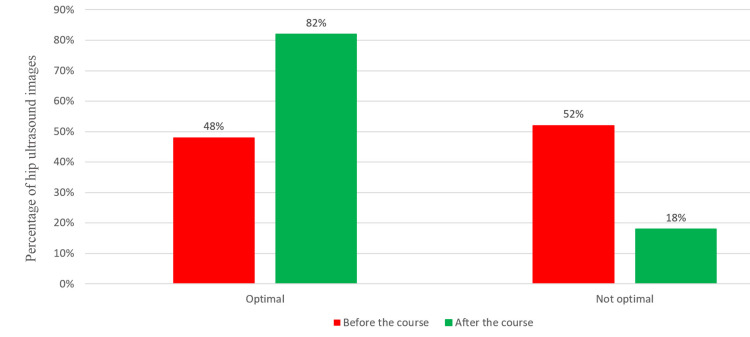
Results: The study evaluated the impact of a focused training course on the quality of infant hip ultrasound images for developmental dysplasia of the hip. Image quality significantly improved after the training, with optimal images increasing from 48% to 82% (P<0.001). Logistic regression confirmed the training's positive effect, highlighting its clinical and statistical significance. The study has also demonstrated excellent agreement among raters for alpha and beta angles, as reflected by ICC statistics. The agreement for alpha angles was notably higher than for beta angles (ICC 0.970 vs. 0.904; P<0.0001). However, inter-rater agreement for hip types, assessed using kappa statistics, was moderate (κ = 0.512) and targeted to address a limited shortfall or gaps in services.
Conclusion: The study confirms the value of focused training in improving the quality of care. This training should be carefully planned and targeted to address limited shortfalls or gaps in services in other areas of service delivery.
Level of evidence: The study is a retrospective cohort with evidence level II.
Keywords: developmental dysplasia of the hip ( ddh ); graf method; hips; neonatal screening; pavlik harness; pediatric orthopedics; pediatric ultrasound; ultrasound.
Copyright © 2024, Alawadhi et al.
Conflict of interest statement
Human subjects: Consent for treatment and open access publication was obtained or waived by all participants in this study. MBRU Institutional Review Board (MBRU-IRB) issued approval SRP-2018-069. Animal subjects: All authors have confirmed that this study did not involve animal subjects or tissue. Conflicts of interest: In compliance with the ICMJE uniform disclosure form, all authors declare the following: Payment/services info: All authors have declared that no financial support was received from any organization for the submitted work. Financial relationships: All authors have declared that they have no financial relationships at present or within the previous three years with any organizations that might have an interest in the submitted work. Other relationships: All authors have declared that there are no other relationships or activities that could appear to have influenced the submitted work.
Figures
References
-
- Gardner ROE, Alshryda S, Kelley SP, Wedge J. Paediatric Orthopaedics. Berlin, Germany: Springer International Publishing; 2016. Evidence-based management of developmental dysplasia of the hip; pp. 27–42.
-
- International interdisciplinary consensus meeting on the evaluation of developmental dysplasia of the hip. O'Beirne JG, Chlapoutakis K, Alshryda S, et al. Ultraschall Med. 2019;40:454–464. - PubMed
-
- Incidence of developmental dysplasia of the hip in Dubai. Moosa NK, Kumar PT, Mahmoodi SM. https://pubmed.ncbi.nlm.nih.gov/19618014/#:~:text=Conclusion%3A%20Twenty... Saudi Med J. 2009;30:952–955. - PubMed
-
- Risk factors for developmental dysplasia of the hip: a meta-analysis. de Hundt M, Vlemmix F, Bais JM, Hutton EK, de Groot CJ, Mol BW, Kok M. Eur J Obstet Gynecol Reprod Biol. 2012;165:8–17. - PubMed
-
- The importance of predicted risk factors in developmental hip dysplasia: an ultrasonographic screening program (Article in Turkish) Karapinar L, Sürenkök F, Oztürk H, Us MR, Yurdakul L. https://pubmed.ncbi.nlm.nih.gov/12510090/ Acta Orthop Traumatol Turc. 2002;36:106–110. - PubMed
LinkOut - more resources
Full Text Sources
Research Materials
Miscellaneous