Review
. 2025 Jan 22;125(2):523-598.
doi: 10.1021/acs.chemrev.4c00216. Epub 2024 Dec 31.

Genetic Code Expansion: Recent Developments and Emerging Applications

Affiliations
Review

Genetic Code Expansion: Recent Developments and Emerging Applications

Yujia Huang et al. Chem Rev. .

Abstract

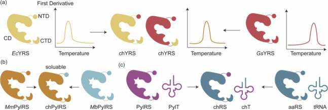
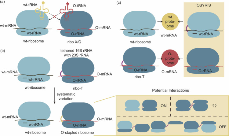
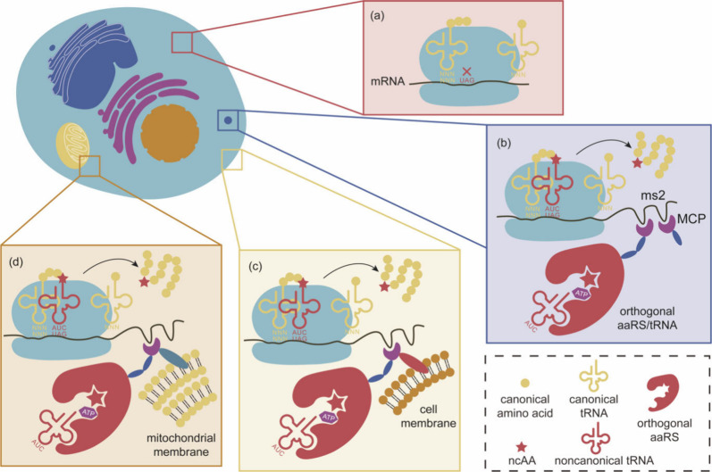
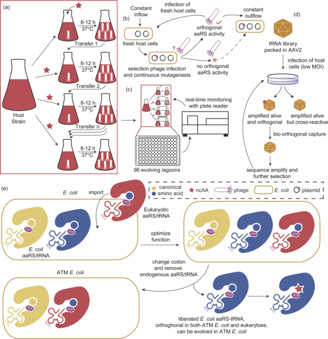
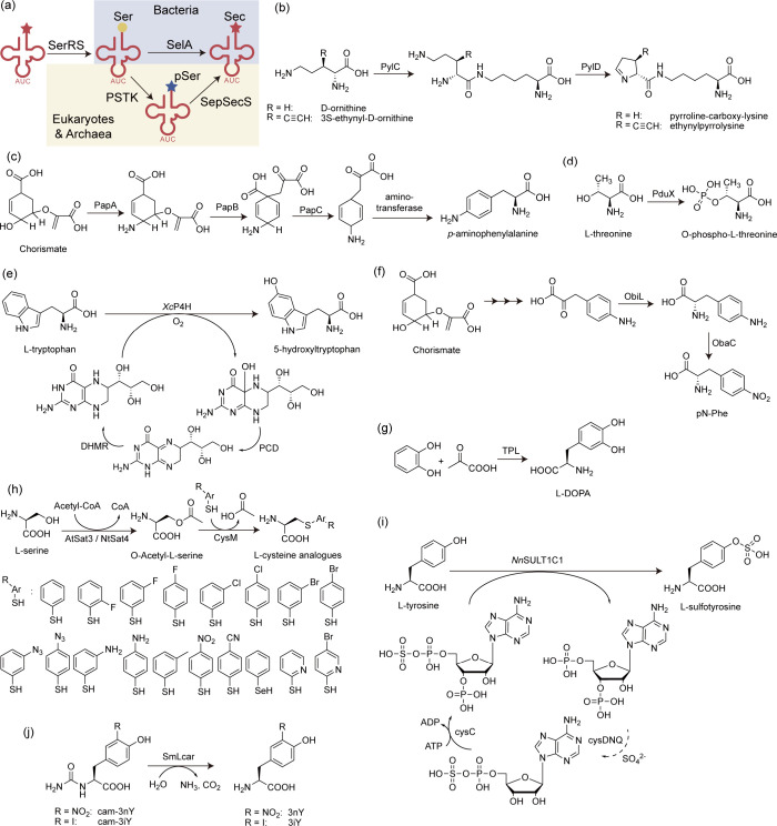
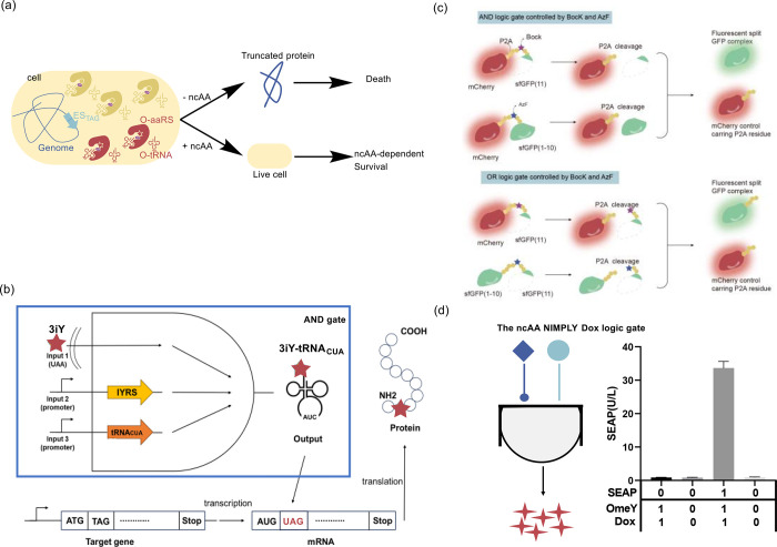
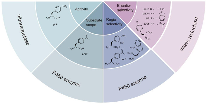
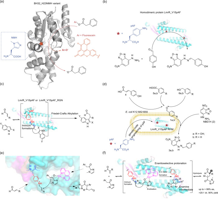
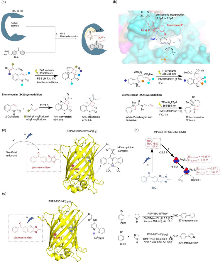
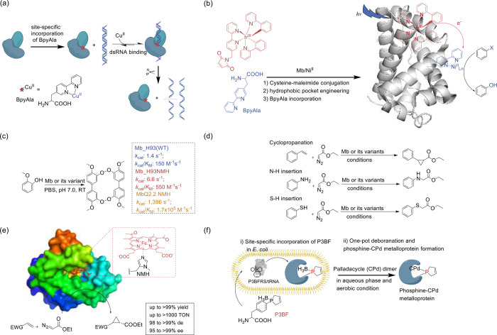
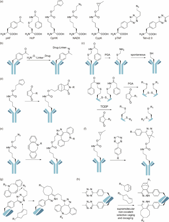
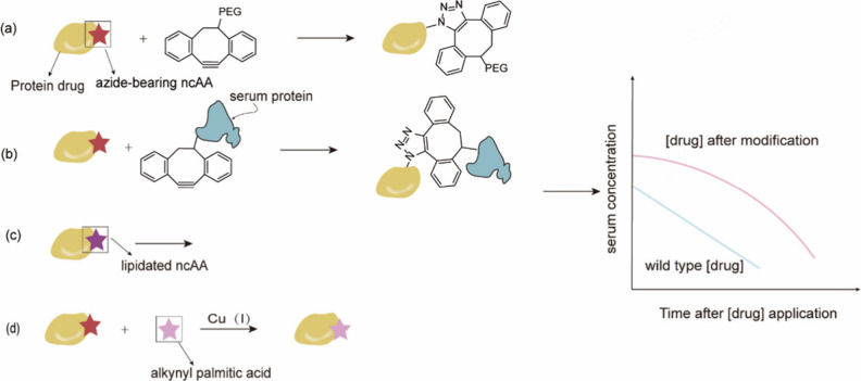
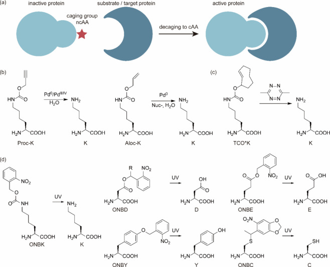
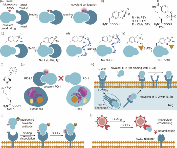
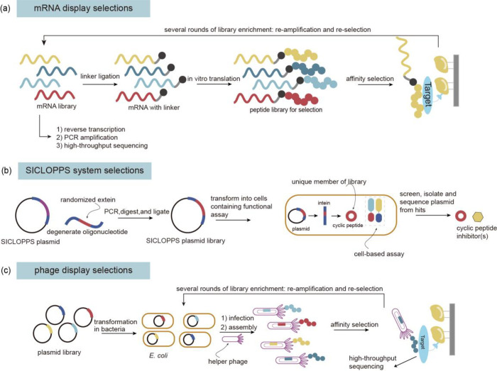
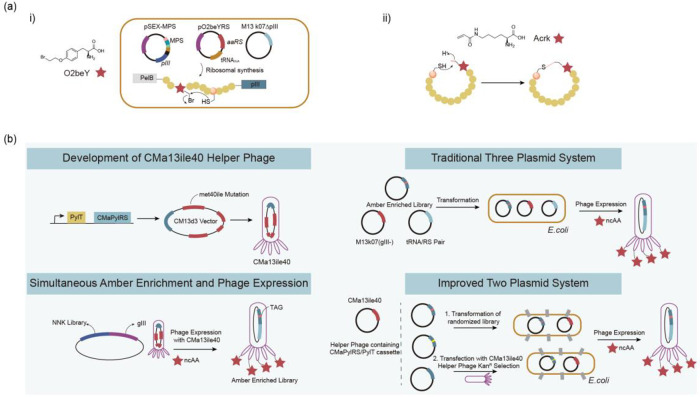
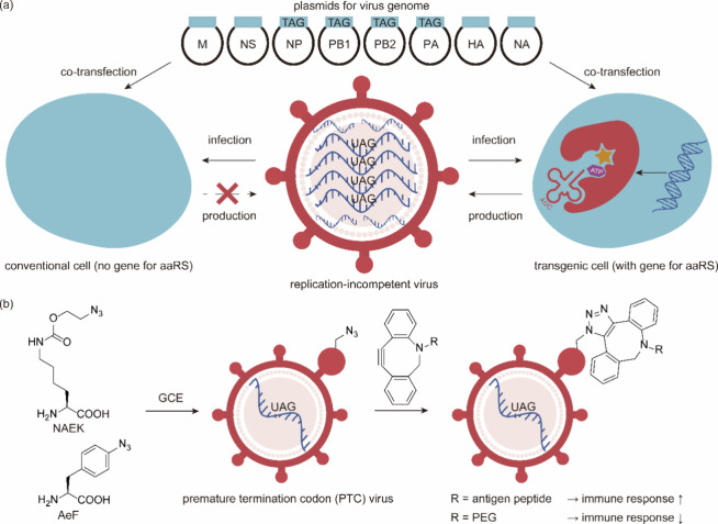
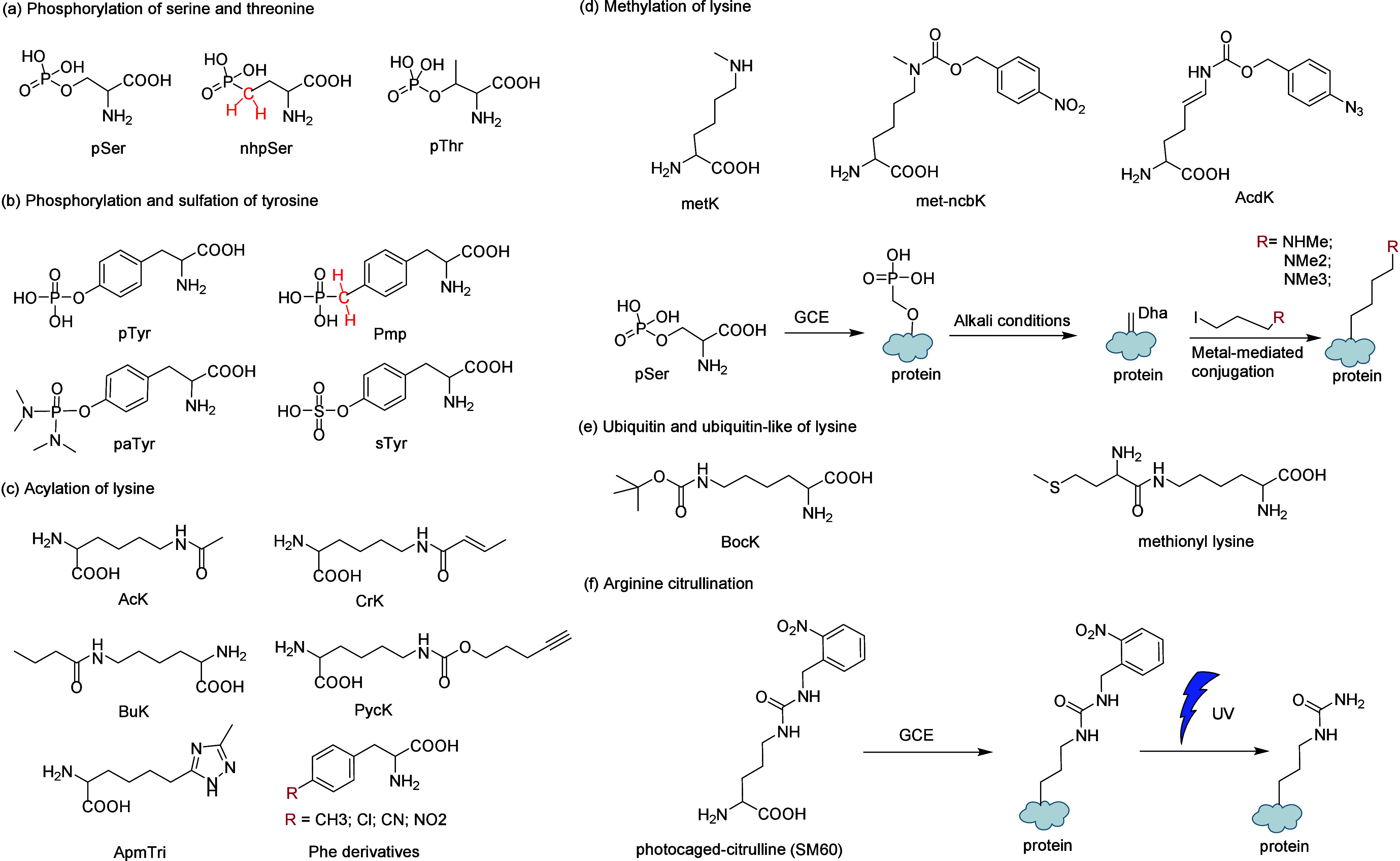
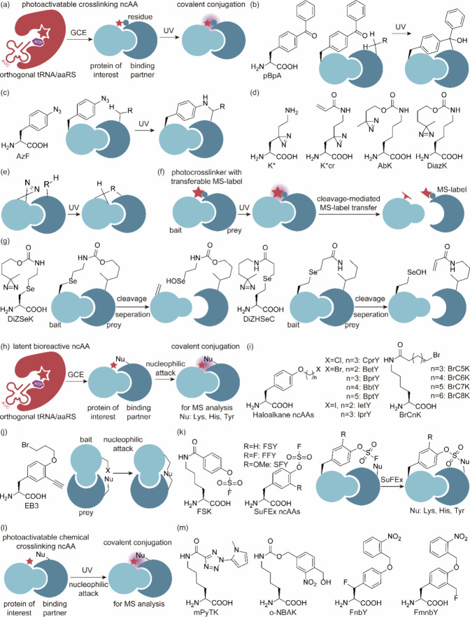
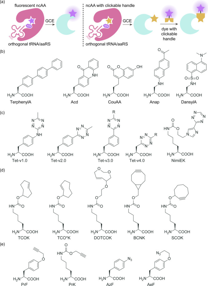
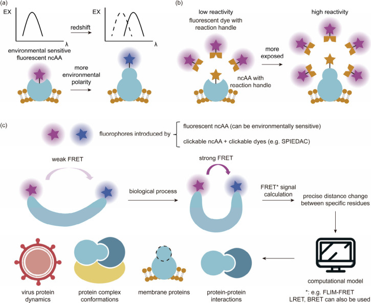
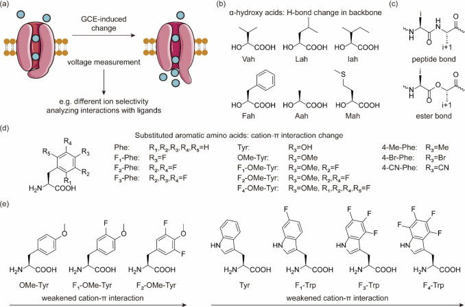
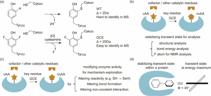
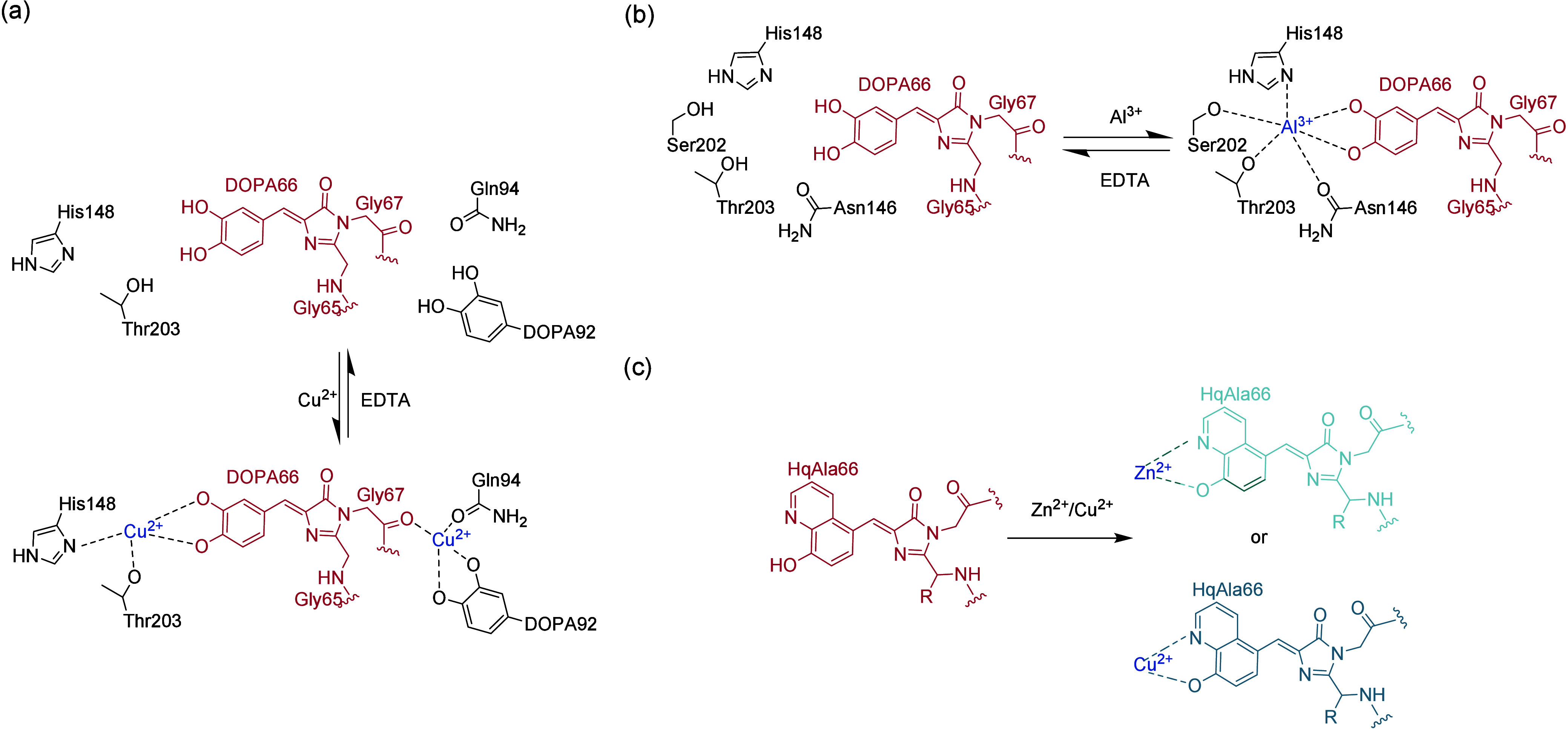
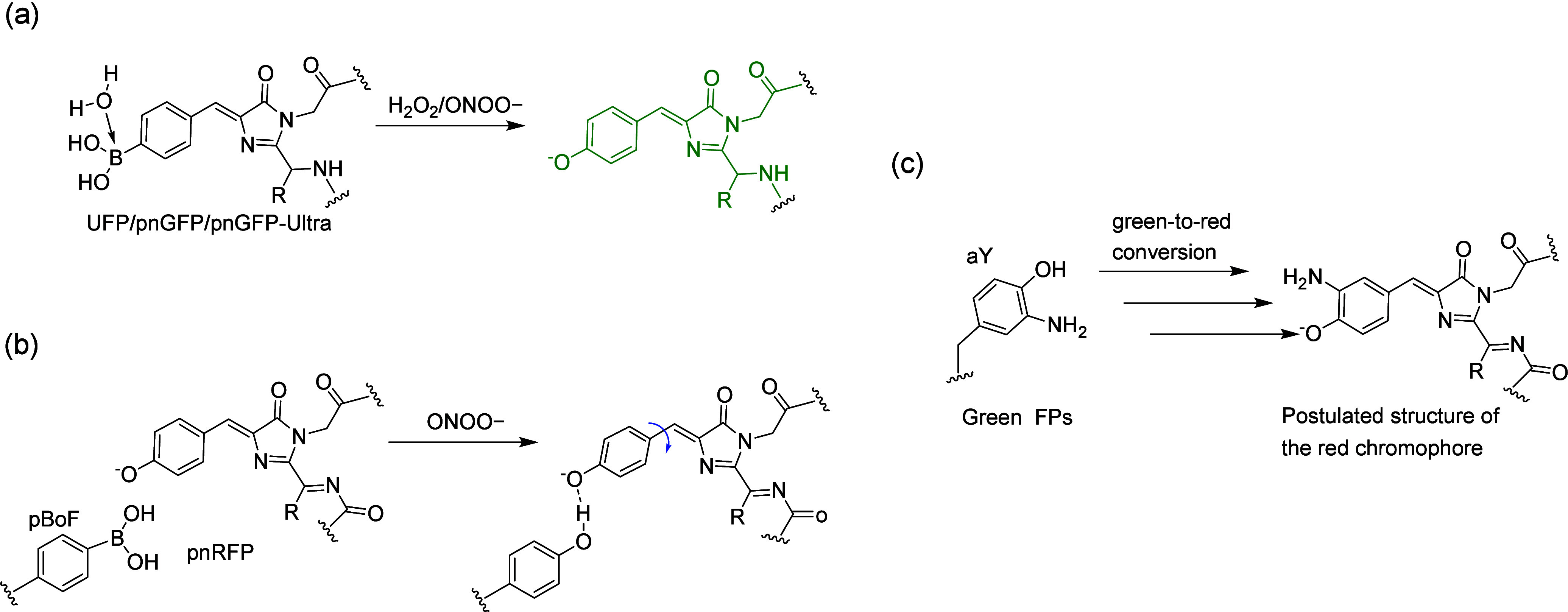
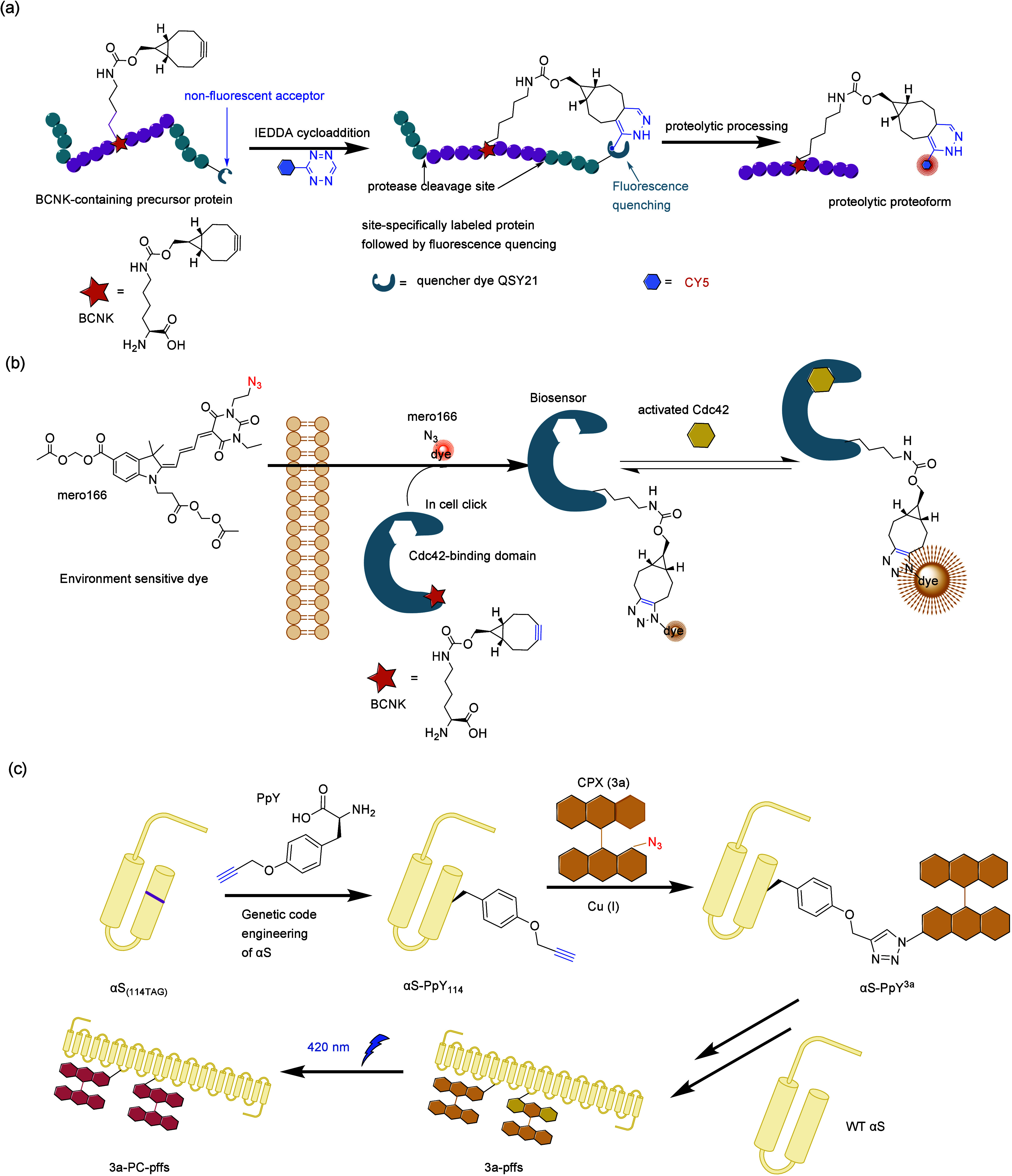
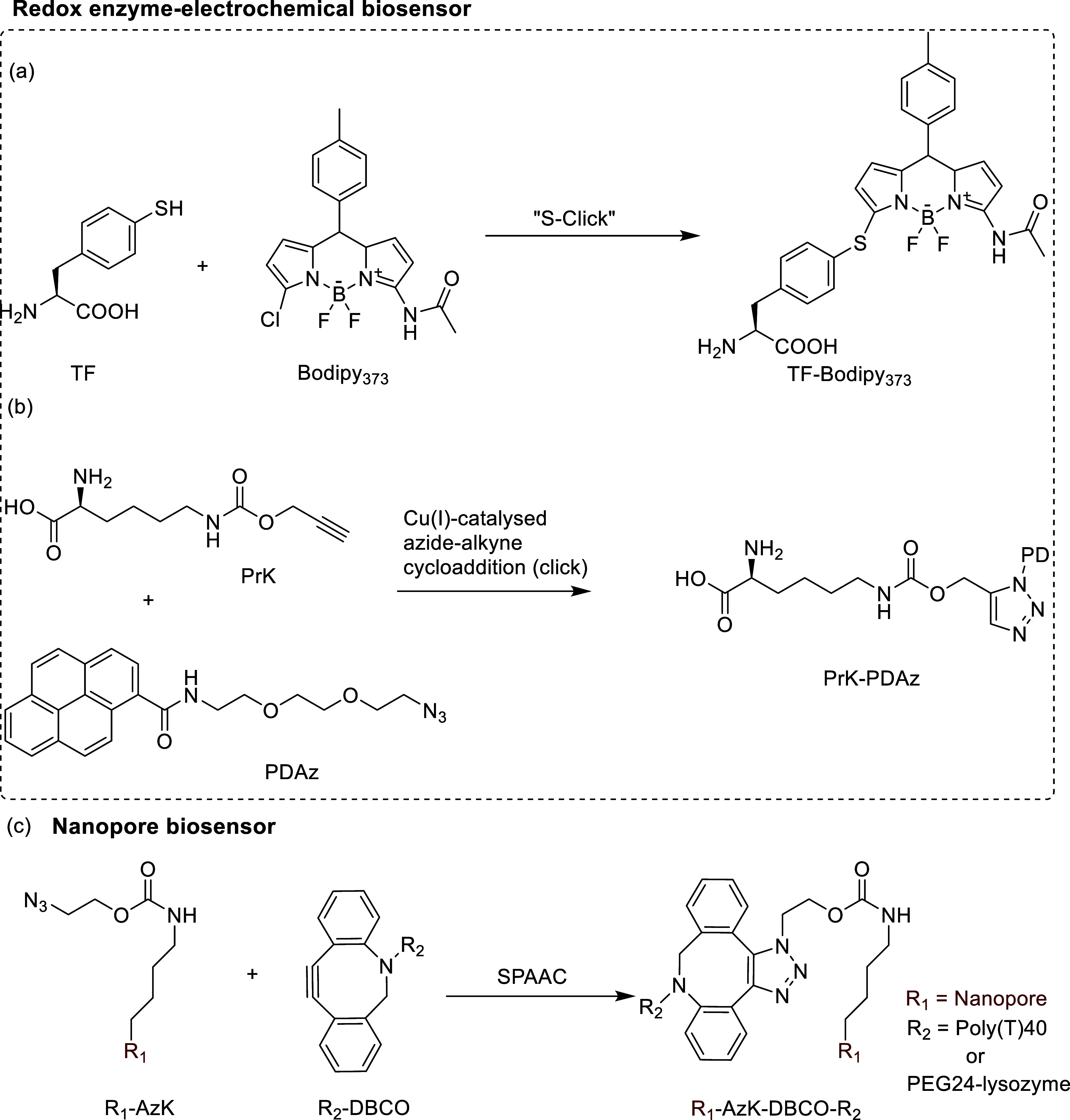
The concept of genetic code expansion (GCE) has revolutionized the field of chemical and synthetic biology, enabling the site-specific incorporation of noncanonical amino acids (ncAAs) into proteins, thus opening new avenues in research and applications across biology and medicine. In this review, we cover the principles of GCE, including the optimization of the aminoacyl-tRNA synthetase (aaRS)/tRNA system and the advancements in translation system engineering. Notable developments include the refinement of aaRS/tRNA pairs, enhancements in screening methods, and the biosynthesis of noncanonical amino acids. The applications of GCE technology span from synthetic biology, where it facilitates gene expression regulation and protein engineering, to medicine, with promising approaches in drug development, vaccine production, and gene editing. The review concludes with a perspective on the future of GCE, underscoring its potential to further expand the toolkit of biology and medicine. Through this comprehensive review, we aim to provide a detailed overview of the current state of GCE technology, its challenges, opportunities, and the frontier it represents in the expansion of the genetic code for novel biological research and therapeutic applications.


Conflict of interest statement

The authors declare the following competing financial interest(s): X.L. has financial interests in Demetrix and Synceres.

Figures

Figure 1
Figure 1
Chimeric aaRS/tRNA pairs. (a) Bacterial TyrRS chimeras created from GsTyrRS and EcTyrRS exhibit higher activity and intermediate thermostability. (b) Fusion of MbPylRS N-terminal domain to the MmPylRS C-terminal domain, creating a novel chimeric PylRS. (c) Transplanting the key orthogonal components from the pyrrolysine system (aaRS and tRNA) to those for canonical amino acid systems can create chimeric aaRS/tRNA pairs. Abbreviations: NTD, N-terminal domain; CD, catalytic domain; CTD, C-terminal domain; ch, chimeric; aaRS, aminoacyl-tRNA synthetase.
Figure 2
Figure 2
Engineering of ribosomes. (a) O-Ribosome and O-mRNA pairs such as ribo-X and ribo-Q consist of an mRNA containing a ribosome-binding site that does not direct translation by the endogenous ribosome, coupled with an orthogonal ribosome capable of efficiently and specifically translating the orthogonal mRNA. (b) In ribo-T and O-stapled ribosome, short RNA linkers integrate sequences from both small and large subunit rRNA. (c) In OSYRIS system, orthogonal proteome was translated by ribosomes composed of the dissociable orthogonal 30S subunit and the wild-type 50S subunit. Meanwhile, the endogenous cellular proteome was translated by ribosomes with tethered subunits. Abbreviations: O-tRNA, orthogonal tRNA; O-mRNA, orthogonal mRNA; O-proteome, orthogonal proteome; OSYRIS, orthogonal translation system based on ribosomes with isolated subunits.
Figure 3
Figure 3
Applications of phase separation in GCE. (a) Traditional GCE technology. (b) Designer membraneless organelles enable codon reassignment of selected mRNAs in eukaryotic cytoplasm. (c) Spatial separation of orthogonal eukaryotic translation on cell membrane. (d) Spatial separation of orthogonal eukaryotic translation on mitochondrial membrane. Abbreviations: MCP, major capsid protein.
Figure 4
Figure 4
Optimization of screening methods. (a) Principle of phage-assisted noncontinuous evolution (PANCE). (b) Principle of phage-assisted continuous evolution (PACE). (c) Principle of phage- and robotics-assisted near-continuous evolution (PRANCE). (d) Principle of virus-assisted directed evolution of tRNAs (VADER). (e) In ATM E. coli strain, the native EcTrpRS/tRNA pair was replaced with an E. coli, optimized counterpart from S. cerevisiae. Abbreviations: AAV, adeno-associated virus; ncAA, noncanonical amino acid; ATM, altered translational machinery.
Figure 5
Figure 5
Biosynthesis of ncAAs. (a) Biosynthesis of selenocysteine in bacteria, eukaryotes, and archaea. (b) Biosynthesis of pyrrolysine and derivatives. (c) Biosynthesis of pAF. (d) Biosynthesis of pThr. (e) Biosynthesis of 5-HTP. (f) Biosynthesis of pN-Phe. (g) Biosynthesis of DOPA. (h) Biosynthesis of ncAAs based on cysteine biosynthetic pathway. (i) Biosynthesis of sTyr. (j) Biosynthesis of 3nY and 3iY. Abbreviations: PSTK, O-phosphoseryl-tRNA kinase; Xc, Xanthomonas campestris; P4H, phenylalanine 4-hydroxylase; DHMR, dihydromonapterin reductase; PCD, pterin-4α-carbinolamine dehydratase; TPL, tyrosine phenol-lyase; 3nY, 3-nitro-l-tyrosine; 3iY, 3-iodo-l-tyrosine; Nn, Nipponia nippon.
Figure 6
Figure 6
Regulation of gene expression. (a) Direct regulation, ncAA-dependent synthetic auxotroph. (b) AND gate that integrates 3 inputs (ncAA plus 2 promoters that respectively drive RS gene and tRNA) and controls ncAA-tRNA formation as an output. (c) AND/OR logic gate controlled by BocK and AzF. The transfection efficiency of vector expressing split GFP and mCherry into mammalian cell was demonstrated by mCherry fluorescence, which maintained consistent intensity across all four conditions: no ncAA, BocK only, AzF only, or both BocK and AzF. The logic operation output was indicated by GFP fluorescence, mCherry fluorescence indicated transfection efficiency. (d) The ncAA NIMPLY Dox logic gate. Designer cells engineered with the transcription-translation combination switch were subjected to various treatments with OmeY and/or Dox, a secreted embryonic alkaline phosphatase (SEAP) was selected as the POI for convenient detection.
Figure 7
Figure 7
Enhancing native catalytic properties of natural enzymes, including enzyme activity, substrate scope, regioselectivity, and enantioselectivity.
Figure 8
Figure 8
Genetic encoding of nucleophilic catalysis. (a) Reaction scheme for the hydrolysis of fluorescein 2-phenylacetate catalyzed by BH32 (His23Me-His), the reaction’s progress was tracked by observing a rise in absorbance at 500 nm, which indicated the formation of fluorescein. (b–f) LmrR-based designer enzymes. LmrR containing pAF catalyzes chromogenic hydrazone formation between 4-hydrazino-7-nitro-2,1,3-benzoxadiazole and 4-hydroxybenzaldehyde, the reaction’s progress was tracked by the following product in absorbance at 472 nm (b). Unlocking iminium catalysis in artificial LmrR variants to create a Friedel–Crafts alkylase (c). In vivo biocatalytic cascades featuring an unnatural hydrazone formation reaction catalyzed by LmrR_V15pAF_RMH (d). An asymmetric Michael addition reaction catalyzed by synergistic combination of two catalytic sites, the pAF residue functions for activation of the enal and Cu(II)-phen for the generation and delivery of the enolate nucleophile (e). A tandem Michael addition/enantioselective protonation reaction catalyzed by LmrR_V15pAF employing two abiological catalytic sites in a synergistic fashion: a genetically encoded pAF and a Lewis acid Cu(II) complex (f).
Figure 9
Figure 9
Genetic encoding of photocatalysis. (a,b) Photoenzyme design. A designed photoenzyme based on DA_20_00 for enantioselective [2 + 2] cycloadditions, photoenzyme development, substrate scope of EnT1.3 and selected variants and biomolecular [2 + 2] cycloadditions (a). Enantioselective [2 + 2]-cycloadditions with triplet photoenzymes. The design of TPe, the substrate scope of TPe variant and biomolecular [2 + 2] cycloadditions (b). (c–e) Light-harvesting metalloenzyme design. PSP was transformed into the resultant photocatalytic CO2-reducing enzyme PSP2–95C93Y97Y by employed the nickel (NiII)–terpyridine complex, which catalyzes the selective CO2 reduction to CO (c). PET pathways in miniature photocatalytic CO2-reducing enzyme mPCE2 (mPCE-C8G-Y30N). A strongly reducing ketyl radical (BpC/ BpC; Epc = −1.47 V) is generated by charge separation in mPCE2, and sequential ET produces the reduced [Fe4S4] cluster (FeA,red/FeA,ox; Epc,2+/1+ = −0.68 V, Epc,1+/0 = −1.25 V, and FeB,red/FeB,ox; Epc,2+/1+ = −0.57 V, Epc,1+/0 = −1.09 V) for CO2 reduction (d). Biocatalytic cross-coupling of aryl halides with a genetically engineered photosensitizer artificial dehalogenase, which features a genetically encoded benzophenone chromophore and site-specifically modified synthetic NiII(bpy) cofactor with tunable proximity to streamline the dual catalysis, which was shown on the left pane. Catalytic C–N bond formation by PSP-95C–NiII(bpy) was shown on the right pane (e).
Figure 10
Figure 10
NcAAs function as noncanonical ligands. (a) Site-specific incorporation of BpyAla in two sites of the VSRS protein p19 dimer introduces a metal binding site. (b) Design scheme of an artificial metallaphotoredox enzyme (AMPE*/C45-Z97) by the introduction of the Ir photocatalyst via bioconjugation and metal binding ncAA, BpyAla, via the active site redesign and GCE method. Photocatalytic C–O coupling enzymes that operate via intramolecular electron transfer. (c) Myoglobin (Mb)-based designer enzyme was made by incorporating Nδ-methylhistidine (NMH) with metal-coordination properties into myoglobin (Mb) and the enzyme activity of the resulting enzyme and its derivates was tested in guaiacol oxidation. (d) Reactivity of Mb variants in different carbene transfer reactions. (e) Stereoselective cyclopropanation of electron-deficient olefins with a cofactor redesigned carbene transferase Mb*. (f) Streamlined in vivo platform to achieve the site-specific genetic incorporation of borane-protected phosphine into proteins followed by one-pot deboronation and palladium coordination.
Figure 11
Figure 11
Protein conjugates. (a) Chemial structures of commonly used ncAAs for bioconjugation. (b) Reaction on the incorporated ncAA pAF for bioconjugation. (c) Reaction on the incorporated ncAA HcP for bioconjugation (upper) and dual-bioconjugation (lower). PGA, penicillin G acylase. For protein with cystine and HcP, PGA can deprotect HcP and the subsequent TCEP can activate cystine, enabling sequential labeling of the protein. TCEP, tris(2-carboxyethyl)phosphine. (d) Reaction on the incorporated ncAA CpHK for bioconjugation. (e) Reaction on the incorporated ncAA NAEK for bioconjugation. (f) Reaction on the incorporated ncAA CypK for bioconjugation. (g) Reaction on the incorporated ncAA pTAF for bioconjugation. (h) A brief schematic of the strategy to prepare protein dual conjugates with ncAA Tet-v2.0. Tetrazine residue positions could be computationally designed so that exposed residues could be reversibly caged by a barrel-shaped supramolecular host, whereas semiburied residues that were resistant to caging retained their reactivity, thus allowing sequential IEDDA labeling.
Figure 12
Figure 12
Applications of the GCE technology using azide groups or lipidated amino acids for site-specific PEGylation or lipidation to tune the pharmacokinetic properties of proteins. (a) Genetically incorporated azide-bearing ncAA enables site-specific PEGylation, increasing the half-life of protein drugs while retaining their bioactivity. (b) Genetically incorporated azide-bearing ncAA allows for the construction of serum protein-drug conjugates, prolonging the serum half-life of the proteins. (c) Genetically incorporated fatty acid-containing ncAA facilitates site-specific fatty acid decoration, extending the protein’s half-life. (d) Genetically incorporated azide-bearing ncAA enables site-specific lipidation, resulting in a tailored half-life.
Figure 13
Figure 13
On-demand activation of proteins. (a) Strategy of on-demand activation of proteins. The ncAA with caging group is incorporated at a key site to inactivate the protein. With decaging methods, the ncAA becomes cAA and activate the protein. cAA, canonical amino acid. (b) Reactions for Proc-Lys and Aloc-Lys to be decaged to lysine by Pd catalyst. (c) Click-to-release reaction for TCO*K to be decaged to lysine by tetrazines. (d) Reactions for ncAAs with nitrobenzyl-based photolabile protecting groups (PPGs) be decaged to cAA.
Figure 14
Figure 14
Covalent protein drugs. (a) Strategy of covalent protein drugs based on proximity-enabled protein cross-linking. The latent reactive ncAA on protein drug can conjugate with target residue on the drug target once the two proteins bind together. (b) Chemical structures of SuFEx-based amino acids. (c) Proximity-enabled protein cross-linking based on SuFEx. The ncAAs can react with Lys, His, and Tyr on the target protein. (d) Proximity-enabled cross-linking with RNA based on SuFEx. The ncAAs can react with 3′-OH on the RNA. (e) Proximity-enabled cross-linking with carbohydrate based on SuFEx. The ncAAs can react with 6′-OH on the RNA. (f) Other amino acids for proximity-enabled cross-linking. The aromatic ring can be varied. (g) Strategy of covalent PD-1. Covalent PD-1 can conjugate with PD-L1 on tumor cells, which prevents PD-L1 to bind with PD-1 on T cells and inhibit T cell activity. (h) Strategy of covalent cytokine IL-2. FSY-bearing IL-2 variants covalently bind to IL-2Rα when in proximity, resulting in persistent recycling of IL-2 and selectively promoting the expansion of Tregs but not effector cells. interleukin-2, IL-2. Treg, regulatory T cell. (i) Strategy of covalent antibody conjugates. The radioactive antibody can covalently bind with receptor, increasing radioisotope levels and extending tumor residence time. (j) Strategy of covalent antibody. The covalent antibody can covalently bind with receptors on virus, preventing the binding between virus and ACE2 on target cells. ACE2, angiotensin-converting enzyme 2.
Figure 15
Figure 15
Comparison of mRNA display, SICLOPPS system, and phage display selections. (a) For mRNA display selections, an initial mRNA library is fused to a linker through various techniques. During in vitro translation, this results in the peptides being covalently attached to their corresponding mRNA coding tags. Affinity selection is then performed, followed by iterative rounds of mRNA/cDNA tag recovery, library reamplification, and reselection until sufficient enrichment is achieved. (b) To generate and screen a SICLOPPS library, start with standard molecular biology techniques to create the library using a degenerate oligonucleotide. This oligonucleotide is designed to specify the ring size of the cyclic peptides, the number of randomized amino acids, and any fixed amino acids. The resulting plasmid library is then transformed into cells equipped with an assay (e.g., FRET, life/death, phenotypic) for screening. Active cyclic peptides are identified by isolating the SICLOPPS plasmids from cells exhibiting the desired phenotype, followed by DNA sequencing to determine their identities. (c) Phage display selections start by transforming a bacterial host with a plasmid library. These bacteria are then infected with helper phages, leading to the production of a phage library where library peptides are displayed on phage coat proteins. Similar to mRNA display selections, this process involves affinity selection followed by multiple cycles of reamplification and reselection until the desired enrichment level is reached.
Figure 16
Figure 16
Application and optimization of phage display. (a) Genetically encoded, phage-displayed ncAA-containing cyclic peptides. (i) Cysteine-reactive O2beY and a proximal cysteine residue, resulting in the formation of macrocyclic peptides constrained by a nonreducible, interside-chain-to-side-chain thioether linkage. (ii) Cyclization of phage-displayed peptides through Michael addition between a cysteine and a genetically incorporated AcrK. (b) An amber-encoding helper phage for more efficient phage display of ncAAs. Comparison of the traditional three-plasmid expression system with the newly designed two-plasmid system for expression of phage libraries containing ncAAs.
Figure 17
Figure 17
Vaccine development. (a) Schematic illustration of the creation of premature termination codon (PTC) influenza viruses. These viruses exhibit replication incompetence in standard cells but exhibit high replication in transgenic cells. The transgenic cells contain integrated cassettes for the expression of orthogonal tRNA (tRNACUA) and tRNA synthase (pylRS). NP, nucleoprotein; PB1 and PB2, polymerase basic proteins 1 and 2; PA, polymerase acidic protein; M, matrix protein; NS, nonstructural protein; HA, hemagglutinin; NA, neuraminidase. (b) Modification for multifunctional virus vaccines. The ncAAs on virus serve as bioorthogonal reaction handles for conjugation with components like antigen peptide or PEG to modify virus and change the functions of virus.
Figure 18
Figure 18
NcAAs used for exploring biological mechanisms of serine/threonine phosphorylation, tyrosine phosphorylation/sulfation, lysine acylation/methylation/ubiquitin, and arginine citrullination, via GCE.
Figure 19
Figure 19
Site-specific cross-linking in situ for research of biological mechanism. (a) Schematic illustration of photoinduced site-specific cross-linking in situ. The photoactivatable cross-linking ncAA on the protein of interest can be activated by UV and covalently onjugate with nearby residue on the binding partner protein. (b) Chemical structure (left) of the photoactivatable cross-linking ncAA pBpA and its cross-linking reaction (right). (c) Chemical structure (left) of the photoactivatable cross-linking ncAA AzF and its cross-linking reaction (right). (d) Chemical structures of diazirine-containing lysine analogues as photoactivatable cross-linking ncAAs. (e) Photoactivated cross-linking reaction for diazirine-containing lysine analogues. (f) Schematic illustration of photoinduced site-specific cross-linking followed by MS-label transfer. After conjugating with the prey protein by UV light, the ncAA can cleave by processing and leave a MS label on the prey protein. MS, mass sprectrum. (g) Chemical structures and their cross-linking reactions of the diazirine-containing self-releasable lysine analogues DiZSeK (left) and DiZHSeC (right). (h) Schematic illustration of chemical cross-linking in situ. The latent bioreactive ncAA on the protein of interest can react with nearby residues such as Lys, His and Tyr. (i) Chemical structures of haloalkane ncAAs as latent bioreactive ncAAs. (j) Chemical structure of the haloalkane ncAA EB3 (left) and the cross-linking reaction on haloalkane ncAAs (right). (k) Chemical structures of SuFEx-based amino acids (left) and their cross-linking reactions (right). (l) Schematic illustration of photoactivated chemical cross-linking. The photoactivatable latent bioreactive ncAA on the protein of interest can react with nearby residues upon activation by UV light. (m) Chemical structures of photoactivated chemical cross-linking amino acids.
Figure 20
Figure 20
Introducing site-specific fluorophores in situ for research of biological mechanisms. (a) Schematic illustration of enlightening proteins with fluorescent ncAAs (left) or ncAAs with reaction handles (right). The ncAAs with reaction handles on proteins can react with fluorescent dyes with reaction handles. (b) Chemical structures of commonly used fluorescent ncAAs. (c) Chemical structures of tetrazine-containing ncAAs used for imaging. (d) Chemical structures of commonly used strained dienophile-containing ncAAs for SPIEDAC reaction. (e) Chemical structures of commonly used ncAAs for SPAAC or CuAAC reaction.
Figure 21
Figure 21
Schematic illustration of strategies of using fluorophores to study biological mechanisms. (a) Schematic illustration of the redshift of fluorescent ncAA induced by enhanced environmental polarity. EX, excitation. (b) Schematic illustration of different reactivities of differently exposed ncAAs with reaction handle. More exposed ncAA have higher reactivity. (c) Schematic illustration of using smFRET on introduced fluorophores to study biological mechanisms. Fluorophores are introduced at specific sites using fluorescent ncAA or clickable ncAAs and dyes. During a biological process, the distance between these residues changes, leading to a change of FRET signal. Inputting these into computational models can help to work out protein or protein complex conformation changes. BRET, bioluminescence resonance energy transfer; LRET, luminescence (or lanthanide-based) resonance energy transfer.
Figure 22
Figure 22
Monitoring voltage changes for study of biological mechanisms. (a) Schematic illustration of GCE-induced change for voltage measurement. Special ncAA is introduced to key sites of proteins like ion channels, leading to changes of protein functions. (b) Chemical structures of α-hydroxy acids. (c) The change of H-bond in backbone when introducing α-hydroxy acids (lower) compared to canonical amino acids (upper). (d) Chemical structures of substituted aromatic amino acids. (e) The change of cation−π interaction when introducing substituted aromatic amino acids.
Figure 23
Figure 23
Exploration of bioinorganic chemistry and organic chemistry. (a) Using mass difference between cAA and ncAA to reveal enzyme mechanisms. More obvious loss of mass provide convenience for MS identification to detect reactions on the enzyme. MS, mass sprectrum. (b) Schematic illustration of stabilizing transient state structure for analysis. The ncAA at key residue cannot cross-link with cofactor as cAA, thus retaining transient uncrosslined state of enzyme for analysis. (c) Schematic illustration of changing enzyme activity for comprehensive mechanistic analysis. Substituting cAA with different ncAAs changes the activity of enzymes, providing insights into the mechanism of this enzyme. (d) Schematic illustration of using protein to stabilize transient state at energy maximum for organic chemistry study. The biphenyl is locked in the protein and keeps a transient state at energy maximum, enabling researchers to study this transient state.
Figure 24
Figure 24
Applications of GCE in biomaterials. (a) The genetically incorporated bidentate bipyridyl-alanine enables controlled protein self-assembly, preserving protein functionality and enhancing thermal stability. (b) The genetically incorporated p-thiolphenylalanine facilitates the biosynthesis of protein and carbon nanotube hybrid biomaterials. (c) The genetically incorporated propargyloxy-phenylalanine allows for site-specific cross-linking, creating pili complexes with gold nanoparticles. (d) The genetically incorporated azide-bearing ncAA enables site-specific cross-linking, forming hybrid protein–DNA cages. (e) The genetically incorporated photoreactive ncAA allows tuning the size of elastin-like polypeptides. (f) The genetically incorporated photoreactive ncAA enhances the stability of elastin-like polypeptides.
Figure 25
Figure 25
Biosensors for transition metal ion detection. (a) The Cu2+ sensor based on L-DOPA. (b) The Al3+ sensor based on L-DOPA. (c) The Zn2+/Cu2+ sensor based on HqAla.
Figure 26
Figure 26
Representative FP-based biosensors for H2O2/ONOOdetection and imaging. (a,b) Biosensor based on pBoF for H2O2/ONOO detection. Introduction of pBoF to the chromophore of UFP, pnGFP, or pnGFP-Ultra allowed it to react with peroxynitrite and form a tyrosine-derived chromophore that enhanced fluorescence (a). pBoF is introduced to an amino acid position near the chromophore of pnRFP, where its reaction with peroxynitrite creates a tyrosine residue, which reduces fluorescence by bending the chromophore through hydrogen bonding (b). (c) Biosensor based on aY for imaging. The postulated structure of the chromophore in the a Y-converted red FPs was shown.
Figure 27
Figure 27
Small-molecule fluorescent probe-based biosensors. (a) Protein labeling and identification of proteolytic proteoform using a GCE-based molecular beacon. Polypeptides containing BCNK are modified with a fluorophore through IEDDA cycloaddition. A corresponding fluorescence quencher connected to the same protein suppresses the fluorescence. The fluorescence signal is produced when the fluorophore and quencher are separated through proteolysis. (b) Membrane-permeant, environment-sensitive dye mero166 generates a biosensor for the small GTPase Cdc42 within living cells. (c) Overview of producing 3a-tagged αS-PpY114 preformed fibrils (3a-pffs) and their subsequent photoconversion to fibrils (3a-PC-pffs).
Figure 28
Figure 28
Redox enzyme-electrochemical biosensor and nanopore biosensor. (a) A schematic illustration of thiol-chlorine nucleophilic substitution reaction (S-click reaction): sulfur atom in TF (S), chlorine atom in Bodipy373 (Cl). (b) A schematic illustration of PrK and pyrene-diethylene glycol-azide (PDAz) with copper(I)-catalyzed azide–alkyne cycloaddition (click) reaction to a pyrene-diethylene glycol-azide (PDAz). (c) DBCO was covalently attached to AzK via a strain-promoted alkyne–azide cycloaddition reaction (SPAAC).

Similar articles

Cited by

References

    1. Liu T. Recent Advances in Genetic Code Expansion: From Cell Engineering to Protein Design. J. Mol. Biol. 2022, 434 (8), 167565.10.1016/j.jmb.2022.167565. - DOI - PubMed
    1. Lee S.; Kim J.; Koh M. Recent Advances in Fluorescence Imaging by Genetically Encoded Non-Canonical Amino Acids. J. Mol. Biol. 2022, 434 (8), 167248.10.1016/j.jmb.2021.167248. - DOI - PubMed
    1. Nguyen T. A.; Cigler M.; Lang K. Expanding the Genetic Code to Study Protein-Protein Interactions. Angew. Chem., Int. Ed. Engl. 2018, 57, 14350.10.1002/anie.201805869. - DOI - PubMed
    1. Huang Y.; Liu T. Therapeutic Applications of Genetic Code Expansion. Synth. Syst. Biotechnol. 2018, 3 (3), 150–158. 10.1016/j.synbio.2018.09.003. - DOI - PMC - PubMed
    1. Sisila V.; Indhu M.; Radhakrishnan J.; Ayyadurai N. Building Biomaterials through Genetic Code Expansion. Trends Biotechnol. 2023, 41 (2), 165–183. 10.1016/j.tibtech.2022.07.003. - DOI - PubMed

LinkOut - more resources