. 2025 Jun;31(6):1789-1799.
doi: 10.1111/odi.15238. Epub 2024 Dec 31.

Changes in Disc Status and Condylar Regeneration After Intracapsular Condylar Fractures in Rabbits

Affiliations

Changes in Disc Status and Condylar Regeneration After Intracapsular Condylar Fractures in Rabbits

Siwei Xu et al. Oral Dis. 2025 Jun.

Abstract

Background: The treatment procedure for intracapsular condylar fractures (ICF) is still being debated. The temporomandibular joint (TMJ) disc is a key factor for treating ICF. The study aims to investigate the changes in TMJ disc status and condylar cartilage regeneration following ICF in a rabbit model, to assist in planning treatment.

Methods: Adolescent and adult rabbits received surgery on the left TMJs: (1) ICF with anterior disc displacement, (2) ICF with the removal of the ICF segment and disc. The animals were euthanized immediately, and at 4, 8, and 12 weeks after surgery. Their left TMJs were collected for histological, SOX 9 immunohistochemical, and micro-CT analyses.

Results: All 36 TMJs (100%) showed anterior disc displacement at 4, 8, and 12 weeks after surgery. Also, condylar cartilage regeneration was observed in all 36 joints. Notably, partial regeneration of condylar cartilage was noted at 4 weeks after removal of the disc and ICF fractured segment in both adolescent and adult groups.

Conclusion: Anterior displaced disc after ICF in adolescent and adult rabbits exhibited sustained disc displacement without therapeutic intervention. TMJ disc and associated attachment are crucial in the condylar cartilage regeneration after ICF.

Keywords: anterior disc displacement; cartilage; intracapsular condylar fracture; rabbit; temporomandibular joint.


Conflict of interest statement

The authors declare no conflicts of interest.

Figures
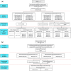
FIGURE 1
FIGURE 1
The flowchart of the study. (a) Experimental ICF with TMJ disc displacement. (b) Removal of the ICF fragment and TMJ disc.
FIGURE 2
FIGURE 2
Surgical procedures on left temporomandibular joint. (a) Exposure of disc (black arrow) and condylar head (white arrow). (b) An oblique osteotomy of the condylar head with anteriorly, inferiorly, and medially displaced fragment and disc.
FIGURE 3
FIGURE 3
The method for defining changes of disc status. Points D1 (D1′) and D2 (D2′) were the anterior and posterior points of the disc, respectively. Changes in disc status were defined as the mean distance between D1–D1′ and D2–D2′. (a) Diagram of a normal disc‐condyle relationship in the rabbit TMJ. (b) Diagram of anteriorly displaced fragment together with disc immediately after surgery in rabbit TMJ.
FIGURE 4
FIGURE 4
Masson's trichrome‐stained sections of rabbit condyles. A blank control in the adolescent group showing a normal disc‐condyle relationship in the rabbit TMJ (a) Immediately after ICF with anteriorly displaced disc (b) Red arrows: Articular disc; Black scale bar: 1.0 mm.
FIGURE 5
FIGURE 5
Changes in body weight in both adolescent and adult rabbits during the experimental period. *p < 0.01; Boxplot: Mean value; Error bar: Standard deviation.
FIGURE 6
FIGURE 6
Coronal and 3D Micro‐CT images of the adolescent and adult groups at 0, 4, 8, and 12 weeks after surgery. (a) In the adolescent group, the condyles show a gradual restoration of the condylar head during the experimental period. (b) In the adult group, the reformed condylar head was smaller and more irregular than that in the adolescent group. (c, d) Quantitative data showed no significant difference of BMD in the operated left condyle compared unoperated right condyle in both adolescent and adult groups at 12 weeks after surgery. White scale bar: 2.0 mm.
FIGURE 7
FIGURE 7
Histological evaluation of left rabbit joints in the adolescent and adult groups at 0, 4, 8, and 12 weeks after surgery. (a) In Masson's trichrome‐stained images of the adolescent group, the discs were still anteriorly displaced. The condyles show a gradual restoration of the condylar head during the experimental period. Red arrows: Articular disc; Black scale bar: 1.0 mm. (b) In Masson's trichrome‐stained images of the adult group, anteriorly displaced discs were still observed at 4, 8, and 12 weeks after surgery. The reformed condylar head was smaller and more irregular than that in the adolescent group. Red arrows: Articular disc; Black scale bar:1.0 mm. (c, d) Quantitative data analyses did not show any significant difference in the change of TMJ disc displacement status at 0, 4, 8, and 12 weeks after surgery in both adolescent and adult groups. Boxplot: Mean value; Error bar: Standard deviation.
FIGURE 8
FIGURE 8
Representative Safranin O staining images and SOX9 immunohistochemistry images in adolescent and adult groups at 0, 4, 8, and 12 weeks after surgery. (a, b) Representative Safranin O staining images of the condyles revealed reformation of the cartilage layers at 4, 8, and 12 weeks after surgery in the adolescent and adult groups. The area contained within each square is enlarged in the images on the right side. White scale bar: 1.0 mm; Black scale bar: 100 μm. (c, d) Representative safranin‐O‐stained images showed significantly decreased thickness of the pre‐hypertrophic and the hypertrophic layers at 8 and 12 weeks after surgery in the adolescent and adult groups, *p < 0.05.
FIGURE 9
FIGURE 9
Representative SOX9 immunohistochemical images in adolescent and adult groups at 4, 8, and 12 weeks after surgery. (a, b) Immunohistochemical data revealed expressions of SOX9 (a chondrocyte marker) in adolescent and adult reformed condylar heads at 4, 8, and 12 weeks after surgery. The area contained within each square is enlarged in the images on the right side. Red arrows labeled Sox9+ cells. White scale bar:1.0 mm; Black scale bar: 100 μm.
FIGURE 10
FIGURE 10
Representative Safranin O staining images and SOX9 immunohistochemistry images in adolescent and adult groups at 4 weeks following removal of TMJ disc and ICF fragment. (a) Representative Safranin O staining images revealed reformation of condylar cartilage layers at 4 weeks following removal of TMJ disc and ICF fragment in the adolescent group. The area contained within the black square is enlarged in the images on the right side. Lower panel images have a higher magnification of the white square box in upper left row image. Black asterisks = associated fibrous tissue attachment. White scale bar: 2.0 mm; Black scale bar: 200 μm. (b) Representative Safranin O staining images revealed reformation of condylar cartilage layers at 4 weeks following removal of TMJ disc and ICF fragment in the adult group. The area contained within the black square is enlarged in the images on the right side. Lower panel images have a higher magnification of the white square box in upper left row image. Black asterisks = associated fibrous tissue attachment. White scale bar: 2.0 mm; Black scale bar: 200 μm. (c) Immunohistochemical data revealed expressions of SOX9 (a chondrocyte marker) in reformed condylar heads at 4 weeks following removal of TMJ disc and ICF fragment of the adolescent and adult groups. The area contained within each black square is enlarged in the images on the right side. Red arrows labeled Sox9+ cells. White scale bar: 2.0 mm; Black scale bar: 200 μm.

Similar articles

Cited by

References

    1. Al‐Moraissi, E. A. , and Ellis E. 3rd. 2015. “Surgical Treatment of Adult Mandibular Condylar Fractures Provides Better Outcomes Than Closed Treatment: A Systematic Review and Meta‐Analysis.” Journal of Oral and Maxillofacial Surgery 73, no. 3: 482–493. 10.1016/j.joms.2014.09.027. - DOI - PubMed
    1. Bi, W. , Deng J. M., Zhang Z., Behringer R. R., and de Crombrugghe B.. 1999. “Sox9 is required for cartilage formation.” Nature Genetics 22, no. 1: 85–89. 10.1038/8792. - DOI - PubMed
    1. Bos, R. R. , Ward Booth R. P., and de Bont L. G.. 1999. “Mandibular condyle fractures: a consensus.” British Journal of Oral & Maxillofacial Surgery 37, no. 2: 87–89. 10.1054/bjom.1998.0014. - DOI - PubMed
    1. Cai, B. L. , Ren R., Yu H. B., Liu P. C., Shen S. G. F., and Shi J.. 2018. “Do Open Reduction and Internal Fixation With Articular Disc Anatomical Reduction and Rigid Anchorage Manifest a Promising Prospect in the Treatment of Intracapsular Fractures?” Journal of Oral and Maxillofacial Surgery 76, no. 5: 1026–1035. 10.1016/j.joms.2017.12.015. - DOI - PubMed
    1. Chrcanovic, B. R. 2012. “Open Versus Closed Reduction: Mandibular Condylar Fractures in Children.” Oral and Maxillofacial Surgery 16, no. 3: 245–255. 10.1007/s10006-012-0344-7. - DOI - PubMed

MeSH terms

Substances

LinkOut - more resources