. 2024 Dec 30;15(1):10727.
doi: 10.1038/s41467-024-54491-9.

Cas12e orthologs evolve variable structural elements to facilitate dsDNA cleavage

Affiliations

Cas12e orthologs evolve variable structural elements to facilitate dsDNA cleavage

Danyuan Li et al. Nat Commun. .

Abstract

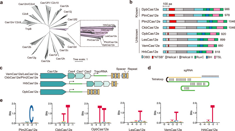
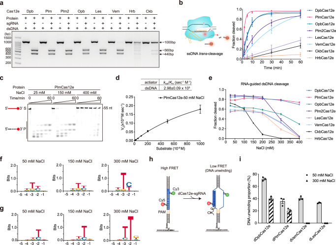
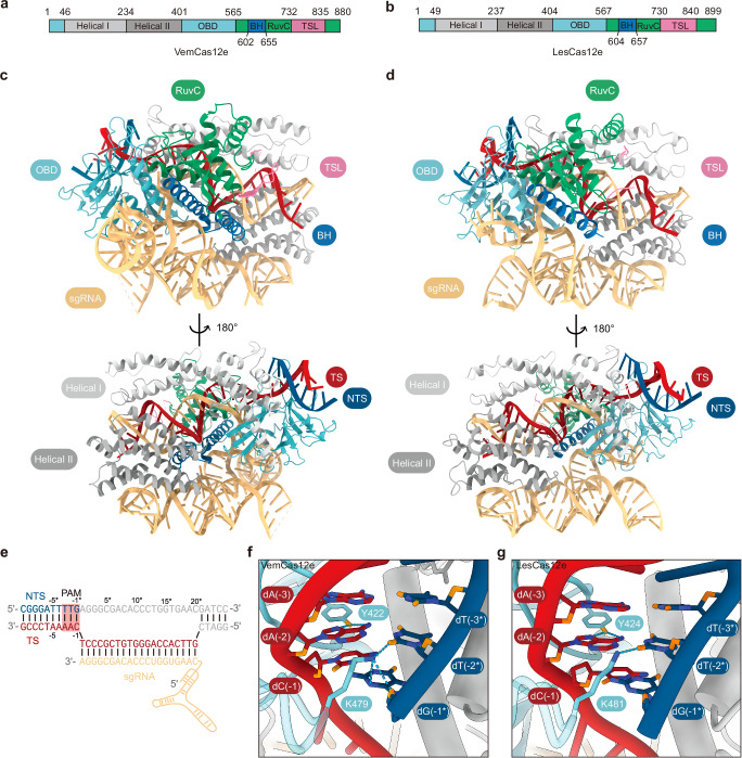
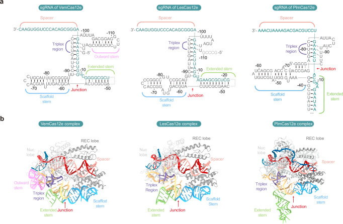
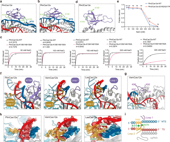
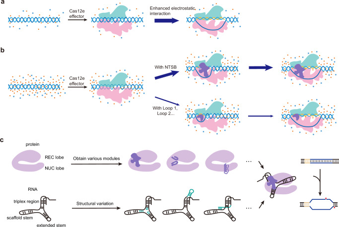
Exceptionally diverse type V CRISPR-Cas systems provide numerous RNA-guided nucleases as powerful tools for DNA manipulation. Two known Cas12e nucleases, DpbCas12e and PlmCas12e, are both effective in genome editing. However, many differences exist in their in vitro dsDNA cleavage activities, reflecting the diversity in Cas12e's enzymatic properties. To comprehensively understand the Cas12e family, we identify and characterize six unreported Cas12e members that vary in their CRISPR-locus architectures, PAM preferences, and cleavage efficacies. Interestingly, among all variants, PlmCas12e exhibits the most robust trans-cleavage activity and the lowest salt sensitivity in cis-cleavage. Further structural comparisons reveal that the unique NTSB domain in PlmCas12e is beneficial to DNA unwinding at high salt concentrations, while some NTSB-lacking Cas12e proteins rely on positively charged loops for dsDNA unwinding. These findings demonstrate how divergent evolution of structural elements shapes the nuclease diversity within the Cas12e family, potentially contributing to their adaptations to varying environmental conditions.


Conflict of interest statement

Competing interests: J.J.G.L., S.Z., and D.L. have submitted patent applications to the China patent office pertaining to the following aspects of this work: (1) DNA manipulating tools using CRISPR-LesCas12e, -VemCas12e, -HrbCas12e, and -CkbCas12e systems. Applicant: Tsinghua University. Status: Granted. Application number: 2022106204929 [P]. (2) DNA manipulating tools using CRISPR-OpbCas12e and -Plm2Cas12e systems. Applicant: Tsinghua University. Status: Filed. The remaining authors declare no competing interests.

Figures

Fig. 1
Fig. 1. Identification of phylogenetic branches of the CRISPR-Cas12e system.
a Phylogenetic analysis of Cas12e orthologs in this study and representative reported type V effectors. The enlarged dashed box illustrates Cas12e proteins positioned in different branches. b Size and domain comparisons of Cas12e effector proteins based on AlphaFold-predicted structures. c In-silico prediction of tracrRNA and crRNA based on base pairing between tracrRNA and crRNA across the CRISPR-Cas locus. Arrows indicate the predicted transcription start sites for tracrRNAs. d A single guide RNA (sgRNA) is obtained by fusing tracrRNA and crRNA with a tetraloop. e PAM screening results of Cas12e orthologs. Source data are provided as a Source Data file.
Fig. 2
Fig. 2. High salt concentration reduces the trans- and cis-cleavage activities of Cas12e orthologs.
a sgRNA-guided 1 kb-dsDNA cleavage assays for Cas12e orthologs. Similar results were obtained in three independent experiments. b Left, schematic of Cas12e (blue)-sgRNA (yellow) RNP complex cleaving target dsDNA (black), resulting in activation of the RuvC nuclease (denoted by a red dot) domain. This schematic is created in BioRender. Liu, J. (2023) BioRender.com/x40b358. Right, comparison of ssDNA trans-cleavage kinetics among Cas12e orthologs at a physiological salt concentration of 150 mM NaCl. Data are shown as mean ± SD from n = 3 independent experiments. c trans-cleavage of 3’-Cy5 labeled 55-nt ssDNA by PlmCas12e under gradients of different salt concentrations. Similar results were obtained in three independent experiments. d Michaelis-Menten kinetic studies of reporter trans-cleavage by PlmCas12e under 50 mM NaCl condition. Data are shown as mean ± SD from n = 3 independent experiments. e Comparison of dsDNA cis-cleavage efficacy among Cas12e orthologs under a gradient of salt concentrations in 0.5 h. Data are shown as mean ± SD from n = 3 independent experiments. Direct lines connecting different points are used to illustrate variations in efficiency. fg PAM screening results of PlmCas12e (f) and VemCas12e (g) at three salt concentrations. Source data are provided as a Source Data file. h Schematic of smFRET assay to quantify dsDNA unwinding with Cy3-labeling on NTS and Cy5-labeling on TS. i DNA unwinding proportion for different Cas12e orthologs after a 5-min incubation of DNA substrate and Cas12e-sgRNA complex. Data are shown as mean ± SEM from n = 3 technical replicates.
Fig. 3
Fig. 3. Cryo-EM structures of VemCas12e and LesCas12e ternary complexes.
a, b Domain architectures of VemCas12e (a) and LesCas12e (b). c, d Overall structures of dVemCas12e (c) and dLesCas12e (d) in complex with sgRNA and target dsDNA. Domains are colored identical to those in a and b. e Partially paired dsDNA target used in both VemCas12e (c) and LesCas12e (d) complexes. Structurally disordered nucleotides are colored gray. f, g PAM recognition patterns of VemCas12e (f ) and LesCas12e (g). Base-specific hydrogen-bonding interactions are indicated with blue dashed lines.
Fig. 4
Fig. 4. sgRNA architectures of VemCas12e and LesCas12e.
a Schematic of guide RNA structures of VemCas12e (left), LesCas12e (middle), and PlmCas12e (right, PDB: 7WB1). The junction connecting the triplex region and two stems is highlighted with a red arrow. The black line indicates the Watson-Crick base pair. The grey line indicates a Non-Watson-Crick base pair. The grey dashed lines indicate disordered regions. b Guide RNA structures of VemCas12e (left), LesCas12e (middle), and PlmCas12e (right, PDB: 7WB1) in ternary complexes. The structure of sgRNA is colored similarly as in a. NTS is colored in blue and TS is colored in red.
Fig. 5
Fig. 5. dsDNA melting mechanisms by NTSB-containing PlmCas12e and NTSB-lacking VemCas12e and LesCas12e.
a, b Positively charged residues involved in DNA duplex interactions in the PlmCas12e NTSB (a PDB: 7WB1) and PlmCas12e NTSB (b, PDB: 7WAZ). The blue dashed lines indicate hydrogen-bonding interactions. c Comparisons of cis-cleavage on dsDNA by wild-type (WT) PlmCas12e and lysine residues-mutated PlmCas12e at varying salt concentrations. The cleaved fraction for each reaction was fit into the One Phase Decay model. k value is the rate constant. Data are shown as mean ± SD from n = 3 independent experiments. d Positively charged residues at corresponding positions to K148 and K150 within the Plm2Cas12e NTSB (predicted). e dsDNA cleavage efficiencies by wild-type Plm2Cas12e and arginine-restored Plm2Cas12e mutant at different salt concentrations. Data are shown as mean ± SD from n = 3 independent experiments. Statistical analysis was performed using a two-way ANOVA test with Sidak’s multiple comparison test. (****p value < 0.0001; “ns” means not significant with the p value > 0.05). The exact p values for the comparison at 25, 50, 100,150, 200, 300, and 400 mM NaCl are 0.9704, 0.8939, 0.1051, 0.9125, 0.1220, <0.0001, and 0.9953, respectively. Source data are provided as a Source Data file. f Comparison of structural elements of NTSB-containing PlmCas12e (PDB: 7WB1) and NTSB-lacking VemCas12e and LesCas12e. NTSB and Loop 1 are colored in purple, and Loop 2 is colored in brown. g Positively charged Loop 1 of VemCas12e. Four lysine residues are denoted. h The electron density map of Loop 2 among three Cas12e orthologs. Residues interacting with dsDNA duplex are denoted. i Schematic diagram showing different structural elements responsible for dsDNA invading in VemCas12e.
Fig. 6
Fig. 6. Modulation of the dsDNA unwinding by Cas12e orthologs at low and high NaCl concentrations.
a Cas12e effector medicated R-loop formation of the dsDNA duplex at low salt concentration. Orange dot, positively charged ion; blue dot, negatively charged ion. Green module, REC lobe; pink module, NUC lobe. b dsDNA unwinding and complete R-loop formation performed by NTSB-containing Cas12e and NTSB-lacking Cas12e at high salt concentration. Purple module, NTSB domain or loops. The thickness of the blue arrowed lines represents the likelihood of reaction occurrence. c The diversities of Cas12e and its cognate sgRNA. Different elements colored dark purple were attained based on the bilobed configuration. The sgRNA scaffolds (in black) with varying details in base pairing (colored green) indicate various sgRNAs. The PAM region on the dsDNA target is colored in yellow. Red triangles indicate the cleavage.

Similar articles

Cited by

References

    1. Makarova, K. S. et al. Evolutionary classification of CRISPR–Cas systems: a burst of class 2 and derived variants. Nat. Rev. Microbiol.18, 67–83 (2020). - PMC - PubMed
    1. Pausch, P. et al. CRISPR-CasΦ from huge phages is a hypercompact genome editor. Science369, 333–337 (2020). - PMC - PubMed
    1. Al-Shayeb, B. et al. Diverse virus-encoded CRISPR-Cas systems include streamlined genome editors. Cell185, 4574–4586. e4516 (2022). - PubMed
    1. Barrangou, R. et al. CRISPR provides acquired resistance against viruses in prokaryotes. Science315, 1709–1712 (2007). - PubMed
    1. Sapranauskas, R. et al. The Streptococcus thermophilus CRISPR/Cas system provides immunity in Escherichia coli. Nucleic Acids Res.39, 9275–9282 (2011). - PMC - PubMed

Publication types

LinkOut - more resources