. 2025 Dec;30(1):2435252.
doi: 10.1080/13510002.2024.2435252. Epub 2024 Dec 31.

MEGF9 prevents lipopolysaccharide-induced cardiac dysfunction through activating AMPK pathway

Affiliations

MEGF9 prevents lipopolysaccharide-induced cardiac dysfunction through activating AMPK pathway

Zhili Jin et al. Redox Rep. 2025 Dec.

Abstract

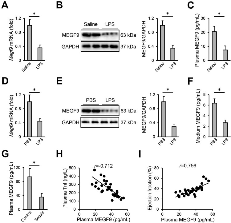
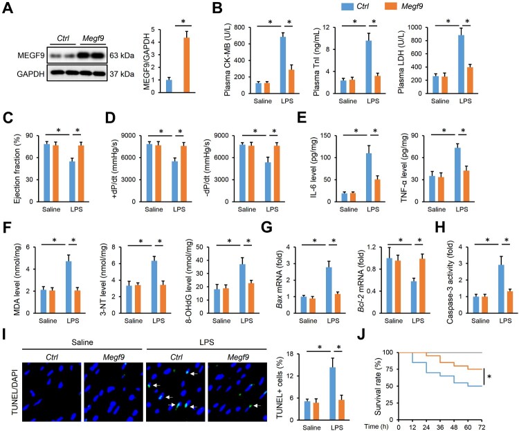
Objective: Inflammation and oxidative damage play critical roles in the pathogenesis of sepsis-induced cardiac dysfunction. Multiple EGF-like domains 9 (MEGF9) is essential for cell homeostasis; however, its role and mechanism in sepsis-induced cardiac injury and impairment remain unclear.

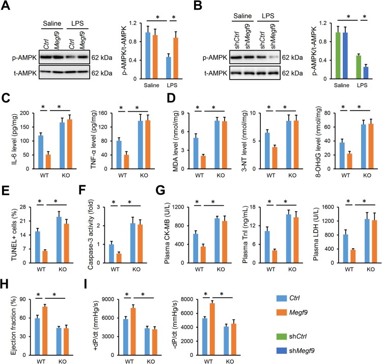
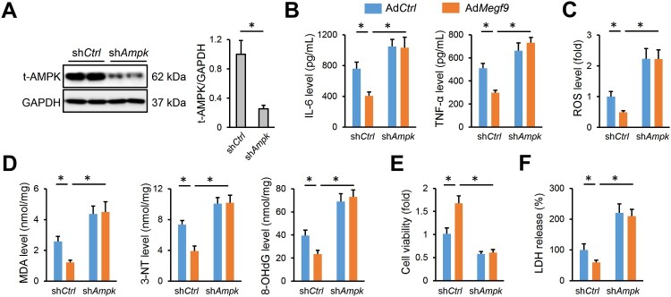
Methods: Adenoviral and adeno-associated viral vectors were applied to overexpress or knock down the expression of MEGF9 in vivo and in vitro. To stimulate septic injury, cardiomyocytes and mice were treated lipopolysaccharide (LPS). To clarify the necessity of AMP-activated protein kinase (AMPK), global AMPK knockout mice were used.

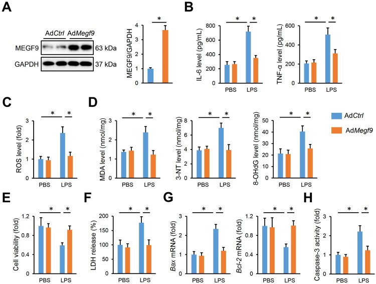
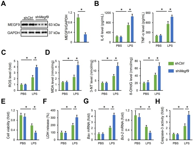
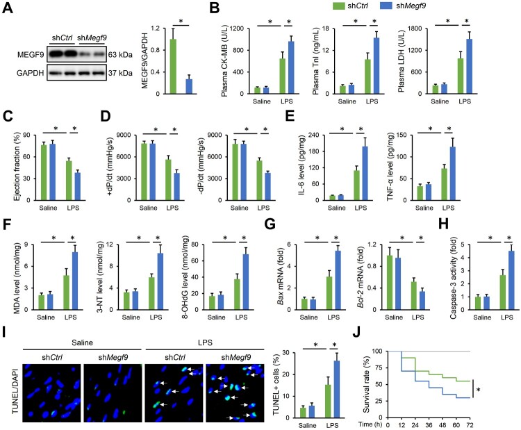
Results: We found that MEGF9 expressions were reduced in cardiomyocytes and mice by LPS stimulation. Compared with negative controls, plasma MEGF9 levels were also decreased in septic patients, and negatively correlated with LPS-induced cardiac dysfunction. In addition, MEGF9 overexpression attenuated, while MEGF9 knockdown aggravated LPS-induced inflammation and oxidative damage in vivo and in vitro, thereby regulating LPS-induced cardiac injury and impairment. Mechanistic studies revealed that MEGF9 overexpression alleviated LPS-induced cardiac dysfunction through activating AMPK pathway.

Conclusion: We for the first time demonstrate that MEGF9 prevents LPS-related inflammation, oxidative damage and cardiac injury through activating AMPK pathway, and provide a proof-of-concept for the treatment of LPS-induced cardiac dysfunction by targeting MEGF9.

Keywords: AMPK; LPS-induced cardiac dysfunction; inflammation; oxidative damage.


Conflict of interest statement

No potential conflict of interest was reported by the author(s).

Figures

Figure 1.
Figure 1.
MEGF9 expression is decreased after LPS stimulation and negatively correlates with sepsis-induced cardiac dysfunction. (A) The mRNA level of MEGF9 in the myocardium from saline- or LPS-treated mice (n = 6). (B) The protein level of MEGF9 in the myocardium from saline- or LPS-treated mice (n = 6). (C) Plasma MEGF9 level in saline- or LPS-treated mice (n = 6). (D) The mRNA level of MEGF9 in NRVMs treated with PBS or LPS (n = 6). (E) The protein level of MEGF9 in NRVMs treated with PBS or LPS (n = 6). (F) MEGF9 level in the medium from PBS- or LPS-treated NRVMs (n = 6). (G) Plasma MEGF9 level in septic patients or negative controls (n = 30). (H) The correlation analysis of plasma MEGF9 level and TnI level in septic patients (n = 30). (I) The correlation analysis of plasma MEGF9 level and ejection fraction in septic patients (n = 30). Results were expressed as means ± S.D., and P < 0.05 was considered to be statistically significant.
Figure 2.
Figure 2.
MEGF9 overexpression attenuates LPS-induced inflammation and oxidative damage in cardiomyocytes. (A) The protein level of MEGF9 in AdCtrl- or AdMegf9-infected NRVMs (n = 6). (B) The levels of IL-6 and TNF-α in the medium from AdCtrl- or AdMegf9-infected NRVMs with or without LPS stimulation (n = 6). (C) ROS generation in AdCtrl- or AdMegf9-infected NRVMs with or without LPS stimulation (n = 6). (D) The levels of MDA, 3-NT and 8-OHdG in AdCtrl- or AdMegf9-infected NRVMs with or without LPS stimulation (n = 6). (E) The viability of AdCtrl- or AdMegf9-infected NRVMs with or without LPS stimulation (n = 6). (F) LDH release to the medium from AdCtrl- or AdMegf9-infected NRVMs with or without LPS stimulation (n = 6). (G) The mRNA levels of Bax and Bcl-2 in AdCtrl- or AdMegf9-infected NRVMs with or without LPS stimulation (n = 6). (H) The activity of caspase-3 in AdCtrl- or AdMegf9-infected NRVMs with or without LPS stimulation (n = 6). Results were expressed as means ± S.D., and P < 0.05 was considered to be statistically significant.
Figure 3.
Figure 3.
MEGF9 knockdown aggravates LPS-induced inflammation and oxidative damage in cardiomyocytes. (A) The protein level of MEGF9 in shCtrl- or shMegf9-infected NRVMs (n = 6). (B) The levels of IL-6 and TNF-α in the medium from shCtrl- or shMegf9-infected NRVMs with or without LPS stimulation (n = 6). (C) ROS generation in shCtrl- or shMegf9-infected NRVMs with or without LPS stimulation (n = 6). (D) The levels of MDA, 3-NT and 8-OHdG in shCtrl- or shMegf9-infected NRVMs with or without LPS stimulation (n = 6). (E) The viability of shCtrl- or shMegf9-infected NRVMs with or without LPS stimulation (n = 6). (F) LDH release to the medium from shCtrl- or shMegf9-infected NRVMs with or without LPS stimulation (n = 6). (G) The mRNA levels of Bax and Bcl-2 in shCtrl- or shMegf9-infected NRVMs with or without LPS stimulation (n = 6). (H) The activity of caspase-3 in shCtrl- or shMegf9-infected NRVMs with or without LPS stimulation (n = 6). Results were expressed as means ± S.D., and P < 0.05 was considered to be statistically significant.
Figure 4.
Figure 4.
Cardiac-specific MEGF9 knockdown exacerbates LPS-induced inflammation, oxidative damage and cardiac dysfunction in mice. (A) The protein level of MEGF9 in shCtrl- or shMegf9-infected hearts (n = 6). (B) Plasma levels of CK-MB, TnI and LDH in shCtrl- or shMegf9-infected mice with or without LPS stimulation (n = 6). (C–D) Cardiac function was evaluated by echocardiographic and hemodynamic analysis (n = 6). (E) Plasma levels of IL-6 and TNF-α in shCtrl- or shMegf9-infected mice with or without LPS stimulation (n = 6). (F) The levels of MDA, 3-NT and 8-OHdG in shCtrl- or shMegf9-infected hearts with or without LPS stimulation (n = 6). (G) The mRNA levels of Bax and Bcl-2 in shCtrl- or shMegf9-infected hearts with or without LPS stimulation (n = 6). (H) The activity of caspase-3 in shCtrl- or shMegf9-infected hearts with or without LPS stimulation (n = 6). (I) Representative images and statistical analysis of TUNEL staining (n = 6). (J) The survival rate in mice treated with a lethal dose of LPS (n = 20). Results were expressed as means ± S.D., and P < 0.05 was considered to be statistically significant.
Figure 5.
Figure 5.
Cardiac-specific MEGF9 overexpression prevents LPS-induced inflammation, oxidative damage and cardiac dysfunction in mice. (A) The protein level of MEGF9 in Ctrl- or Megf9-infected hearts (n = 6). (B) Plasma levels of CK-MB, TnI and LDH in Ctrl- or Megf9-infected mice with or without LPS stimulation (n = 6). (C–D) Cardiac function was evaluated by echocardiographic and hemodynamic analysis (n = 6). (E) Plasma levels of IL-6 and TNF-α in Ctrl- or Megf9-infected mice with or without LPS stimulation (n = 6). (F) The levels of MDA, 3-NT and 8-OHdG in Ctrl- or Megf9-infected hearts with or without LPS stimulation (n = 6). (G) The mRNA levels of Bax and Bcl-2 in Ctrl- or Megf9-infected hearts with or without LPS stimulation (n = 6). (H) The activity of caspase-3 in Ctrl- or Megf9-infected hearts with or without LPS stimulation (n = 6). (I) Representative images and statistical analysis of TUNEL staining (n = 6). (J) The survival rate in mice treated with a lethal dose of LPS (n = 20). Results were expressed as means ± S.D., and P < 0.05 was considered to be statistically significant.
Figure 6.
Figure 6.
MEGF9 overexpression alleviates LPS-induced cardiac dysfunction through activating AMPK pathway in vivo. (A) The protein levels of p-AMPK and t-AMPK in Ctrl- or Megf9-infected hearts with or without LPS stimulation (n = 6). (B) The protein levels of p-AMPK and t-AMPK in shCtrl- or shMegf9-infected hearts with or without LPS stimulation (n = 6). (C) Plasma levels of IL-6 and TNF-α in shCtrl- or shMegf9-infected mice with or without AMPK deficiency in response to LPS stimulation (n = 6). (D) The levels of MDA, 3-NT and 8-OHdG in shCtrl- or shMegf9-infected hearts with or without AMPK deficiency in response to LPS stimulation (n = 6). (E) Statistical analysis of TUNEL staining (n = 6). (F) The activity of caspase-3 in shCtrl- or shMegf9-infected hearts with or without AMPK deficiency in response to LPS stimulation (n = 6). (G) Plasma levels of CK-MB, TnI and LDH in shCtrl- or shMegf9-infected mice with or without AMPK deficiency in response to LPS stimulation (n = 6). (H–I) Cardiac function was evaluated by echocardiographic and hemodynamic analysis (n = 6). Results were expressed as means ± S.D., and P < 0.05 was considered to be statistically significant.
Figure 7.
Figure 7.
MEGF9 overexpression alleviates LPS-induced cardiac dysfunction through activating AMPK pathway in vitro. (A) The protein level of t-AMPK in shCtrl- or shAmpk-infected NRVMs (n = 6). (B) The levels of IL-6 and TNF-α in the medium from AdCtrl- or AdMegf9-infected NRVMs with or without AMPK knockdown in response to LPS stimulation (n = 6). (C) ROS generation in shCtrl- or shMegf9-infected NRVMs with or without AMPK knockdown in response to LPS stimulation (n = 6). (D) The levels of MDA, 3-NT and 8-OHdG in shCtrl- or shMegf9-infected NRVMs with or without AMPK knockdown in response to LPS stimulation (n = 6). (E) The viability of shCtrl- or shMegf9-infected NRVMs with or without AMPK knockdown in response to LPS stimulation (n = 6). (F) LDH release to the medium from shCtrl- or shMegf9-infected NRVMs with or without AMPK knockdown in response to LPS stimulation (n = 6). Results were expressed as means ± S.D., and P < 0.05 was considered to be statistically significant.

Similar articles

Cited by

  • miR-200c inhibition and catalase accelerate diabetic wound healing.
    D'Agostino M, Sileno S, Lulli D, De Luca N, Scarponi C, Teson M, Torcinaro A, De Santa F, Cirielli C, Furgiuele S, Morrell CH, Dellambra E, Odorisio T, Lakatta EG, Avitabile D, Capogrossi MC, Magenta A. D'Agostino M, et al. J Biomed Sci. 2025 Feb 14;32(1):21. doi: 10.1186/s12929-024-01113-7. J Biomed Sci. 2025. PMID: 39948670 Free PMC article.

References

    1. Landesberg G, Gilon D, Meroz Y, et al. . Diastolic dysfunction and mortality in severe sepsis and septic shock. Eur Heart J. 2012;33(7):895–903. doi:10.1093/eurheartj/ehr351 - DOI - PMC - PubMed
    1. Parrillo JE, Parker MM, Natanson C, et al. . Septic shock in humans. Advances in the understanding of pathogenesis, cardiovascular dysfunction, and therapy. Ann Intern Med. 1990;113(3):227–242. doi:10.7326/0003-4819-113-3-227 - DOI - PubMed
    1. Wang R, Xu Y, Fang Y, et al. . Pathogenetic mechanisms of septic cardiomyopathy. J Cell Physiol. 2022;237(1):49–58. doi:10.1002/jcp.30527 - DOI - PubMed
    1. Carbone F, Liberale L, Preda A, et al. . Septic cardiomyopathy: from pathophysiology to the clinical setting. Cells. 2022;11(18):2833. doi:10.3390/cells11182833 - DOI - PMC - PubMed
    1. Zhu XX, Wang X, Jiao SY, et al. . Cardiomyocyte peroxisome proliferator-activated receptor alpha prevents septic cardiomyopathy via improving mitochondrial function. Acta Pharmacol Sin. 2023;44(11):2184–2200. doi:10.1038/s41401-023-01107-5 - DOI - PMC - PubMed

LinkOut - more resources