. 2024 Dec 30;15(1):10738.
doi: 10.1038/s41467-024-54914-7.

Design of an equilibrative nucleoside transporter subtype 1 inhibitor for pain relief

Affiliations

Design of an equilibrative nucleoside transporter subtype 1 inhibitor for pain relief

Nicholas J Wright et al. Nat Commun. .

Abstract

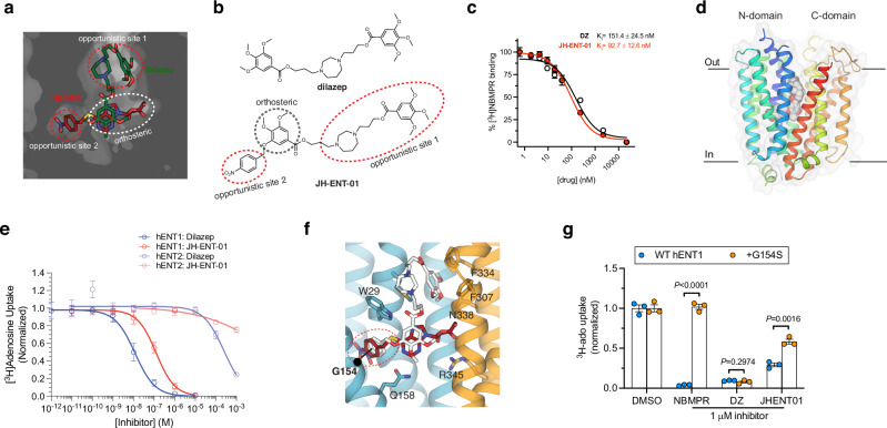
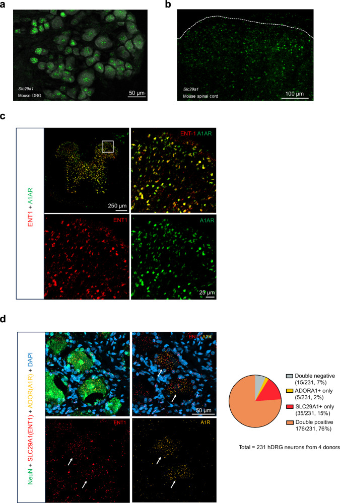
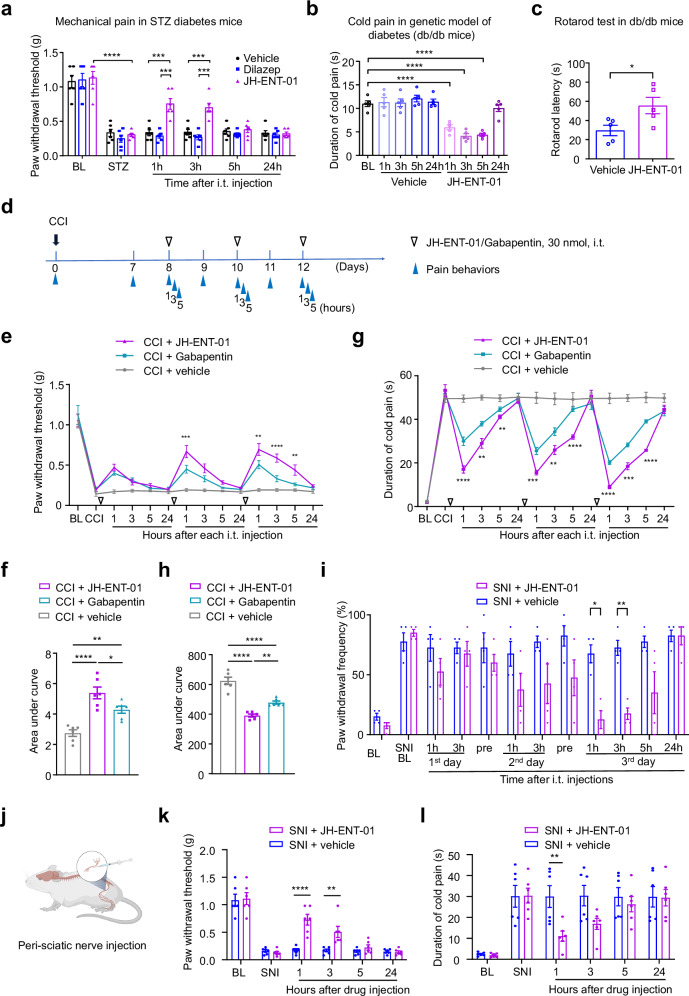
The current opioid crisis urgently calls for developing non-addictive pain medications. Progress has been slow, highlighting the need to uncover targets with unique mechanisms of action. Extracellular adenosine alleviates pain by activating the adenosine A1 receptor (A1R). However, efforts to develop A1R agonists have faced obstacles. The equilibrative nucleoside transporter subtype 1 (ENT1) plays a crucial role in regulating adenosine levels across cell membranes. We postulate that ENT1 inhibition may enhance extracellular adenosine levels, potentiating endogenous adenosine action at A1R and leading to analgesic effects. Here, we modify the ENT1 inhibitor dilazep based on its complex X-ray structure and show that this modified inhibitor reduces neuropathic and inflammatory pain in animal models while dilazep does not. Notably, our ENT1 inhibitor surpasses gabapentin in analgesic efficacy in a neuropathic pain model. Additionally, our inhibitor exhibits less cardiac side effect than dilazep via systemic administration and shows no side effects via local/intrathecal administration. ENT1 is colocalized with A1R in mouse and human dorsal root ganglia, and the analgesic effect of our inhibitor is linked to A1R. Our studies reveal ENT1 as a therapeutic target for analgesia, highlighting the promise of rationally designed ENT1 inhibitors for non-opioid pain medications.


Conflict of interest statement

Competing interests: N.J.W., J.H., R.-R.J., and S.-Y.L. are inventors of the patent application that was filed (028193-0022-WO01). The rest of the authors declare no competing interests.

Figures

Fig. 1
Fig. 1. Structure-based ENT1 inhibitor design.
a Structural overlay of NBMPR and dilazep in the central cavity of hENT1cryst, with shared and distinct inhibitor binding sites labeled. b Chemical structure of dilazep and the rationally designed adenosine reuptake inhibitor, JH-ENT-01. The new inhibitor contains chemical moieties analogous to features of both dilazep and NBMPR. c Inhibitory constants (Ki) from cold-competition displacement of [3H]-NBMPR from hENT1cryst by dilazep of JH-ENT-01 (scintillation proximity assay; n = 6 technical replicates across biological duplicates; mean ± s.e.m. shown). d X-ray crystal structure of JH-ENT-01 complexed to hENT1cryst. e hENT1 and hENT2 dependent [3H]-adenosine (ado) uptake and their inhibition by dilazep and JH-ENT-01 (n = 2–3 biological replicate titrations with technical triplicates, mean ± s.e.m. shown). f Structural overlay of JH-ENT-01 and NBMPR shows a difference of para-nitrobenzyl group binding poses within opportunistic site 2. g Comparison of [3H]-adenosine uptake block in oocytes expressing WT or G154S hENT1 by NBMPR, dilazep, or JH-ENT-01 (n = 3 biological replicates, mean ± s.e.m.; P values indicated from non-paired parametric t-test).
Fig. 2
Fig. 2. ENT1 and A1R expression in DRG and spinal cord neurons.
a, b RNA scope in situ hybridization showing ENT1 transcript (SLC29A1) expression in the DRG (a) and spinal cord dorsal horn (b) of mice. The dorsal horn top edge is indicated by the dotted line. Scales, 50 μm in a and 100 μm in b. c Double immunostaining showing co-localization of ENT1 and A1R in the dorsal horn and ventral horn (box size, 250 × 250 μm). Right panels, enlarged images from the boxes. Scale, 25 μm. Experiments in ac were repeated 2–3 times. d Left, RNAscope in situ hybridization images showing co-localization of SLC29A1 and ADOR1 (A1R) in human DRG (hDRG) neurons. The same DRG sections were co-stained with NeuN antibody (neuronal marker) and DAPI (nuclei marker). Arrows indicate neurons that are positive for both ENT1 and A1R. Scale, 50 μm. Right, quantification showing the ratio and percentage of SLC29A1 or/and ADOR1 positive and negative DRG neurons from four human donors.
Fig. 3
Fig. 3. Effects of ENT1 inhibitors on cardiovascular function.
a, b Effects of intravenous (i.v.) injection of dilazep and JH-ENT-01 (30 mg/kg) on heart rates in ECG test, with representative ECG traces before the treatment and at 10 min, 30 min, and 60 min after the vehicle and drug treatment in a, quantification of heart rates as beats per min in b (n = 6 animals from both sexes). Two-way ANOVA followed by Bonferroni posthoc comparison. *P < 0.05, **P < 0.01, ***P < 0.001; ns, not significant. Figure 3b was created in BioRender. He, W. (2024) BioRender.com/f39n75. c, d Effects of intraperitoneal (i.p.) injection of dilazep and JH-ENT-01 (10 and 30 mg/kg) on heart rates in ECG test, with representative ECG traces before the treatment and at 10 min, 30 min, and 60 min after the vehicle and drug treatment in c, and quantification of heart rates as beats per min in d (n = 3 animals from both sexes). All data were expressed as mean ± s.e.m. Figure 3d was created in BioRender. He, W. (2024) BioRender.com/o52y585.
Fig. 4
Fig. 4. JH-ENT-01 reduces formalin-induced acute inflammatory pain.
ac Effects of intrathecal (i.t.) injection of the ENT1 inhibitors JH-ENT-01 (1, 10, and 100 nmols) and dilazep (100 nmol) on formalin-induced inflammatory pain in phase 1 (0–10 min) and phase 2 (10–45 min) in male mice (a), female mice (b), and males and females combined (c). Top panels show the time course of formalin-induced pain. n = 4 per sex; n = 8 for both sexes. The inhibitors were given 30 min prior to the intraplantar injection of formalin (5%, 20 μl). d Rotarod tests before and 60 min after i.t. delivery of 100 nmol of JH-ENT-01 and dilazep. n = 6 animals per group with mixed sexes. Two-way ANOVA followed by Bonferroni posthoc comparison. *P < 0.05, **P < 0.01, ***P < 0.001, ****P < 0.0001; ns, not significant. All data were expressed as mean ± s.e.m.
Fig. 5
Fig. 5. Diverse routes of JH-ENT-01 administration effectively reduce CFA-induced persistent inflammatory pain.
ac Effects of intravenous injections of JH-ENT-01 (10 mg/kg, i.v.). a Schematic of drug injections and behavioral testing with PWT shown in b and PWF in c. n = 6 mice/group, equal number male and female). Figure 5a was created in BioRender. He, W. (2024) BioRender.com/f39n675. df Effects of subcutaneous injections of JH-ENT-01 (10 mg/kg, s.c.). d Schematic of drug injections and behavioral testing. Figure 5d was created in BioRender. He, W. (2024) BioRender.com/f05p763. e, f PWT shown in e and PWF in f. n = 6 mice/group equal number male and female mice. gi Effects of intraplantar injections of JH-ENT-01 (10 μg). g Schematic of drug injections and behavioral testing in the CFA model. Created in BioRender. He, W. (2024) BioRender.com/j51a415. h, i PWT (h) and PWF (i) in the CFA model. n = 6 mice/group with equal number male and female mice. Two-way ANOVA followed by Bonferroni posthoc comparison (b, c, e, f, h, i). *P < 0.05, **P < 0.01, ***P < 0.001. All data were expressed as Mean ± SEM. BL baseline; CFA complete Freund’s adjuvant; PWF paw withdrawal frequency; PWT paw withdrawal threshold.
Fig. 6
Fig. 6. Effects of ENT1 inhibitors on neuropathic pain in different models.
a Mechanical pain (PWT) after intrathecal (i.t., 10 nmol) injections of dilazep (n = 6), JH-ENT-01 (n = 6), and vehicle control (n = 6) in mice with STZ-induced diabetic neuropathy. b Cold pain in acetone test after intrathecal (i.t., 10 nmol) injections of JH-ENT-01 (n = 5) and vehicle control (n = 5) in db/db mice. c Rotarod test showing improved motor function by JH-ENT-01 at 3 hours after the same treatment as shown in b. d, schematic of drug injection and behavioral testing for CCI-induced neuropathic pain and comparison of JH-ENT-01 with gabapentin (30 nmol, i.t.). e, g PWT (e) and cold pain (g) after i.t. injection of JH-ENT-01 and gabapentin. f, h AUC analysis of e, g. (n = 6 mice with equal number male and female; e, g **P < 0.01, ***P < 0.001, **** P < 0.0001, gabapentin vs. JH-ENT-01; f, h *P < 0.05, **P < 0.01, ****P < 0.001, as indicated. i Effects of intrathecal injections (i.t., 30 nmol) of JH-ENT-01 (n = 4) and vehicle (n = 4) in the SNI model. jl Effects of peri-sciatic injection (j) of JH-ENT-01 (10 nmol) on SNI-induced mechanical pain (k) and cold pain (l). Figure 6j was created in BioRender. He, W. (2024) BioRender.com/a70b516. **P < 0.01, **** P < 0.0001. Two-way ANOVA followed by post-hoc comparison (a, e, g, i, k, l). One-way ANOVA followed by post-hoc comparison (b, f, h); Student’s t test (c, two-sided). Both males and females are included in each group. BL baseline; CCI chronic constriction injury; SNI spared nerve injury; STZ streptozotocin; PWF paw withdrawal frequency; PWT paw withdrawal threshold. All data were expressed as mean ± s.e.m.
Fig. 7
Fig. 7. Pharmacokinetic properties of ENT1 inhibitors following subcutaneous administration.
a PWT after subcutaneous (s.c.) injections of dilazep (n = 9, 5 males and 4 females, 3 × 30 mg/kg) and JH-ENT-01 (n = 7, 5 males and 2 females, 3 × 30 mg/kg) in mice with STZ-induced diabetic neuropathy. Arrows indicate three s.c.. injections on days 8, 10, and 12. Note that all mice survived after the s.c. injections. Two-way ANOVA followed by Bonferroni posthoc comparison (P = 0.027 for overall comparison between JH-ENT-01 and dilazep. * P < 0.05, JH-ENT-01 vs. dilazep at 1 h after 3rd injection). b Pharmacokinetic profile of JH-ENT-01 and dilazep in plasma, following a single s.c. injection of JH-ENT-01 (30 mg/kg) and equimolar amount of dilazep (n = 3 male mice per time point). All data were expressed as mean ± s.e.m.
Fig. 8
Fig. 8. The antinociceptive effects of JH-ENT-01 depend on A1R.
ac Patch clamp recordings showing the effects of JH-ENT-01 on synaptic transmission (sEPSCs) in spinal cord slices of STZ-treated in mice at 1 week. a Schematic of patch clamp recordings in spinal cord slice. Created in BioRender. He, W. (2024) BioRender.com/d34r338. b sEPSC traces before, during, and after the inhibitor perfusion (3 μM, 3 min). The lower traces are shorter segments of the upper trace, marked as 1 (before the perfusion) and 2 (after perfusion). c Quantification of sEPSC frequency (top) and amplitude (bottom) before and after JH-ENT-01 treatment (n = 13 neurons from 3 male mice, paired Student’s t test, two-sided). d, e Reversal of JH-ENT-01’s analgesia by A1R antagonist DPCPX in STZ-treated mice (d) and CFA-treated mice (e) via intrathecal route. DPCPX (10 nmol) was given 30 min prior to the injection of ENT1 inhibitor (10 nmol). f The reversal of JH-ENT-01’s analgesia on the CFA-induced inflammatory pain is observed only with the A1R-specific antagonist DPCPX (10 nmol), not the A2AR-specific inhibitor ZM241385 (10 nmol), the A2BR-specific inhibitor PSB603 (10 nmol), and the A3R-specific inhibitor MRS3777 (10 nmol). *P < 0.05, **P < 0.01, ***P < 0.001, ****P < 0.0001. Two-Way ANOVA followed by Bonferroni posthoc comparison. n = 6 males. g sEPSC traces before, during, and after the A1R antagonist DPCPX perfusion (1 μM, green) and DPCPX together with JH-ENT-01 (3 μM, blue). h Quantification of sEPSC frequency (left) and amplitude (right) of g (n = 16 neurons from 3 mice–2 males and 1 female; paired Student’s t test, two-sided). All data were expressed as mean ± s.e.m.

Similar articles

References

    1. Sawynok, J. Adenosine and ATP receptors. Handb. Exp. Pharmacol. 309–328 (2007). - PubMed
    1. Sawynok, J. Adenosine receptor targets for pain. Neuroscience338, 1–18 (2016). - PubMed
    1. Sawynok, J. & Sweeney, M. I. The role of purines in nociception. Neuroscience32, 557–569 (1989). - PubMed
    1. Vincenzi, F., Pasquini, S., Borea, P. A. & Varani, K. Targeting adenosine receptors: a potential pharmacological avenue for acute and chronic pain. Int. J. Mol. Sci.21, 8710 (2020). - PMC - PubMed
    1. Zylka, M. J. Pain-relieving prospects for adenosine receptors and ectonucleotidases. Trends Mol. Med.17, 188–196 (2011). - PMC - PubMed

Publication types

MeSH terms

Associated data

LinkOut - more resources