. 2024 Dec 30;15(1):10768.
doi: 10.1038/s41467-024-54934-3.

A single mutation in dairy cow-associated H5N1 viruses increases receptor binding breadth

Affiliations

A single mutation in dairy cow-associated H5N1 viruses increases receptor binding breadth

Marina R Good et al. Nat Commun. .

Abstract

Clade 2.3.4.4b H5N1 is causing an unprecedented outbreak in dairy cows in the United States. To understand if recent H5N1 viruses are changing their receptor use, we screened recombinant hemagglutinin (HA) from historical and recent 2.3.4.4b H5N1 viruses for binding to distinct glycans bearing terminal sialic acids using a glycan microarray. We find that H5 from A/Texas/37/2024, an isolate from the dairy cow outbreak, has increased binding breadth to core glycans bearing terminal α2,3 sialic acids, the avian receptor, compared to historical and recent 2.3.4.4b H5N1 viruses. We do not observe any binding to α2,6 sialic acids, the receptor used by human seasonal influenza viruses. Using molecular dynamics and a cryo-EM structure of A/Texas/37/2024 H5, we show A/Texas/37/2024 H5 is more flexible within the receptor-binding site compared to a 2.3.4.4b H5 from 2022. We identify a single mutation outside of the receptor binding site, T199I, is responsible for increased binding breadth, as it increases receptor binding site flexibility. Together, these data show recent H5N1 viruses are evolving increased receptor binding breadth which could impact the host range and cell types infected with H5N1.


Conflict of interest statement

Competing interests: The authors declare no competing interests.

Figures

Fig. 1
Fig. 1. Phylogenetic tree of highly pathogenic avian H5N1.
The Neighbor-Joining (NJ) phylogenetic analysis of 94 hemagglutinin gene sequences. Clade 2.3.4.4 is subdivided into distinct subclades, including 2.3.4.4e, 2.3.4.4 h, 2.3.4.4 g, 2.3.4.4c, and the currently dominant 2.3.4.4b clade. The new group belonging to clade 2.3.4.4b includes strains isolated from domestic dairy cows and humans and animals linked to H5N1-positive dairy farms (highlighted with a blue region and animal symbols) in the United States in 2024. Distinct clades of the virus are labeled on the right side of the figure. Tips are labeled with H5Nx strain names, host species, and isolation dates. Nodes represent inferred common ancestors of the grouped tips. Branch lengths are proportional to the number of nucleotide substitutions per site, indicating the divergence between nodes. Sequences used for analysis are in Supplementary Data 1. Symbols depicting various host animals was in part created in BioRender. Ji, W. (2024) https://BioRender.com/f70l446.
Fig. 2
Fig. 2. A dairy cow associated H5N1 virus exhibits increased glycan binding breadth.
A rH5 binding to distinct Neu5Ac glycans. Green checkmarks indicate a positive binding result for the corresponding glycan. Normalized relative fluorescence unit (RFU) values ± standard deviation are indicated below checkmarks. Value above each glycan indicates the glycan number in the array. Symbols represent mannose (man), N-acetylglucosamine (GlcNAc), galactose (Gal), L-Fucose (L-Fuc), and N-acetylneuraminic acid (Neu5Ac). Glycans studied are in Supplementary Data 2. Source data are provided in the source data file. BD MD simulations of A/Colorado/18/2022 and A/Texas/37/2024 to characterize the LSTa binding site properties. B Representative structure obtained from MD simulations showing interactions of LSTa (burgundy) with A/Colorado/18/2022 and A/Texas/37/2024 in solution. C Conformational states of A/Colorado/18/2022 and A/Texas/37/2024 binding to LSTa. Each shade of blue represents a distinct conformation. Molecular coordinates are provided in Supplementary Data 3. D Residue-wise B-factor, as a measure of flexibility, mapped on the respective A/Colorado/18/2022 and A/Texas/37/2024 structure.
Fig. 3
Fig. 3. Cryo-EM structure of A/Texas/37/2024 HA in complex with LSTa and CR9114.
A Cryo-EM structure of rH5 from A/Texas/37/2024 bound with the sialic acid and first galactose-2 of LSTa (magenta) and CR9114 (gray). Each HA protomer is a distinct shade of blue. B Ribbon structure of rH5 from A/Texas/37/2024 highlighting the major H5 contacts with LSTa (maroon). C HA contacts and interactions with LSTa.
Fig. 4
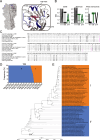
Fig. 4. 2.3.4.4b H5N1 viruses in the Americas recently acquired T199I.
A Structural depiction on A/Texas/37/2024 of the RBS and recent mutations. Blue residues indicate those found within the 130-loop, 190-helix, or 220-loop. Red residues indicate mutations of interest. B Logo plots of positions 111, 199, and 214 based on geographical location. The Americas logo plot does not include sequences from the dairy cow outbreak. C Amino acid alignment of HA1 from H5N1 viruses in this study. Residues in magenta are positions, 111, 199, and 214. D Frequency of T199 (blue) and I199 (orange) in circulating 2.3.4.4b H5N1 viruses in the Americas, including the dairy cow outbreak, between March 2022 and May 2024. Source data are provided in the source data file. E Maximum Clade Credibility tree of 2.3.4.4b clade H5N1 viruses in the Americas with T199 or I199 from 2022 to 2024. Posterior probabilities were marked with black dots at the nodes, with the main clades labeled by number. The size of the dots corresponds to the posterior probability values. The larger the black dot, the higher the value it represents. The scale bar at the bottom represents 0.3 substitutions per site.
Fig. 5
Fig. 5. T199I is responsible for increased glycan binding breadth in A/Texas/37/2024.
A Glycan binding profiles of WT A/Texas/37/2024 and the I199T mutant of A/Texas/37/2024 to distinct Neu5Ac glycans. Green checkmarks indicate a positive binding result for the corresponding glycan. Normalized relative fluorescence unit (RFU) values ± standard deviation are indicated below checkmarks. Only glycans above the background signal are shown. Symbols represent mannose (man), N-acetylglucosamine (GlcNAc), galactose (Gal), L-Fucose (L-Fuc), and N-acetylneuraminic acid (Neu5Ac). B Number and type of glycans bound by each H5. C MD model of residues involved in direct or water mediated hydrogen bonds with T199 in the MD simulation. Molecular coordinates are provided in Supplementary Data 3. D B-factor analysis of A/Texas/37/2024 I199T. E Summary of individual residues’ B-factor for the 130- and 220-loops and the 190-helix of A/Colorado/18/2022, A/Texas/37/2024, and the I199T mutant of A/Texas/37/2024 H5. Source data are provided in the source data file.

Similar articles

Cited by

References

    1. Aguero, M. et al. Highly pathogenic avian influenza A(H5N1) virus infection in farmed minks, Spain, October 2022. Euro. Surveill.28, 10.2807/1560-7917.ES.2023.28.3.2300001 (2023). - PMC - PubMed
    1. Lindh, E. et al. Highly pathogenic avian influenza A(H5N1) virus infection on multiple fur farms in the South and Central Ostrobothnia regions of Finland, July 2023. Euro. Surveill. 28, 10.2807/1560-7917.ES.2023.28.31.2300400 (2023). - PMC - PubMed
    1. European Food Safety, A. et al. Avian influenza overview December 2022 - March 2023. EFSA J.21, e07917 (2023). - PMC - PubMed
    1. USDA. Highly Pathogenic Avian Influenza (HPAI) Detections in Livestock, https://www.aphis.usda.gov/livestock-poultry-disease/avian/avian-influen... (2024).
    1. Burrough, E. R. et al. Highly Pathogenic Avian Influenza A(H5N1) Clade 2.3.4.4b Virus Infection in Domestic Dairy Cattle and Cats, United States, 2024. Emerg. Infect. Dis.30, 10.3201/eid3007.240508 (2024). - PMC - PubMed

Publication types

MeSH terms

Associated data

LinkOut - more resources