. 2024 Dec 30;15(1):10784.
doi: 10.1038/s41467-024-55002-6.

Anti-CTLA4 treatment reduces lymphedema risk potentially through a systemic expansion of the FOXP3+ Treg population

Affiliations

Anti-CTLA4 treatment reduces lymphedema risk potentially through a systemic expansion of the FOXP3+ Treg population

Stefan Wolf et al. Nat Commun. .

Abstract

Secondary lymphedema is a common sequel of oncologic surgery and presents a global health burden still lacking pharmacological treatment. The infiltration of the lymphedematous extremities with CD4+T cells influences lymphedema onset and emerges as a promising therapy target. Here, we show that the modulation of CD4+FOXP3+CD25+regulatory T (Treg) cells upon anti-CTLA4 treatment protects against lymphedema development in patients with melanoma and in a mouse lymphedema model. A retrospective evaluation of a melanoma patient registry reveals that anti-CTLA4 reduces lymphedema risk; in parallel, anti-CTLA4 reduces edema and improves lymphatic function in a mouse-tail lymphedema model. This protective effect of anti-CTLA4 correlates with a systemic expansion of Tregs, both in the animal model and in patients with melanoma. Our data thus show that anti-CTLA4 with its lymphedema-protective and anti-tumor properties is a promising candidate for more diverse application in the clinics.


Conflict of interest statement

Competing interests: NL acts as Scientific Advisor and Consultant for Medical Microinstruments (MMI). RD has intermittent, project-focused consulting and/or advisory relationships with Novartis, Merck Sharp & Dhome (MSD), Bristol-Myers Squibb (BMS), Roche, Amgen, Takeda, Pierre Fabre, Sun Pharma, Sanofi, Catalym, Second Genome, Regeneron, Alligator, T3 Pharma, MaxiVAX SA, Pfizer, Simcere and touchIME outside the submitted work. All other authors declare that they have no competing interests.

Figures

Fig. 1
Fig. 1. Anti-CTLA4 therapy reduces lymphedema risk in melanoma patients.
A Study flow diagram: 1464 melanoma patients were reviewed, out of which 479 underwent lymphadenectomy (LAD). 266 of the LAD treated patients received adjuvant immunotherapy, divided into 4 subgroups: anti-CTLA4 treatment (68 patients), combination treatment with anti-CTLA4 and anti-PD1 (92 patients), anti-PD1 treatment (58 patients) and interferon therapy (48 patients). B Melanoma patients undergoing LAD show a significantly higher lymphedema risk than patients receiving only sentinel lymph node biopsies. C Immunotherapy (IT) cumulatively reduces the lymphedema risk in melanoma patients undergoing LAD. D Anti-CTLA4 therapy reduces significantly the risk of lymphedema development upon LAD, whereas the risk in patient receiving anti-PD1 and interferon treatment is comparable to the group receiving only LAD and patient numbers are provided in (E). Significance was determined via Fisher’s Exact Test (B, C) and for the comparison between the treatment groups a generalized linear model was used (D).
Fig. 2
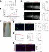
Fig. 2. Increased infiltration with CD4+, CD25+ and FOXP3+ cells, as well as increased CD4, CD25 and CTLA4 expression characterizes lymphedema tissue.
AC Representative immunohistological images and quantification of histological analysis in paraffin skin tissue sections. An increased infiltration of CD4+ (A), CD25+ (B) and FOXP3+ (C) cells was detected in skin sections from lymphedema patients. Arrows indicate positive cells. D The evaluation of the CD4, CTLA4, CD25 and FOXP3 mRNA expression in human subcutaneous fat tissue revealed increased CD4, CD25 and CTLA4 expression levels in patients with lymphedema. N(C) = 9 patients and N(LE) = 12 patients (AD). Line represent mean ± SD of each group. Significance was determined via Welch’s t-test (AC) and ANOVA and the pairwise multiple comparison analysis was corrected for multiple comparisons the method of Benjamini, Krieger and Yekutieli (D). Scale bars: 200 μm.
Fig. 3
Fig. 3. Anti-CTLA4 treatment exhibits efficacy in the lymphedema mouse tail model.
A Surgery was performed on day 0 (D0), treatment was given on day 3, 6, 9 and 12 (D3, D6, D9, D12) and evaluation/endpoint was in day 14 (D14). B Anti-CTLA4 administration (aCTLA4) following the surgical induction of lymphedema leads to significantly reduced edema 1 and 2 weeks (W) after surgery. (N(C) = 10 and N(aCTLA4)=10) C Representative photographs of tails of control and anti-CTLA4 (aCTLA4)–treated mice two weeks postoperatively. D Representative images of near-infrared intravital microscopy of the tail lymphatic network ~1.5 cm distally to the surgical site, visualized through the uptake and transport of a lymphatic-specific fluorescent tracer 10 and 20 min after infusion near the tip of the tail. Quantification of lymphatic vascular transport based on fluorescence intensity revealed significantly increased lymphatic vessel function in the anti-CTLA4 treatment group. Scale bar: 2000 μm E Representative microscopical pictures of the lymphatic vascular morphology, indicating a normalized lymphatic phenotype in the anti-CTLA4–treated group. Scale bar: 250 μm F Graphical representation of lymphatic vessel pulsations in the tail collecting vessels of mice treated with or without aCTLA4 2 weeks. Quantification of pulsation frequency and amplitude is shown to the right G Representative pictures illustrating the significantly increased Foxp3+ cell infiltration in the anti-CTLA4–treated group. Scale bar: 125 μm. All experiments show data from the same 10 control mice (C) and 10 aCTLA4 treated mice (aCTLA4). Line represent mean ± SD of each group. Significance was determined via a 2- way ANOVA followed by a Bonferroni’s multiple comparisons test (B) and Welch’s t-test (D, E, F).
Fig. 4
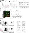
Fig. 4. Anti-CTLA4 treatment does not influence the Th1/Th2 balance but results in a distinct serum cytokine milieu.
A, B The evaluation of the Th1 (Il2, Infγ, Tnfα and Cxcr3) and Th2 (Il10, Il13, Ccr4 and Ccr8) marker mRNA expression in lymphedematous tail skin suggests that the Th1/Th2 immune response balance is not affected after 2 weeks of anti-CTLA4 treatment (aCTLA4) in lymphedema. C Quantification of altered tissue cytokines, indicating that only Il1-α increases locally in the lymphedematous tissue in response to anti-CTLA4 treatment. D Normalized heatmap of serum cytokines. Quantification of significantly altered serum cytokines, indicating the increased levels of Tnfα, Ccl3, Ccl4 and Il5 while the levels of Ccl5 decrease in response to the treatment. (N(C) = 10 mice and N(aCTLA4)=9 mice). E Representative flow cytometry analysis graphs of the phenotypic characterization performed to assess the circulating lymphoid compartment using PBMCs (gated on CD45+/CD11b) in mice treated with anti-CTLA4 versus controls. The analysis indicates comparable CD4+ and CD8+ cell frequencies between the two groups. Representative flow cytometry analysis graphs of the Foxp3+ Treg compartment in murine PBMCs (gated from CD45+CD11bCD4+) upon anti-CTLA4 treatment versus controls. The quantification of the Foxp3+ population between the two groups (gated on CD45+/CD11b/CD4+) revealed increased circulating levels of Foxp3+ Tregs in response to anti-CTLA4 treatment. F Foxp3+ Treg cell number was also increased within the total alive single cell population. N(C) = 10 mice and N(aCTLA4) = 10 mice (AC), N(C) = 10 mice and N(aCTLA4) = 9 mice (D), N(C) = 8 mice and N(aCTLA4) = 8 mice (E, F). Line represent mean ± SD of each group. Significance was determined via a Welch’s t-test (F). and ANOVA and the pairwise multiple comparison analysis was corrected for multiple comparisons with the method of Benjamini, Krieger and Yekutieli (AE).
Fig. 5
Fig. 5. Anti-CTLA4 treatment drives Foxp3+ Treg expansion and triggers distinct immune response pathways in edematous mouse tail tissue.
A, B Representative graphs of the T cell phenotypic characterization using flow cytometry. Upon initial gating for the CD45+CD19-CD11b- populations, the frequency of the CD4+ and CD8+ cells were assessed, indicating an isolated CD4+ cell increase in response to anti-CTLA4 treatment. C Representative graphs of the flow cytometry analysis of the gated CD4+ cells. The phenotypic characterization of the Treg suppressive function using the markers CD25, CTLA4 and CD44 cells revealed a significantly increased proportion of Foxp3+CD25+, Foxp3+CD44+ and Foxp3+CTLA4+ immunosuppresive Tregs within the otherwise expanded CD4+ compartment (N(C) = 7 mice and N(aCTLA4) = 7 mice). D Foxp3+CD25+ Treg cell number was also increased within the total live single cell population. E An increased expression of Ctla4 and Foxp3 is present upon anti-CTLA4 treatment while the CD25 expression levels remained unaltered. F Network plots of Gene Set Enrichment Analysis showed differentially expressed genes associated with an increased immune response and reduced keratinization. G Analysis of activated biological processes in the anti-CTLA4 treated-group, based on Gene Set Enrichment Analysis of the differentially expressed genes. Red represents upregulated biological processes and blue are downregulated biological processes. H List of the 10 most significantly downregulated “toxic pathologies” as described by MetaCore enrichment analysis in the anti-CTLA4 treaded group (bulk RNA Seq: N(C) = 10 mice and N(aCTLA4) = 10 mice). Line represent mean ± SD of each group. Significance was determined via a Welch’s t-test (D, E) and ANOVA and the pairwise multiple comparison analysis was corrected for multiple comparisons with the method of Benjamini, Krieger and Yekutieli (B, C).
Fig. 6
Fig. 6. Anti-CTLA4 treatment leads to the systemic expansion of FOXP3+ Treg cells in humans.
A Representative flow cytometry analysis graphs of the phenotypic characterization performed to assess the circulating lymphoid compartment from human PBMCs (gated on CD45+/CD11b cells) in melanoma patients at Day 0 (Pre-Treatment) and B up to 8 weeks after anti-CTLA4 treatment start (Post-Treatment). C Quantification of the circulating CD4+, CD8+, FOXP3+/CD25+ and CTLA4+ cells in melanoma patients at Day 0 versus Day 40 indicate an isolated systemic expansion of the FOXP3+ Tregs, induced by the anti-CTLA4 treatment. D Increased percentage of FOXP3+CD25+ within all alive single cells upon aCTLA4 treatment. E, F Quantification of Treg-related serum cytokines indicated increased levels of TNFα, IL10, IL17A and IL16, while the levels of MIP1b, IL6, INFγ and CCL2 remained unaltered in response to the treatment. PBMCs from 7 patients pre and post treatment (AD) and serum from 16 patients pre and post treatment (E, F) were analysed. Significance was determined via Welch’s t-test (D) and RM one way ANOVA and the pairwise multiple comparison analysis was corrected for multiple comparisons with the method of Benjamini, Krieger and Yekutieli (C, E, F).

References

    1. Paskett, E. D., Dean, J. A., Oliveri, J. M. & Harrop, J. P. Cancer-related lymphedema risk factors, diagnosis, treatment, and impact: a review. J. Clin. Oncol.30, 3726–3733 (2012). - PubMed
    1. Beaulac, S. M., McNair, L. A., Scott, T. E., LaMorte, W. W. & Kavanah, M. T. Lymphedema and quality of life in survivors of early-stage breast cancer. Arch. Surg.137, 1253–1257 (2002). - PubMed
    1. Cowher, M. S. et al. Conservative axillary surgery in breast cancer patients undergoing mastectomy: long-term results. J. Am. Coll. Surg.218, 819–824 (2014). - PubMed
    1. Lin, Y. et al. Manual lymphatic drainage for breast cancer-related lymphedema: a systematic review and meta-analysis of randomized controlled trials. Clin. Breast Cancer22, e664–e673 (2022). - PubMed
    1. Chang, D. W. et al. Surgical treatment of lymphedema: a systematic review and meta-analysis of controlled trials. results of a consensus conference. Plast. Reconstr. Surg.147, 975–993 (2021). - PubMed

Publication types