. 2024 Dec 30;15(1):10825.
doi: 10.1038/s41467-024-55101-4.

The structural basis of protective and nonprotective human monoclonal antibodies targeting the parainfluenza virus type 3 hemagglutinin-neuraminidase

Affiliations

The structural basis of protective and nonprotective human monoclonal antibodies targeting the parainfluenza virus type 3 hemagglutinin-neuraminidase

Rose J Miller et al. Nat Commun. .

Abstract

Parainfluenza virus 3 (PIV3) infection poses a substantial risk to vulnerable groups including infants, the elderly, and immunocompromised individuals, and lacks effective treatments or vaccines. This study focuses on targeting the hemagglutinin-neuraminidase (HN) protein, a structural glycoprotein of PIV3 critical for viral infection and egress. With the objective of targeting these activities of HN, we identified eight neutralizing human monoclonal antibodies (mAbs) with potent effects on viral neutralization, cell-cell fusion inhibition, and complement deposition. Three epitopes on PIV3 HN were delineated and one epitope, Site 2, elicits a mAb with cross-neutralizing ability against PIV1 and PIV3. Cryo-EM revealed the cross-neutralizing mAb utilizes a long CDR3 loop to bind inside the pocket of the sialic acid binding site. Additionally, we resolved the structure of a non-protective mAb binding to Site 1 near the HN:F-interaction site. The potent Site 2-directed mAb demonstrated clinical efficacy in hamsters, reducing viral replication prophylactically and therapeutically. These findings advance our understanding of PIV3 immunity and underscore the significance of targeting HN for clinical therapeutic development against PIV3.


Conflict of interest statement

Competing interests: J.J.M. is an inventor on a patent application related to anti-parainfluenza virus monoclonal antibodies.

Figures
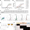
Fig. 1
Fig. 1. Assessment of potent neutralizing mAbs against PIV3 and epitope discovery on the head domain of the PIV3 HN protein.
A ELISA binding curves data displays mAb binding with high affinity to the head domain of HN. Data points are the average of four technical replicates from one experiment and are representative of two biological replicates. Data are presented as mean values +/− SD. B Plaque reduction neutralization test curves for the eight mAbs. Data points are the average of three technical replicates from one experiment and are representative of two biological replicates. Data are presented as mean values +/− SD. C Summary of the half maximal effective concentration (EC50) values and half maximal inhibitory concentration (IC50) values calculated from (A) and (B). D PIV3-infected LLC-MK2 cells were stained with anti-PIV3 HN mAbs PIV3HN-05, and PIV3HN-09 conjugated to PE. Anti-F mAb PIA174 was used as a positive control, and a mAb against human metapneumovirus, MPV467, was used as a negative control. Both anti-HN mAbs and anti-F mAbs were detected in virally infected cells. Data represent the results from one technical replicate and are representative of two biological replicates. E Epitope binning was performed via biolayer interferometry (BLI) by loading His-tagged HN protein onto an anti-HIS sensor, and then associating the sensor with one mAb followed by a second mAb. Created in BioRender. Miller, R. (2024) https://BioRender.com/x68o050. F Three distinct epitopes on the PIV3 HN protein were discovered from the competitive binding to HN by the mAbs. Data indicate the binding of the second antibody to the sensor in the presence of the first antibody. Values in black boxes describe mAbs with high levels of competition (≤ 30), whereas values in white boxes describe mAbs that do not compete for binding (≥ 70).
Fig. 2
Fig. 2. Fusion inhibition of mAbs.
Fusion inhibition of mAbs from each site was assessed by transfecting Vero-SLAM cells with either HN or F and GFP 3 h prior to adding mAb. Syncytia formation was visualized 24 h following transfection and fusion inhibition was evaluated by comparison to the PBS-treated control. Previously identified anti-F mAb PIA174 was used as a positive control. PBS-treated cells were used as a negative control to show standard levels of syncytia. The results shown are images from one technical replicate from one experiment and are representative of 3 biological replicates. The scale bar represents 100 µm.
Fig. 3
Fig. 3. Complement recruitment and PIV1 cross-neutralization capabilities of mAbs.
A Complement deposition was assessed due to its importance in aiding mAbs in preventing viral replication. Complement has several functions, including formation of the membrane attack complex (MAC), and initiation of cell phagocytosis or cellular cytotoxicity. Created in BioRender. Miller, R. (2024) https://BioRender.com/l30r012. B HN-coated beads were stained with individual mAbs before introducing the C3 complement protein. Deposition of the C3b protein to HN-coated beads stained with mAbs was measured by comparison to C3b deposition alone. Data points are the results of three technical replicates from one experiment, and the data are representative of two biological replicates. Data are presented as mean values +/− SD. Statistical significance was calculated using a one-sided ANOVA for multiple comparisons (**P < 0.0086). C PRNT was performed with added guinea pig complement at a 1:1000 dilution. All except two mAbs had increased neutralization potency with added complement, indicated by a decreased IC50 value. Each data point is the IC50 value from one neutralization experiment with or without complement, and the data are representative of two biological replicates. D One mAb from each epitope was evaluated for hemagglutination inhibition (HI) using guinea pig red blood cells. Data are the results from one experiment representative of two biological replicates. E One mAb, PIV3HN-05, cross-neutralized parainfluenza virus type 1 (PIV1) in vitro with an IC50 value of 180 ng/mL. The PIV1/3 cross-neutralizing mAb, 3 × 1, was used as a positive control. Data points are the average of three technical replicates from one experiment and are representative of two biological replicates. Data are presented as mean values +/− SD.
Fig. 4
Fig. 4. Structural insights into the non-protective Site 1 epitope of the PIV3 HN protein.
A X-ray crystal structure displaying PIVHN-13 Fab binding to the dimer interface of the head domain of the PIV3 HN protein was determined to be 2.4 Å. PIV3 HN monomers are shown in steel blue and sea green. The PIVHN-13 heavy chain is shown in brick red, while the light chain is shown in salmon. The previously proposed interaction site between the PIV3 F and PIV3 HN proteins is highlighted in gold and the sialic acid active site is highlighted in magenta. B Structures of the PIVHN-13 Fab, the PIV3 HN dimer, and the pre-fusion PIV3 F (PDB: 6MJZ) protein were fit into the PIV3 F-HN interaction map (EMB: 27550). Site 1 on PIV3 HN contains no interactions with the PIV3 F-PIV3 HN interaction site. C Interactions between the heavy and light chain of PIVHN-13 Fab and both PIV3 HN monomers were identified. Top: CRDH3 forms a hydrophobic pocket surrounded by hydrogen bonds between CRD2 residues Gly58 and Ser64 with PIV3 HN residues Ser122 and Leu124, respectively. Hydrophobic regions are highlighted with yellow translucent surfaces, with decreasing hydrophobicity depicted in teal. Middle: CDRH2 and FRH3 in the Fab heavy chain form various hydrogen bonds with the PIV3 HN monomer. All hydrogen bonds are formed with one monomer except for one between His63 on the heavy chain and Asn139 on the second HN monomer. Bottom: Hydrogen bonds between the light chain of the Fab and one HN monomer are found in the CDRL3 and FRL1. Hydrogen bonds are shown in black dotted lines. Oxygen atoms are colored red, and nitrogen atoms are colored blue.
Fig. 5
Fig. 5. Structural insights into the protective Site 2-specific mAb PIV3HN-05.
A Cryo-EM map of PIV3HN-05 Fab complexed with HN, with PIV3HN-05 Fab binding to the apical surface of HN. B When fit into a map of F (PDB: 6MJZ) and HN interaction (EMB: 27550), the PIV3HN-05 CDRH3 does not spatially interact with the active site of HN but fits directly within the HN active site. C Overlay of the Alphafold3 predicted structure (gray) and experimentally determined structure (HC: indigo, LC: mauve) of PIV3HN-05 Fab complexed to HN. HN monomers are shown in steel blue and sea green. D Interactions of the PIV3HN-05 CDRH3 with the PIV3 HN protein in the sialic acid binding pocket. Two hydrogen bonds are indicated with black dotted lines. Hydrophobic regions are highlighted with yellow translucent surfaces, with decreasing hydrophobicity depicted in teal. HN residues associated with sialic acid receptor interactions are labeled. Difluorosialic acid atoms fit into the HN model (PDB: 4WEF) and are depicted in black.
Fig. 6
Fig. 6. mAb treatment of Syrian hamsters before and after PIV3 challenge.
A Syrian hamsters (n = 10) were treated with 10 mg/kg of one mAb binding to each identified site on HN 24 h prior to the intranasal PIV3 challenge. Lungs were collected four days following the viral challenge. Created in BioRender. Miller, R. (2024) https://BioRender.com/w46b621. B Plaque assays revealed decreased viral load in the lungs with prophylactically PIV05- and PIV09-treated hamsters. Data are presented as mean values. Statistical significance was calculated using a one-sided ANOVA for multiple comparisons (***P = 0.0002, *P = 0.02). C Syrian hamsters (n = 5) were treated 24 hours after the intranasal PIV3 challenge. Lungs were collected four days following the viral challenge. Created in BioRender. Miller, R. (2024) https://BioRender.com/y64f007. D Viral load in the lungs of PIV05 was significantly lower than in the mock-treated group. Data are presented as mean values. Statistical significance was calculated using a one-sided ANOVA for multiple comparisons (**P = 0.007, *P = 0.04).

References

    1. DeGroote, N. P. et al. Human parainfluenza virus circulation, United States, 2011–2019. J. Clin. Virol.124, 104261 (2020). - PMC - PubMed
    1. Human Parainfluenza National Trends - NREVSS | CDC [Internet]. https://www.cdc.gov/surveillance/nrevss/human-paraflu/natl-trend.html (2023).
    1. Branche, A. R. & Falsey, A. R. Respiratory viral infections: Parainfluenza virus infection. Semin Respir. Crit. Care Med.37, 538 (2016). - PMC - PubMed
    1. Cortez, K. J. et al. Outbreak of human parainfluenza virus 3 infections in a hematopoietic stem cell transplant population. J. Infect. Dis.184, 1093–1097 (2001). - PubMed
    1. Matsuse, H. et al. Naturally occurring Parainfluenza virus 3 infection in adults induces mild exacerbation of asthma associated with increased sputum concentrations of cysteinyl Leukotrienes. Int. Arch. Allergy Immunol.138, 267–272 (2005). - PubMed

Publication types

MeSH terms

Associated data

LinkOut - more resources